New study shows bidirectional link between inflammatory bowel disease and depression

Inflammatory bowel disease (IBD) is a chronic condition involving inflammation of the digestive tract, affecting some 1.6 million Americans. Depression affects more than 16 million Americans.
A new study from Keck Medicine of USC shows that patients diagnosed with IBD were nine times as likely to develop depression than the general population. In addition, their siblings who did not suffer from IBD were almost two times as likely to develop depression.
Conversely, patients with depression were two times as likely to develop IBD, and their siblings without depression were more than one and a half times as likely to develop IBD.
“This research reveals a clinical overlap between both conditions, and is the first study to investigate the two-way association between IBD and depression in siblings,” said Bing Zhang, MD, a gastroenterologist with Keck Medicine and co-lead author of the study.
Zhang and his fellow researchers analyzed the data of more than 20 million people from Taiwan’s National Health Insurance Research Database, which contains comprehensive medical information on more than 99% of Taiwanese residents.
For 11 years, they tracked patients with either IBD or depression and their siblings without either condition, comparing onset of depression or IBD with a control group of people without either condition, but with similar age, sex and socioeconomic status.

Read more →

Doctors Transplant 3-D Printed Ear Made of Human Cells

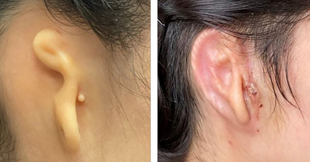
3DBio Therapeutics, a biotech company in Queens, said it had for the first time used 3-D printing to make a body part with a patient’s own cells.A 20-year-old woman who was born with a small and misshapen right ear has received a 3-D printed ear implant made from her own cells, the manufacturer announced on Thursday. Independent experts said that the transplant, part of the first clinical trial of a successful medical application of this technology, was a stunning advance in the field of tissue engineering.The new ear was printed in a shape that precisely matched the woman’s left ear, according to 3DBio Therapeutics, a regenerative medicine company based in Queens. The new ear, transplanted in March, will continue to regenerate cartilage tissue, giving it the look and feel of a natural ear, the company said.“It’s definitely a big deal,” said Adam Feinberg, a professor of biomedical engineering and materials science and engineering at Carnegie Mellon University. Dr. Feinberg, who is not affiliated with 3DBio, is a co-founder of FluidForm, a regenerative medicine company that also uses 3-D printing. “It shows this technology is not an ‘if’ anymore, but a ‘when,’” he said.The results of the woman’s reconstructive surgery were announced by 3DBio in a news release. Citing proprietary concerns, the company has not publicly disclosed the technical details of the process, making it more difficult for outside experts to evaluate. The company said that federal regulators had reviewed the trial design and set strict manufacturing standards, and that the data would be published in a medical journal when the study was complete.The clinical trial, which includes 11 patients, is still ongoing, and it’s possible that the transplants could fail or bring unanticipated health complications. But since the cells originated from the patient’s own tissue, the new ear is not likely to be rejected by the body, doctors and company officials said.3DBio’s success, seven years in the making, is one of several recent breakthroughs in the quest to improve organ and tissue transplants. In January, surgeons in Maryland transplanted a genetically modified pig’s heart into a 57-year-old man with heart disease, extending his life by two months. Scientists are also developing techniques to extend the life of donor organs so they do not go to waste; Swiss doctors reported this week that a patient who received a human liver that had been preserved for three days was still healthy a year later.United Therapeutics Corp., the company that provided the genetically engineered pig for the heart procedure, is also experimenting with 3-D printing to produce lungs for transplants, a spokesman said. And scientists from the Israel Institute of Technology reported in September that they had 3-D printed a network of blood vessels, which would be necessary to supply blood to implanted tissues.Dr. Arturo Bonilla, a pediatric ear reconstructive surgeon in San Antonio who performed the surgery. “If everything goes as planned, this will revolutionize the way this is done,” he said.Sergio Flores for The New York TimesCompanies have previously used 3-D printing technology to produce custom-fit prosthetic limbs made of plastic and lightweight metals. But the ear implant, made from a tiny glob of cells harvested from the woman’s misshapen ear, appears to be the first known example of a 3-D printed implant made of living tissues.The patient, who is from Mexico, was born with microtia, a rare birth defect that causes the auricle, or external part of the ear, to be small and malformed (it also can affect hearing in the ear). With more research, company executives said, the technology could be used to make many other replacement body parts, including spinal discs, noses, knee menisci, rotator cuffs and reconstructive tissue for lumpectomies. Further down the road, they said, 3-D printing could even produce far more complex vital organs, like livers, kidneys and pancreases.“This is so exciting, sometimes I have to temper myself a little bit,” said Dr. Arturo Bonilla, a pediatric ear reconstructive surgeon in San Antonio who performed the woman’s implant surgery. The trial was funded by 3DBio Therapeutics, but Dr. Bonilla does not have any financial stake in the company. “If everything goes as planned, this will revolutionize the way this is done,” he said.James Iatridis, who heads a spine bioengineering laboratory at Mount Sinai’s Icahn School of Medicine, said that other 3-D printed tissue implants were in the pipeline, but that he was unaware of any other products being tested in a clinical trial.“The 3-D ear implant is then a proof of concept to evaluate biocompatibility, and shape matching and shape retention, in living people,” Dr. Iatridis said.Still, the external part of the ear is a relatively simple appendage that is more cosmetic than functional, said Dr. Feinberg of Carnegie Mellon. He cautioned that the path toward solid organs — like livers, kidneys, hearts and lungs — was still a long one. “Just going from an ear to a spinal disc is a pretty big jump, but it’s more realistic if you’ve got the ear,” he said.The 3-D printing manufacturing process creates a solid, three-dimensional object from a digital model. The technology generally involves a computer-controlled printer depositing material in thin layers to create the precise shape of the object.The new ear implant from 3DBio Therapeutics integrates several proprietary technologies, executives said, beginning with a method for turning a small sample of a patient’s cells into billions of cells. The company’s 3-D printer uses a collagen-based “bio ink” that is safe in the body and that keeps all of the materials sterile.A worker at 3DBio Therapeutics in Long Island City, Queens, applied nutrients to a sample ear implant in a clean room.Andres Kudacki for The New York TimesAll of the research and manufacturing takes place under one roof, in a new, unassuming brick building in Queens equipped with hygienic “clean rooms” that are staffed by masked technicians wearing bunny suit coveralls, gloves and bootees.“It comes in as a biopsy from the patient, and it leaves a living ear,” Daniel Cohen, 3DBio’s chief executive and co-founder, explained during a tour of the facility last week.The patient who received the new ear was one of the first to have had a successful transplant as part of the clinical trial led by Dr. Bonilla. (Trial volunteers are also being enrolled at Cedars-Sinai Medical Center in Los Angeles.)The surgeon began by removing half of a gram of cartilage from the woman’s microtia ear remnant. He then shipped that, along with a 3-D scan of her healthy ear, from San Antonio to the 3DBio building in Long Island City, Queens.At the facility, the patient’s chondrocytes — cells responsible for cartilage formation — were isolated from the tissue sample and grown in a proprietary slurry of nutrients, proliferating into billions of cells.The living cells were then mixed with the company’s collagen-based bio-ink, “like chocolate chips mixed into cookie dough ice cream,” according to Nathaniel Bachrach, 3DBio’s chief scientific officer.The collagen was inserted through a syringe into the specialized 3-D bio-printer, which squirted out the material from a nozzle in a steady, thin stream, zipping around to create a small oblong shape that was a mirror replica of the patient’s healthy ear. The entire printing process lasted less than 10 minutes.The printed ear shape was then encased in a protective biodegradable shell and shipped overnight in cold storage to Dr. Bonilla. He then implanted the ear under the patient’s skin, just above her jawbone. When the skin was tightened around the implant, the shape of an ear emerged.A sample ear implant made by 3DBio Therapeutics.Andres Kudacki for The New York TimesRoughly 1,500 babies born in the United States each year have either microtia or a related condition, anotia, in which the entire external ear is missing. The clinical trial so far has included 11 volunteers, ages 6 to 25, who will be followed for five years to evaluate long-term safety and aesthetic outcomes.Another option for microtia reconstruction — which is often done while the patients are young so they aren’t subjected to bullying or ridicule when they start school — requires inpatient surgery to harvest cartilage from the patient’s ribs, which is then carved into an approximate shape of the ear, Dr. Bonilla said.But the new procedure can be done in a few hours, and outside of a hospital. 3DBio did not specify what it would charge for the implant, but a spokeswoman said the pricing would be in line with the cost of the current standard of care.“I’ve always felt the whole microtia world has been waiting for a technology where we wouldn’t have to go into the chest, and patients would heal from one day to the next,” Dr. Bonilla said.The patient, Alexa, who asked to be identified only by her first name because of privacy concerns, said she was excited about the new ear, even though it was still covered by a bandage. Though many children with microtia are teased by their peers, which can lead to anxiety, depression and hostility, Alexa said she was never very bothered by it until her teen years, when she became more self-conscious about her appearance.“You care a little more for your image when you’re a teenager,” she said. “Some people said things that were not thoughtful, and it started bothering me.”Alexa said she had perfected the art of covering her right ear by wearing her hair long and loose, and most people could not even tell that she was missing an ear. But now, she said, she is looking forward to having fun with her hair, putting it back in pigtails or up in a bun.“I think my self-esteem will go up,” she said.

Read more →

Reprogrammed Cells Attack and Tame Pancreatic Cancer in One Woman

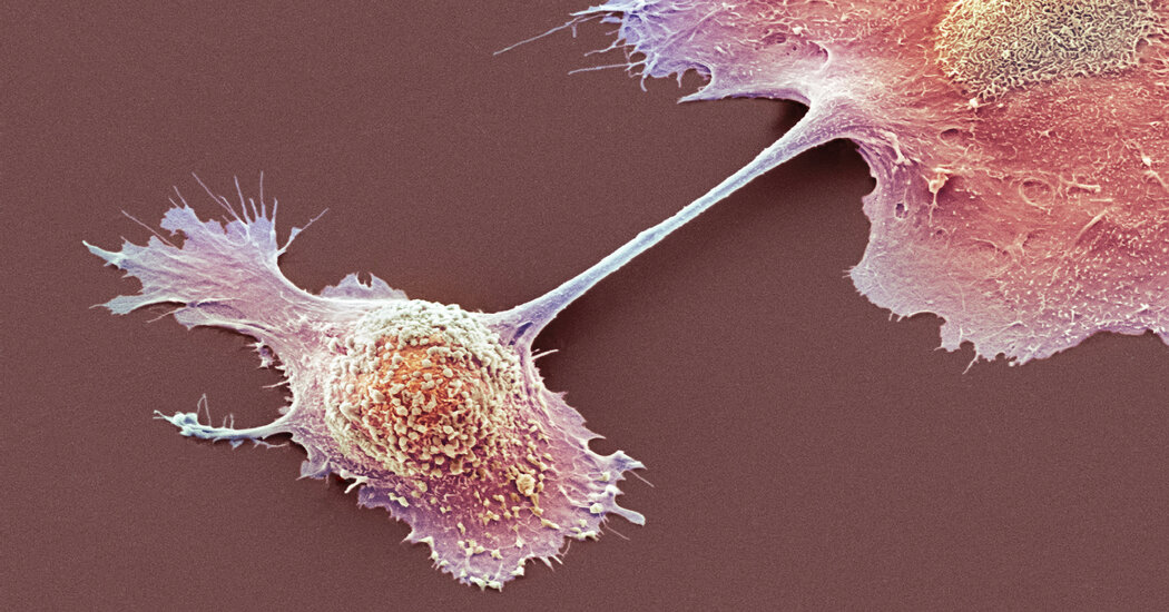
Another patient who had the same treatment did not survive. But the demonstration of the technique could help with other cancers.Researchers have managed to tame pancreatic cancer in a woman whose cancer was far advanced and after other forms of treatment had failed.The experiment that helped her is complex and highly personalized and is not immediately applicable to most cancer patients. Another pancreatic cancer patient, who received the same treatment, did not respond and died of her disease.Nonetheless, a leading journal — The New England Journal of Medicine — published a report of the study on Wednesday.Dr. Eric Rubin, the journal’s editor in chief called the proof of concept experiment “an important step along the way” to devising similar treatments that might be applicable to lung, colon and other cancers.The experiment involved genetically reprogramming the patient’s T cells, a type of white blood cell of the immune system, so they can recognize and kill cancer cells. The technique was developed by Eric Tran and Dr. Rom Leidner of the Earle A. Chiles Research Institute, a division of Providence Cancer Institute in Portland, Ore.To turn a cancer patient’s T cells into a living drug, the researchers had to overcame serious challenges. Pancreatic cancer is one of the most difficult to treat. While new treatments have allowed patients with other cancers to live longer and to have a better quality of life, pancreatic cancer has stubbornly resisted these advances. Less than 10 percent of patients live past five years.For most patients, said Dr. William Jarnagin, a pancreatic cancer specialist at Memorial Sloan Kettering Cancer Center, who was not involved in the current experiment, the cancer has already spread by the time it is discovered. Even when the tumors are caught in the pancreas and surgically removed, about 85 percent of patients have recurrences.“Our treatments are not doing the job,” Dr. Jarnagin said.The technique described in the new paper, “is not off-the-shelf,” Dr. Tran said. He added that “it takes specialized facilities and expertise to manufacture the T cells.”But, Dr. Leidner said, “the beauty of it” is that the reprogrammed T cells will only attack cancer cells. Other cells will be left alone.The first problem in trying to entice T cells to kill cancer cells is that mutated proteins that drive the growth of cancer are hidden inside cells.There is, though, a hint to the immune system that the cancer cells are abnormal. They contain fragments of mutated cancer proteins on their surface, “kind of like molecular bread crumbs,” Dr. Leidner said. The challenge was to get T cells to see those crumbs.The solution employed was to collect the patient’s own T cells and genetically modify them in the lab to recognize and attach to those bits of mutated proteins. Then the T cells were infused back into the patient.In this case the target was KRAS, a mutated protein implicated in 25 percent of all cancers, including about 95 percent of pancreas cancers, 40 percent of colon cancers and a third of lung cancers.“Folks have been trying to target KRAS immunologically for more than 20 years,” said Dr. Robert Vonderheide, a pancreatic cancer specialist and director of the University of Pennsylvania’s Abramson Cancer Center.The mutated KRAS gene “is such a bull’s-eye,” Dr. Vonderheide said, that killing cancer cells by attacking cells with KRAS mutations has “major implications.”But the encouraging result comes with some real caveats. For starters, it is not clear why the other patient who died did not respond to the therapy.Dr. Elizabeth Jaffee, a pancreatic cancer specialist at Johns Hopkins Medicine also highlighted the location of the patient’s metastases, or where the cancer had spread to. Metastases arose only in the patient’s lungs. Most pancreatic cancer patients have metastases in their liver that are more difficult to treat.“I would like to see liver lesions go away,” Dr. Jaffee said.Kathy Wilkes, the patient who was successfully treated, is 71 and lives in Ormond-by-the-Sea, Fla. It is too soon to know if the cancer will come roaring back.New Developments in Cancer ResearchCard 1 of 6Progress in the field.

Read more →

Brain cell activity plays critical role in central nervous system disorder outcomes

Investigators at Cedars-Sinai have comprehensively mapped molecular activity in the brain and spinal cord that is responsible for regulating the body’s response to central nervous system (CNS) disorders such as Alzheimer’s, Huntington’s disease and spinal cord injuries.
The research focused on cellular changes in astrocytes, a specialized support cell type in the brain and spinal cord. These cellular changes, known collectively as “reactivity,” play a critical role in regulating outcomes for central nervous system disorders.
This is the first time a team of scientists has provided evidence demonstrating that astrocytes use specialized collections of molecules called transcriptional regulators to shape disorder-specific changes in their molecular profiles.
The discovery, detailed in the peer-reviewed journal Nature, can help lead to the development of a broad range of new therapies that target specific astrocyte activity to help treat a variety of central nervous system conditions, including multiple sclerosis and stroke.
“There is a growing interest in targeting astrocyte reactivity as treatment strategies for CNS disorders,” said Joshua Burda, PhD, lead and co-corresponding author of the study, and assistant professor in the Department of Biomedical Sciences and the Department of Neurology. “Understanding how different kinds of astrocyte responses are coordinated and the consequences of manipulating those responses not only will help us better understand diseases of the central nervous system but can provide crucial insights that enable the development of better therapies for these conditions.”
Astrocyte reactivity is a hallmark of virtually all nervous system injuries and diseases. Yet, there is still little understanding of what astrocyte reactivity is, what causes it, how it differs across disorders, and how these differences are regulated.

Read more →

Investigators ID gene critical to human immune response

Cedars-Sinai investigators have identified a genethat plays an essential role in the innate human immune system. The gene, NLRP11, helps activate the inflammatory response that tells the body’s white blood cells to go on the attack against a foreign presence.
The findings, published in Nature Immunology, bring medical science closer to understanding a biological process that can both help and harm the body.
“Chronic inflammation is an underlying cause of innumerable human diseases,” said Christian Stehlik, PhD, a co-senior author of the study and director of Pathology Research at Cedars-Sinai. “If you study the molecular mechanisms involved in how inflammation occurs and how it is regulated, you find something that can be applied very broadly.”
When the immune system senses a bacteria, virus, toxin or other foreign presence in the body, it sends white blood cells to surround the unwanted substance and release chemicals to attack it. This response leads to inflammation, which causes redness, pain, warmth and swelling in the affected area as the body heals itself. Sometimes this defensive response lasts longer than it should, resulting in chronic inflammation. Or, the immune system may mistakenly attack healthy cells, leading to autoimmune disease.
“Acute inflammation is necessary and beneficial to eradicate infection and initiate wound healing,” said Andrea Dorfleutner, PhD, co-senior author of the study and associate professor in the departments of Academic Pathology and Biomedical Sciences at Cedars-Sinai. “Chronic, long-term, uncontrolled inflammation, however, is detrimental and can damage the body’s organs and tissues.”
The key to controlling the inflammatory response and preventing chronic inflammation may lie in being able to influence the expression of the NLRP11 gene.
The investigators used a gene-editing system called CRISPR/Cas9 to remove genes or introduce gene mutations in human white blood cells called macrophages. They observed that when they deleted NLRP11, it prevented an immune system sensor called the NLRP3 inflammasome from being activated and launching the inflammatory response.
When the investigators restored the NLRP11 gene, the NLRP3 inflammasome sent its attack signals, which triggered the typical inflammatory process. The investigators chose to focus on this gene in particular because it is not expressed in mice, which led them to hypothesize that it was integral to the complex immune system that exists in humans.
“Now that we have a better picture of the mechanisms behind inflammation, we can come up with completely new strategies to target it that have not been possible before,” Dorfleutner said.
The first authors of the study are Anu Gangopadhyay, Savita Devi, PhD, and Shivendra Tenguria, PhD, all investigators in the Stehlik and Dorfleutner Laboratory.
Funding: The study was funded by the National Institutes of Health (award numbers AI099009, AR064349, AI134030, AI140702, AI165797, and AI120625) and the American Heart Association (award number 834502).
Story Source:
Materials provided by Cedars-Sinai Medical Center. Note: Content may be edited for style and length.

Read more →

A stem cell model could help unravel the complex biology behind some psychiatric disorders

Researchers from the Icahn School of Medicine at Mount Sinai have applied a novel stem cell model to map disease risk variants in human neurons, which could help provide insights into the biological mechanisms that underlie neuropsychiatric disorders such as autism and schizophrenia.
The team’s in vitro cellular model, described in the May 31 issue of Cell Reports, is designed to enable future researchers to elucidate the disease mechanisms involving genome-wide association studies (GWAS) that characterize different risk alleles (common genetic variants conferring risk) for psychiatric disorders. This research could potentially lead to improved diagnostics for the detection of psychiatric disorders years before symptoms appear in patients.
The study focuses on mapping cis-regulatory elements in human neurons that can be linked to psychiatric disease heritability. Cis-regulatory elements, such as promoters and enhancers, are non-coding DNA sequences regulating gene expression and are thus vital components of the genetic regulatory network. Previous genetic studies have revealed a significant enrichment of common variants in the cis-regulatory elements, including those associated with autism spectrum disorder, schizophrenia, and bipolar disorder.
“While common risk variants can shed light on the underlying molecular mechanism, identifying causal variants remains challenging for scientists,” says Nan Yang, PhD, Assistant Professor of Neuroscience at the Icahn School of Medicine of Mount Sinai, and senior author of the study. “That’s because cis-regulatory elements, particularly the enhancers, vary across cell types and activity states. Typically, researchers can only use postmortem brain samples where the neurons are no longer active. As a result, they are likely to miss enhancers that only respond to stimulation. Our approach is to map cis-regulatory elements in human neurons derived from pluripotent stem cells. That allows us to replicate neurons in the human brain that can be affected by different types of neuropsychiatric disease, and conduct mechanistic studies of human genetic variants that are inaccessible from other types of human samples.”
In recent years, GWAS have identified hundreds of gene regions associated with psychiatric disease, though understanding disease pathophysiology has been elusive. The functional genomics approach Dr. Yang and her team developed uses stem cell models that can help resolve the impact of patient-specific variants across cell types, genetic background, and environmental conditions. This unique approach effectively lays a foundation to translate risk variants to genes, genes to pathways, and pathways to circuits that reveal the synergistic relationship between disease risk factors within and between the cell types in the brain.
“Our research attempts to decode and transfer highly complex genetic insights into medically actionable information,” says Dr. Yang, who is a member of the Black Family Stem Cell Institute, The Friedman Brain Institute, and The Ronald M. Loeb Center for Alzheimer’s Disease within the Mount Sinai Health System. “That means improving our diagnostic capabilities, predicting clinical trajectories, and identifying presymptomatic points of therapeutic intervention for psychiatric disorders.”
By characterizing cell-type specific and activity-regulated gene expression patterns in human cell-derived neurons, Dr. Yang believes her team’s study can greatly benefit the research community. “Our data can guide choosing relevant cell types of experimental conditions to further elucidate molecular mechanisms of disease across the genome,” she points out. “And that could lead to the development of biomarkers that might detect neuropsychiatric disorders years before they manifest themselves in patients, while there is still time to delay or possibly prevent them.”

Read more →

High fat diet, unregulated athletic exercise endurance enhancers linked to risk of pancreatic cancer

Researchers at the University of Michigan Rogel Cancer Center have found a cell nuclear receptor activated by high fat diets and synthetic substances in unregulated athletic performance enhancers fuels the progression of precancerous pancreas lesions into pancreatic cancer.
Pancreatic ductal adenocarcinoma is a highly lethal form of cancer with rising occurrence, and strategies to prevent and treat the disease are urgently needed. Most cases of pancreatic cancer arise from pre-cancerous lesions called pancreatic intraepithelial neoplasia; about 55-80% of adults over 40 are estimated to have these low grade pre-cancerous silent pancreatic lesions. A study published in Nature Communications, led by Imad Shureiqi, M.D., shows that, pre-cancerous pancreatic lesions in mice, similar to those found in humans, contain higher levels of the transcriptional receptor peroxisome proliferator activated receptor-delta (PPARδ).
PPARδ regulates the expression of a wide spectrum of key genes that influences biological processes like lipid metabolism and cancer formation. Activation of PPARδ dramatically accelerates the progression of pre-cancerous lesions into pancreatic cancer. Shureiqi previously worked at MD Anderson Cancer Center at the University of Texas where he conducted much of this study, specifically in partnership with Xiangsheng Zuo, M.D., Ph.D., before moving his research to the cancer center in 2020.
“We became interested in studying the effects of PPARδ on pancreatic carcinogenesis because our prior observations showed that PPARδ strongly promoted other gastrointestinal cancers. But there’s very limited information about PPARδ’s role in pancreatic cancer’s development,” said Shureiqi.
Activation of PPARδ correlates with excessive exposure to certain ligands, both natural and synthetic. Some ligands naturally occur in high fat diets, which have been associated with increased risk for pancreatic cancer in humans and animal models. High fat diets are enriched with fatty acids that are natural ligands of PPARδ.
Other synthetic forms of PPARδ ligands, like Cardarine (GW501516), are found in exercise supplements, aimed to boost physical performance and endurance. GW501516 was originally designed by pharmaceutical companies to encourage the body to use more fat and treat noncancerous conditions like obesity and hyperlipemia. Pharmaceutical development of GW501516 and other similar potent PPARδ agonists for medical use has long been discontinued given their potential procancerous side effects. Though studies on how PPARδ affects colorectal cancer date back to 1999, and pharmaceutical companies have halted synthetic PPARδ ligand development, unregulated internet outlets still sell substances like Cardarine. Ads are largely marketed to young people, claiming it will help them build muscle endurance and burn fat.
Shureiqi explains that, initially, researchers found that these synthetic ligands reduced fatigue in mice. This news made its way to major media outlets, who nicknamed it “exercise in a pill.” “Unfortunately, what the media didn’t address was the dark side of PPARδ. Like muscle cells, synthetic PPARδ ligands also help cancer cells get more energy from fats as a fuel source,” he said.
“It’s shocking to me,” Shureiqi continued. “Animal models repeatedly show the strong relationship between PPARδ and cancer promotion in the case of colorectal cancer and stomach cancer. Now we’re gaining more information about how it affects pancreatic cancer.”
Critical factors that promote the progression of silent pancreatic precancerous lesions to pancreatic cancer remain poorly defined, especially those that are easy to target. While most of these pre-cancerous lesions don’t develop into cancer, understanding how they progress is still crucial to finding interventions to address the rising rate of pancreatic cancer. Findings from this study indicate that people who have silent precancerous lesions, even those that are low grade, could increase their risk of developing pancreatic cancer by consuming PPARδ natural activators, like in high fat diets, or synthetic ones, like Cardarine. Future development of effective agents to block PPARδ activation could be a new approach to prevent the progression of precancerous lesions into pancreatic cancer. Limiting exposure to high fat diets could also be considered for those with a high prevalence of pre-cancerous pancreatic lesions. But for now, the prevalent sales and use of those athletic boosting synthetic PPARδ activating substances causes the most pressing concern.
“This new information should alert individuals to the potential serious health risks from using synthetic PPARδ agonists,” Shureiqi said. “We’re trying to spread the message that’s using those substances is not a good idea. It might enhance muscle endurance, but it also enhances cancer’s ability to use energy and grow.”
Funding: National Cancer Institute grants R01CA266223, R01CA142969, R01CA195686, R01-CA206539, R01CA236905 R03CA235106, K08CA234222; the Cancer Prevention and Research Institute of Texas grants RP150195 RP140224;, DDC seed fund. This study made use of the MD Anderson Cancer Center Genetically Engineered Mouse Facility, Functional Genomics Core, Flow Cytometry and Cellular Imaging Facility, the Next Generation Sequencing Core and Research Animal Support Facility — Smithville Laboratory Animal Genetic Services, supported by Cancer Center Support Grant P30CA016672. The Next Generation Sequencing Core was also supported by CPRIT Core Facility Support Grant RP120348.

Read more →

New blood test can help doctors diagnose tuberculosis and monitor treatment

Researchers at Tulane University School of Medicine have developed a new highly sensitive blood test for tuberculosis (TB) that screens for DNA fragments of the Mycobacterium tuberculosis bacteria that causes the deadly disease.
The test could give doctors a new tool to both quickly identify TB and then gauge whether drug treatments are effective by monitoring levels of DNA from the pathogen circulating through the bloodstream, according to a new study published in the journal The Lancet Microbe.
Tuberculosis is now the second most deadly infectious disease in the world, behind only COVID-19. In 2020, an estimated 10 million people contracted TB and 1.5 million people died from it, according to the World Health Organization.
Most TB tests rely on screening sputum, a thick type of mucus from the lungs. But collecting sputum from patients suspected of having TB can be difficult, especially for children. TB can also be harder to diagnose in immunocompromised HIV patients and others where the infection migrates outside of the lungs into other areas of the body. In these extrapulmonary cases, patients can have little bacteria in the sputum, which leads to false negatives using current testing methods, said lead study author Tony Hu, PhD, Weatherhead Presidential Chair in Biotechnology Innovation at Tulane University.
“This assay may be a game-changer for TB diagnoses that not only provides accurate diagnosis results but also has the potential to predict disease progression and monitor treatment,” Hu said. “This will help doctors rapidly intervene in treatment and reduce the risk of death, especially for children living with HIV.”
The study evaluated a CRISPR-based assay that screened for cell-free DNA from live Mycobacterium tuberculosis bacilli. The screening target is released into the bloodstream and cleared quite rapidly, providing a real-time snapshot of active infection.

Read more →

Why ketamine is a speedster antidepressant

Ketamine is the speedster of antidepressants, working within hours compared to more common antidepressants that can take several weeks. But ketamine can only be given for a limited amount of time because of its many side effects.
Now, a new Northwestern Medicine study identifies for the first time exactly how ketamine works so quickly, and how it might be adapted for use as a drug without the side effects.
The study in mice shows ketamine works as a rapid antidepressant by increasing the activity of the very small number of newborn neurons, which are part of an ongoing neurogenesis in the brain.
New neurons are always being made at a slow rate. It’s been known that increasing the number of neurons leads to behavioral changes. Other antidepressants work by increasing the rate of neurogenesis, in other words, increasing the number of neurons. But this takes weeks to happen.
By contrast, ketamine produces behavioral changes simply by increasing the activity of the existing new neurons. This can happen immediately when the cells are activated by ketamine.
“We narrowed down the population of cells to a small window that is involved,” said lead study author Dr. John Kessler, a professor of neurology at Northwestern University Feinberg School of Medicine and the Ken and Ruth Davee Professor of Stem Cell Biology. “That’s important because when you give ketamine to patients now, it affects multiple regions of the brain and causes a lot of adverse side effects. But since we now know exactly which cells we want to target, we can design drugs to focus only on those cells.”
The side effects of ketamine include blurred or double vision, nausea, vomiting, insomnia, drowsiness and addiction.

Read more →

Consumers embrace milk carton QR codes, may cut food waste

The “use-by” and “best-by” dates printed on milk cartons and gallon jugs may soon become a thing of the past, giving way to more accurate and informative QR codes. A new Cornell University study finds that consumers will use the QR codes — to better depict how long the milk is drinkable and create substantially less agricultural and food waste.
In the U.S., dairy products are among the top three food groups with the largest share of wasted food, said Samantha Lau, a doctoral student in food science who works in the lab of Martin Wiedmann, the professor of food safety in the College of Agriculture and Life Sciences.
In the early spring semester, Lau, also working with Cornell’s Milk Quality Improvement Program, connected with the Cornell Dairy Bar — which sells fluid milk in addition to ice cream on campus. She wanted to assess consumer acceptance for QR code technology that may one day replace the static best-by or sell-by dates commonly found on food products.
Customers had a choice: purchasing milk with printed best-by dates or buying containers with QR codes, which, when scanned by a smart phone, would display the best-by date.
In the same Cornell Dairy Bar study, Lau placed a dynamic pricing element where consumers were encouraged to purchase milk with a shorter remaining shelf life — by offering a price discount as the best-by date approached.
“During two-month study, over 60% of customers purchased the milk with the QR code, showing a considerable interest in using this new technology,” Lau said. “This revealed that the use of QR codes on food products can be an innovative way to address the larger issue of food waste.”
For fluid milk, ensuring quality and accurately portraying an expected shelf life is key, but microbial spoilage is a major contributor to food loss and waste globally, said Lau, the lead author of a related scientific paper published earlier this spring.

Read more →