Covid’s risks are concentrated among Americans of Biden’s age.

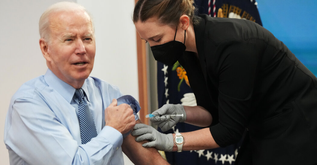
President Biden is 79, and Americans his age and older have made up larger and larger shares of those dying from Covid in recent months. The virus has taken advantage of falling immunity caused by long delays since older people’s last vaccinations, and the Omicron variant has evolved a growing ability to skirt the body’s defenses.Covid has been killing substantially fewer Americans of all ages this summer than it did during the peak of the wintertime Omicron wave. Still, older people remain at significantly higher risk.As of early June, four times as many Americans aged 75 to 84 were dying each week from the virus, compared with people two decades younger, according to data from the Centers for Disease Control and Prevention. (Those death counts are provisional, the C.D.C. cautioned, because they were based on death certificates and did not account for all deaths in those age groups.)That is an even bigger age gap than existed at the peak of the Omicron wave this winter. Then, the number of people aged 75 to 84 killed by Covid each week was twice as high as the number aged 55 to 64.The president received a second booster shot in late March, significantly reducing his risk of severe illness. This spring, people aged 50 and older who had received a single booster were dying from Covid at four times the rate of those with two booster doses, the C.D.C. has reported.In 2022, Covid deaths, though always concentrated in older people, have skewed toward older people more than they did at any point since vaccines became widely available. Many older people were vaccinated early in 2021, and among those who have not yet received a booster shot, immune defenses generated by the shots have significantly waned.In contrast, middle-aged Americans, who suffered a large share of pandemic deaths last summer and fall, are benefiting from greater stores of immune protection from both vaccination and prior infections.While Covid deaths remain far lower than in the winter, they are climbing again among older people as the immune-evasive Omicron subvariant known as BA.5 causes more infections, according to the latest C.D.C. data. From early May to early June, the number of Americans aged 75 to 84 dying from Covid each week increased by nearly 50 percent.

Read more →

Why Biden’s Second Booster Wasn’t Enough to Prevent Infection

President Biden’s coronavirus infection is a stark illustration that the Covid vaccines, powerful as they are, are far from the bulletproof shields that scientists once hoped for.Mr. Biden has received multiple doses of the Pfizer-BioNTech vaccine; his most recent shot, a second booster, was on March 30. Studies suggest that those doses will provide a powerful bulwark against severe illness — and indeed, the president has only mild symptoms so far after testing positive on Thursday, according to the White House.But even booster doses offer little defense against infection, particularly with the most recent versions of the virus. What little protection they do offer wanes sharply and quickly, several studies have shown. In the president’s case, the booster shot he received nearly four months ago is likely to have lost most of its potency at preventing infection.Earlier in the pandemic, experts believed that the vaccines would be enough to forestall not just severe disease, but also the vast majority of infections. And that was true when earlier versions of the virus, including the Delta variant, swept the globe.But the Omicron variant upended those hopes. As more of the population gained some immunity, whether from infection or vaccines, the virus evolved to dodge those defenses. BA.1, the subvariant of Omicron that circulated over the winter, was adept at causing infections even in those who had received a booster dose of vaccine just weeks earlier.Each subsequent avatar of the virus has become still better at sidestepping immunity. BA.5, which now accounts for nearly 80 percent of cases in the United States, is the most wily yet. Detailed data collected in Qatar suggests that immunity from previous infection and vaccines is weakest against BA.5 compared with its predecessors.BA.5 is also highly contagious. The nation is recording roughly 130,000 cases per day on average; that number is likely to be a huge underestimate, because most people test at home or do not test at all.The number of hospitalizations has also spiked over the past few weeks, although BA.5 does not appear to cause more severe disease than other forms of Omicron.Given how much the virus has changed, the administration has been debating the value of authorizing additional shots of the original vaccine in the fall, and offering second boosters to adults younger than age 50. An advisory panel of the Food and Drug Administration said last month that the vaccine manufacturers should make shots tailored to the newest variants.But it’s unclear whether those shots will arrive in time to forestall a fall surge, and whether the virus will have once again evolved beyond their reach.

Read more →

Early Alzheimer's detection up to 17 years in advance

A sensor identifies misfolded protein biomarkers in the blood. This offers a chance to detect Alzheimer’s disease before any symptoms occur. Researchers intend to bring it to market maturity.
Alzheimer’s disease has a symptom-free period of 15 to 20 years before the first clinical symptoms emerge. Using an immuno-infrared sensor developed in Bochum, a research team is able to identify signs of Alzheimer’s disease in the blood up to 17 years before the first clinical symptoms appear. The sensor detects the misfolding of the protein biomarker amyloid-beta. As the disease progresses, this misfolding causes characteristic deposits in the brain, so-called plaques.
“Our goal is to determine the risk of developing Alzheimer’s dementia at a later stage with a simple blood test even before the toxic plaques can form in the brain, in order to ensure that a therapy can be initiated in time,” says Professor Klaus Gerwert, founding director of the Centre for Protein Diagnostics (PRODI) at Ruhr-Universität Bochum. His team cooperated for the study with a group at the German Cancer Research Centre in Heidelberg (DKFZ) headed by Professor Hermann Brenner.
The team published the results obtained with the immuno-infrared sensor in the journal Alzheimer’s & Dementia: The Journal of the Alzheimer’s Association on 19 July 2022. This study is supported by a comparative study published in the same journal on 2 March 2022, in which the researchers used complementary single-molecule array (SIMOA) technology.
Early detection of symptom-free people with a high risk of Alzheimer’s disease
The researchers analysed blood plasma from participants in the ESTHER study conducted in Saarland for potential Alzheimer’s biomarkers. The blood samples had been taken between 2000 and 2002 and then frozen. At that time, the test participants were between 50 and 75 years old and hadn’t yet been diagnosed with Alzheimer’s disease. For the current study, 68 participants were selected who had been diagnosed with Alzheimer’s disease during the 17-year follow-up and compared with 240 control subjects without such a diagnosis. The team headed by Klaus Gerwert and Hermann Brenner aimed to find out whether signs of Alzheimer’s disease could already be found in the blood samples at the beginning of the study.

Read more →

Natural food more mouth-watering to children than processed fare

Children are more likely to prefer foods they believe to be natural to human-made options, rating them higher for tastiness, safety and desirability, a study shows.
Researchers say the tendency in adults to prefer natural food is well documented. However, the latest findings found this food bias exists in early and middle childhood as well.
Researchers at the Universities of Edinburgh and Yale studied the preferences of more than 374 adults and children in the United States when presented with apples and orange juice and told of their origins.
In one study, 137 children aged six to 10 years old were shown three apples. They were told one was grown on a farm, one was made in a lab, and another grown on a tree inside a lab.
The team used questionnaires and statistical models to assess the children’s apple preferences in terms of perceived tastiness, perceived safety and desire to eat. Adults took part in the same study to compare age groups.
Both children and adults preferred apples they believed were grown on farms to those grown in labs, researchers found.

Read more →

Software program allows simultaneous viewing of tissue images through dimensionality reduction

Imaging of tissue specimens is an important aspect of translational research that bridges the gap between basic laboratory science and clinical science to improve the understanding of cancer and aid in the development of new therapies. To analyze images to their fullest potential, scientists ideally need an application that enables multiple images to be viewed simultaneously. In an article published in the journal Patterns, Moffitt Cancer Center researchers describe a new open-source software program they developed that allows users to view many multiplexed images simultaneously.
There have been significant improvements in the approaches to study cancer over the past decade, including new techniques to study tissue samples. For example, machines can now be programmed to stain hundreds of slides simultaneously, or alternatively, up to 1,000 different tissue sample cores can be placed on a single slide and stained for biomarkers at the same time. With the advent of these approaches comes a wealth of possibilities to generate new data and information. Due to the magnitude of this information and the complex nature of cancer itself, computational modeling and software are needed to view and study the cancer biomarkers, tissue architecture, and cellular interactions among these samples.
As researchers in Moffitt’s Integrated Mathematical Oncology Department (IMO) were working on a project, they realized that the currently available software for image viewing was not amenable to their needs.
“We were interested in understanding the underlying spatial patterns between tumor and immune cells and how the tumors were organized. This required us to compare multiple images simultaneously and we realized there was no software, free or commercial, enabling this,” said Sandhya Prabhakaran, Ph.D., lead author and applied research scientist at Moffitt.
The IMO team decided to create a software program that would enable them to view multiple images at the same time and extract data through additional analyses that could be used for a variety of purposes, including identifying biomarkers and understanding tissue architecture and the spatial organization of different cell types. Their program, called Mistic, takes information from multidimensional images and uses dimensionality reduction methods called t-distributed stochastic neighbor embedding (t-SNE) to abstract each image to a point in reduced space. Mistic is an open-source software that can be used with images from Vectra, CyCIF, t-CyCIF and CODEX.
In their publication, the researchers describe the creation of Mistic and some of the applications that it could be used for. For example, they demonstrated that the software could be used to view 92 images from patients with non-small cell lung cancer and deduce how biomarkers cluster across patients with different responses to treatment. In another example, the researchers used Mistic combined with statistical analysis to assess the spatial colocalization and coexpression of immune cell markers in 210 endometrial cancer samples.
The team is excited about the potential applications for Mistic and have plans to improve the software.
“We will enhance Mistic to use biologically meaningful regions of interest from the multiplexed image to render the overall image t-SNE. We also have plans to augment Mistic with other visualization software and build a cross-platform viewer plugin to improve the adoption, usability and functionality of Mistic in the biomedical research community,” said Sandy Anderson, Ph.D., author and chair of Moffitt’s IMO Department.
In addition to Mistic, the Patterns featured the IMO team in a People of Data article titled “Developing tools for analyzing and viewing multiplexed images.” Here, the IMO team gets to introduce themselves, discuss their research passion and the challenges and opportunities relevant to imaging in mathematical oncology.

Read more →

Medical face mask membrane that can capture, deactivate SARS-CoV-2 spike protein on contact

A team of University of Kentucky researchers led by College of Engineering Professor Dibakar Bhattacharyya, Ph.D., and his Ph.D. student, Rollie Mills, have developed a medical face mask membrane that can capture and deactivate the SARS-CoV-2 spike protein on contact.
At the beginning of the COVID-19 pandemic in 2020, Bhattacharyya, known to friends and colleagues as “DB,” along with collaborators across disciplines at UK, received a grant from the National Science Foundation (NSF) to create the material. Their work was published in the Nature journal Communications Materials on May 24.
SARS-CoV-2 is covered in spike proteins, which allow the virus to enter host cells once in the body. The team developed a membrane that includes proteolytic enzymes that attach to the protein spikes and deactivate them.
“This new material can filter out the virus like the N95 mask does, but also includes antiviral enzymes that completely deactivate it. This innovation is another layer of protection against SARS-CoV-2 that can help prevent the virus from spreading,” said DB, the director of UK’s Center of Membrane Sciences. “It’s promising to the development new products that can protect against SARS-CoV-2 and a number of other human pathogenic viruses.”
DB’s team included J. Todd Hastings, Ph.D., Thomas Dziubla, Ph.D., and Kevin Baldridge, Ph.D. from the College of Engineering; Yinan Wei, Ph.D., a former professor in the College of Arts and Sciences’ Department of Chemistry; and Lou Hersh, Ph.D., in the College of Medicine’s Department of Molecular and Cellular Biochemistry. College of Engineering doctoral student Rollie Mills (NSF Graduate Fellow and first author of the article), and undergraduate students Ronald Vogler, Matthew Bernard and Jacob Concolino contributed extensively to the project.
The team developed the membrane, which was fabricated through an existing collaboration with a large-scale membrane manufacturer. It was then tested using SARS-CoV-2 spike proteins that were immobilized on synthetic particles. Not only could the material filter out coronavirus-sized aerosols, but it was also able to destroy the spike proteins within 30 seconds of contact.
The study reports that the membrane provided a protection factor above the Occupational Safety and Health Administration’s standard for N95 masks, meaning that it could filter at least 95% of airborne particles.
“These membranes have been proven to be a promising system of advancement toward the new generation of respiratory face masks and enclosed-environment filters that can significantly reduce coronavirus transmission by virus protein deactivation and enhanced aerosol particle capture,” the study reports.
The new membrane builds upon the center’s National Institute of Environmental Health Sciences (NIEHS) and NSF-funded activities, which have developed various functionalized membranes for environmental remediation. In contrast to passive membranes, functionalized membranes provide additional benefits by interacting with undesired particles like viruses through selective binding or deactivation.
Story Source:
Materials provided by University of Kentucky. Original written by Elizabeth Chapin. Note: Content may be edited for style and length.

Read more →

Why some flu viruses cause more severe infections

Avian influenza viruses, or “bird flu,” normally infects, well, birds. But when the virus does infect humans, it can cause more severe illness than other similar influenza viruses.
Research led by the University of Pittsburgh’s Jason Shoemaker uses computational modeling to try to understand the body’s immune response to avian flu. His latest work, published in the journal Viruses, finds that the levels of interferon may be responsible for its more severe presentation — and may also be the key to treating it.
“It’s hard to see what’s happening in the human body when it’s infected with a virus, but our computational modeling can help us understand the immune system’s reaction, and where we might be able to help it do a better job,” said Shoemaker, who is an assistant professor of chemical and petroleum engineering at the Swanson School of Engineering. “We need more modeling to really understand what happens to high-risk individuals when they’re infected to make them high-risk. Then we can figure out how to better treat them.”
Shoemaker and his coauthors Emily Ackerman and Jordan Weaver, graduate students in Shoemaker’s ImmunoSystems Lab, used data from mice infected with either H5N1, the high-pathogenic (or disease-causing) avian flu, or H1N1, the low-pathogenic swine flu. They then used an engineering-based approach to model and predict virus replication and key immune responses based on the mice’s infections, including the levels of interferon and immune cell activity.
By exploring the different biological responses, the researchers were able to determine that the production rate of interferon drives the strain-specific immune responses observed in the mice. In other words, the high viral load and the resulting interferon production by cells in the lungs after H5N1 infection seems to be the main reason for differing infection outcomes.
“This modeling provides more evidence for the theory that interferon is induced earlier and more severely by the high-pathogenic H5N1 strain than by other influenza viruses,” said Shoemaker. “Interferon then appears to be a main factor in determining how severe the infection will be and explains the distinctive immune response we see in H5N1 infections.”
Though the recent paper did not specifically look at SARS-CoV-2, the virus that causes COVID-19, the findings could still give researchers a path to developing better treatments. The group’s modeling work has revealed other factors that may be at play, as well. For example, the lab is also using agent-based modeling to understand why women often experience a more severe immune responses to the flu.
Story Source:
Materials provided by University of Pittsburgh. Note: Content may be edited for style and length.

Read more →

AI speeds sepsis detection to prevent hundreds of deaths

Patients are 20% less likely to die of sepsis because a new AI system developed at Johns Hopkins University catches symptoms hours earlier than traditional methods, an extensive hospital study demonstrates. The system, created by a Johns Hopkins researcher whose young nephew died from sepsis, scours medical records and clinical notes to identify patients at risk of life-threatening complications. The work, which could significantly cut patient mortality from one of the top causes of hospital deaths worldwide, is published today in Nature Medicine and Nature Digital Medicine.
“It is the first instance where AI is implemented at the bedside, used by thousands of providers, and where we’re seeing lives saved,” said Suchi Saria, founding research director of the Malone Center for Engineering in Healthcare at Johns Hopkins and lead author of the studies, which evaluated more than a half million patients over two years. “This is an extraordinary leap that will save thousands of sepsis patients annually. And the approach is now being applied to improve outcomes in other important problem areas beyond sepsis.” Sepsis occurs when an infection triggers a chain reaction throughout the body. Inflammation can lead to blood clots and leaking blood vessels, and ultimately can cause organ damage or organ failure. About 1.7 million adults develop sepsis every year in the United States and more than 250,000 of them die.
Sepsis is easy to miss since symptoms such as fever and confusion are common in other conditions, Saria said. The faster it’s caught, the better a patient’s chances for survival. “One of the most effective ways of improving outcomes is early detection and giving the right treatments in a timely way, but historically this has been a difficult challenge due to lack of systems for accurate early identification,” said Saria, who directs the Machine Learning and Healthcare Lab at Johns Hopkins.
To address the problem, Saria and other Johns Hopkins doctors and researcher developed the Targeted Real-Time Early Warning System. Combining a patient’s medical history with current symptoms and lab results, the machine-learning system shows clinicians when someone is at risk for sepsis and suggests treatment protocols, such as starting antibiotics. The AI tracks patients from when they arrive in the hospital through discharge, ensuring that critical information isn’t overlooked even if staff changes or a patient moves to a different department. During the study, more than 4,000 clinicians from five hospitals used the AI in treating 590,000 patients. The system also reviewed 173,931 previous patient cases. In 82% of sepsis cases, the AI was accurate nearly 40% of the time.
Previous attempts to use electronic tools to detect sepsis caught less than half that many cases and were accurate 2% to 5% of the time. All sepsis cases are eventually caught, but with the current standard of care, the condition kills 30% of the people who develop it. In the most severe sepsis cases where an hour delay is the difference between life and death, the AI detected it an average of nearly six hours earlier than traditional methods. “This is a breakthrough in many ways,” said co-author Albert Wu, an internist and director of the Johns Hopkins Center for Health Services and Outcomes Research.
“Up to this point, most of these types of systems have guessed wrong much more often than they get it right. Those false alarms undermine confidence.” Unlike conventional approaches, the system allows doctors to see why the tool is making specific recommendations. The work is extremely personal to Saria, who lost her nephew as a young adult to sepsis. “Sepsis develops very quickly and this is what happened in my nephew’s case,” she said. “When doctors detected it, he was already in septic shock.” Bayesian Health, a company spun-off from Johns Hopkins, led and managed the deployment across all testing sites. The team also partnered with the two largest electronic health record system providers, Epic and Cerner, to ensure that the tool can be implemented at other hospitals. The team has adapted the technology to identify patients at risk for pressure injuries, commonly known as bed sores, and those at risk for sudden deterioration caused by bleeding, acute respiratory failure, and cardiac arrest.
“The approach used here is foundationally different,” Saria said. “It’s adaptive and takes into consideration the diversity of the patient population, the unique ways in which doctors and nurses deliver care across different sites, and the unique characteristics of each health system, allowing it to be significantly more accurate and to gain provider trust and adoption.”
Co-authors of the three studies in Nature Medicine and Nature Digital Medicine include Katharine Henry, Roy Adams, Cassandra Parent, David Hager, Edward Chen, Mustapha Saheed, and Albert Wu of Johns Hopkins University; Hossein Soleimani of University of California, San Francisco; Anirudh Sridharan of Howard County General Hospital; Lauren Johnson, Maureen Henley, Sheila Miranda, Katrina Houston, and Anushree Ahluwalia of The Johns Hopkins Hospital; Sara Cosgrove and Eili Klein of Johns Hopkins University School of Medicine; Andrew Markowski of Suburban Hospital; and Robert Linton of Howard County General Hospital.
The work was funded by the Gordon and Betty Moore Foundation (No. 3926 and 3186.01), the National Science Foundation Future of Work at the Human-technology Frontier (No. 1840088), and the Alfred P. Sloan Foundation research fellowship (2018).
Story Source:
Materials provided by Johns Hopkins University. Original written by Laura Cech. Note: Content may be edited for style and length.

Read more →

Cancer cells make unique form of collagen, protecting them from immune response

Cancer cells produce small amounts of their own form of collagen, creating a unique extracellular matrix that affects the tumor microbiome and protects against immune responses, according to a new study by researchers at The University of Texas MD Anderson Cancer Center. This abnormal collagen structure is fundamentally different from normal collagen made in the human body, providing a highly specific target for therapeutic strategies.
This study, published today in Cancer Cell, builds upon previously published findings from the laboratory of Raghu Kalluri, M.D., Ph.D., chair of Cancer Biology and director of operations for the James P. Allison Institute, to bring a new understanding of the unique roles of collagen made by fibroblasts and by cancer cells.
“Cancer cells make an atypical collagen to create their own protective extracellular matrix that helps their proliferation and their ability to survive and repel T cells. It also changes the microbiome in a way that helps them thrive,” said Kalluri, senior author on the study. “Uncovering and understanding this unique adaptation can help us target more specific treatments to combat these effects.”
Type I collagen, the most abundant protein in the body, is produced by fibroblasts and found mostly in bones, tendons and skin. Previously, collagen in tumors was believed to promote cancer development, but Kalluri’s laboratory showed that it likely plays a protective role in suppressing pancreatic cancer progression.
In its normal form, collagen is a heterotrimer consisting of two α1 chains and one α2 chain, which assemble to form a triple-helix structure as part of the extracellular matrix. However, when studying human pancreatic cancer cell lines, the researchers discovered the cells expressed only the α1 gene (COL1a1), whereas fibroblasts expressed both genes.
Further analysis revealed that cancer cells have silenced the α2 gene (COL1a2) through epigenetic hypermethylation, resulting in a cancer-specific collagen ‘homotrimer’ made up of three α1 chains.

Read more →

An anti-bacterial liaison

Macrophages are key cells of our innate immune response. By populating almost all tissues in our body, these cells have an essential role in maintaining our organs in a healthy state, as they constantly remove dying cells or eliminate microbes that have invaded tissues. As cells specialized in eating and devouring, macrophages are exceptionally well adapted to take up, digest and destroy foreign material.
However, certain microorganisms and bacteria such as Salmonella have developed strategies to protect themselves from the macrophages’ digesting attempts, causing severe Typhoid infections and inflammations. Scientists from the MPI of Immunobiology and Epigenetics in Freiburg now report in their latest study published in the scientific journal Nature Metabolism how the inter-organellar crosstalk between phago-lysosomes and mitochondria restricts the growth of such bacteria inside macrophages.
Signals from the digestion cell organelle
The interior of a macrophage, like most other cells, is subdivided into several distinct compartments. These so-called “organelles” each take over specific functions inside the cell, in analogy to the organ systems of humans, which fulfilling specific roles in our body. As professional scavenger cells, macrophages have a very prominent digestion organelle, the phago-lysosome, where engulfed microorganisms are commonly degraded into pieces and become inactivated. “It has long been known that the molecule TFEB (Transcription factor EB) is important for the regulation of the phago-lysosomal system. More recent evidence also suggested that TFEB supports the defense against bacteria,” says Max Planck group leader Angelika Rambold.
She and her team wanted to understand how exactly TFEB mediates its anti-bacterial role in macrophages. They confirmed earlier findings showing that a broad range of microbes, bacterial and inflammatory stimuli activate TFEB and thus the phago-lysosomal system. “It made sense that pathogen signals trigger TFEB as macrophages need a more active digestion system quickly after they devour a meal of bacteria. But, interestingly, the experiments also revealed an additional strong effect of TFEB activation on another intracellular organelle system — mitochondria. This was completely unexpected and novel to us,” says Angelika Rambold.
Instructing mitochondria to increase anti-microbial activity
Mitochondria are best known as “powerhouses of the cell.” Composed of an inner and outer mitochondrial membrane, these organelles are the primary sites of cellular respiration and release energy from nutrients. Moreover, the mitochondria in immune cells were recently identified as sources of anti-microbial metabolites.
By using a broad experimental tool set, including metabolomics, molecular biology, and imaging techniques, the Max Planck researchers identified the pathway controlling an unexpected crosstalk between lysosomes and mitochondria. “Macrophages make use of extensive inter-organellar communication: the lysosome activates TFEB, which shuttles into the nucleus where it controls the transcription of a protein called IRG1. This protein is imported into mitochondria, where it acts as a major enzyme to produce the anti-microbial metabolite itaconate,” explains Angelika Rambold.
Exploiting organelle communication to control bacterial infections
The researchers explored whether they could exploit this newly identified pathway to control bacterial growth. “We speculated that activating this pathway could be used to target certain bacterial species, such as Salmonella,” says Angelika Rambold. “Salmonella can escape the degradation by the phago-lysosomal system. They manage to grow inside macrophages, which can lead to the spreading of these bacteria to several organs in an infected body,” explains Alexander Westermann, collaborating scientist from the University of Würzburg.
When the researchers activated TFEB in infected macrophages in mice, the TFEB-Irg1-itaconate pathway inhibited the growth of Salmonella inside the cells. These data show that the lysosome-to-mitochondria interplay represents an antibacterial defense mechanism to protect the macrophage from being exploited as a bacterial growth niche.
In light of the increasing emergence of multi-drug resistant bacteria, with more than 10 million expected deaths per year by 2050 according to the various expert groups, it becomes important to identify new strategies to control bacterial infections that escape immune mechanisms. Utilizing the TFEB-Irg1-itaconate pathway or itaconate itself to treat infections caused by itaconate-sensitive bacteria might be a promising path. According to the scientists from Freiburg and Würzburg, more work, however, is needed to assess whether these new intervention points can be successfully applied to humans.

Read more →