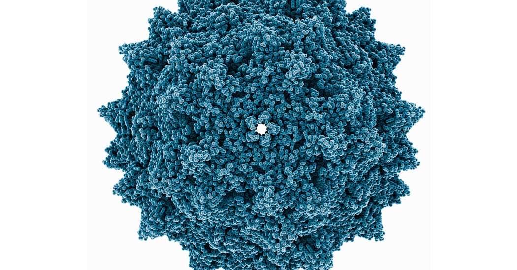
Human lung proteins can advance or thwart SARS-CoV-2 infections

Researchers have taken an important step toward understanding the microscopic battle that plays out between our lung cells and the SARS-CoV-2 virus that causes COVID-19. A UC Berkeley-led study has identified specific proteins within our bodies that can promote or protect us from SARS-CoV-2 infections, potentially opening the door to new antiviral therapies.
In the study, published this week in Nature Genetics, researchers used CRISPR technology to test the impact of every human gene on SARS-CoV-2 infections in human lung cells. Their findings revealed new pathways that the virus relies on to infect cells, as well as the antiviral pathways that help protect against viral infection. Notably, they showed that mucins — the main component of mucus found in the lungs — seem to help block the SARS-CoV-2 virus from entering our cells.
“Our data suggest that mucins play a key role in restricting SARS-CoV-2 infection by acting as a barrier to viruses that are attempting to access our lung epithelial cells,” said Scott Biering, the study’s co-lead author and a postdoctoral researcher in Eva Harris’s lab at UC Berkeley’s School of Public Health. “Further, our data suggest that mucin expression levels in an individual’s lungs may impact COVID-19 disease progression.”
Patrick Hsu — cofounder of the Arc Institute and Berkeley assistant professor of bioengineering, Deb Faculty Fellow and Innovative Genomics Institute Investigator — is the principal investigator of the study, which brought together researchers from 10 institutions. Harris, professor of public health at UC Berkeley, and Silvana Konermann, assistant professor at Stanford University School of Medicine, were co-senior authors of the study. Other collaborators contributed from Stanford University, the University of North Carolina at Chapel Hill, Yale School of Medicine and Cornell University, and spanned disciplines ranging from immunology, bioengineering, epidemiology, molecular biology and genetics.
Together, the researchers were trying to determine how the SARS-CoV-2 virus enters human cells and replicates so efficiently during illness. They also wanted to identify specific defense mechanisms in human cells that might be able to fight infection, which could inspire new therapeutic strategies.
Researchers discovered that MUC1 and MUC4, types of mucins found in lung cell membranes, defend lung cells from infection. This finding is important because previous studies had suggested that an accumulation of mucus could be the reason why some people became seriously ill with COVID-19 — since the mucus can make it difficult for people to breathe — and proposed using drugs to deplete mucus. This Berkeley-led study suggests that such a strategy could interfere with mucins that provide a valuable defense mechanism against SARS-CoV-2 infection.

Read more →

Teva Reaches Tentative $4.25 Billion Settlement Over Opioids

The deal would end thousands of lawsuits against one of the largest producers of opioid painkillers during the height of the opioid epidemic.Teva Pharmaceuticals, one of the country’s biggest manufacturers of generic opioids, announced a settlement in principle with some 2,500 local governments, states and tribes over the company’s role in the deadly, ongoing opioid epidemic.The deal — worth up to $4.25 billion — came after a series of blistering trials and previous settlements in individual cases across the country over the past year.Though much lesser-known, Teva, an Israeli company, and its affiliates produced far more prescription opioids during the peak years of the crisis than marquee-name opioid manufacturers such as Johnson & Johnson did. Its production of both generic and branded painkillers dwarfed the output of Purdue Pharma, the maker of OxyContin, the medication most immediately associated with setting off an avalanche of overdoses and deaths.Under the deal, Teva would make payouts over 13 years, directed to state, local and tribal programs to ease the opioid crisis, which has only deepened during the coronavirus pandemic. The $4.25 billion total included the nearly $550 million in settlements the company had already struck as trials got underway in San Francisco as well as in Florida, West Virginia, Texas, Louisiana and Rhode Island.States and communities can choose to accept a portion of their payouts in overdose reversal medications, rather than cash.Fentanyl Overdoses: What to KnowCard 1 of 5Devastating losses.

Read more →

Viral Infections and Gene Variant Are Linked to Child Hepatitis Cases

Some of the children were infected with adeno-associated virus 2 and shared a relatively uncommon gene variant, according to two small studies.A complex combination of factors could be responsible for the pediatric hepatitis cases that have been puzzling doctors in recent months, according to two small, new studies.The studies are based on just a few dozen cases and have not yet been peer-reviewed or published in scientific journals. Still, they suggest that the children who have developed severe, unexplained cases of liver inflammation may have been simultaneously infected with two different viruses, including one known as adeno-associated virus 2 (A.A.V.2), a typically benign virus that requires a second “helper” virus in order to replicate.Adenoviruses, which have previously been found in many of the children with the mysterious hepatitis reported within the last year, are common helper viruses for A.A.V.2.Many of the children studied also had a relatively uncommon version of a gene that plays an important role in the immune response, the scientists found.Together, the findings suggest a possible explanation for the hepatitis cases: In a small subset of children with this particular gene variant, dual infections with A.A.V.2. and a helper virus, often an adenovirus, trigger an abnormal immune response that damages the liver.Understanding HepatitisAn inflammation of the liver generally caused by a virus, hepatitis carries a host of complicating factors, side effects and stigma.Symptoms in Children: Understand the warning signs of hepatitis parents should watch out for as a series of rare, but unusual cases are reported across the United States.Searching for Answers: Recent studies suggest that a common childhood virus might be involved in the hepatitis cases in children, but many questions remain.Preventing Transmission: With infections on the rise, most adults should be getting screened for hepatitis.Pandemic’s Effect: A wave of diagnostics ushered in by Covid could help revive flagging efforts to eliminate hepatitis C, one of the most common forms of the disease.Still, the researchers acknowledged that the studies are based on a small number of children in just one region of the world (the United Kingdom) and that a causal link had not been proven.“There’s a lot that we still don’t know,” said Dr. Antonia Ho, a clinical senior lecturer at the MRC-University of Glasgow Centre for Virus Research and an author of one of the new studies.But, she added: “We felt — because there’s been very little in the way of answers of what are the causes — that we needed to release these findings so that other people can start looking for A.A.V.2. and investigate this in more detail.”The findings are intriguing but preliminary, said Dr. Saul Karpen, a pediatric hepatologist at Emory University and Children’s Healthcare of Atlanta, who was not involved in the research. “This is not a definitive study,” he said. “Thematically, it certainly can make sense, but there is not full support for it.”The pediatric hepatitis cases are exceedingly rare but can be severe. As of July 8, 1,010 probable cases had been reported from 35 countries, according to the World Health Organization. Five percent of those children have required liver transplants, and 2 percent have died.Several early studies have found that many of the children were infected by an adenovirus, one of a group of common viruses that typically cause cold or flulike symptoms. The new studies suggest that if adenoviruses are involved in the hepatitis cases, they may just be part of the story.In one of the new studies, scientists compared nine Scottish children with unexplained hepatitis to 58 children in control groups. The researchers used genomic sequencing to identify any viruses present in blood, liver and other samples from the children.The scientists found adeno-associated virus 2 in the blood of all nine affected children and in liver samples from all four of the children from whom such samples were available. They also found an adenovirus in six of the children and a common herpes virus in three.On the other hand, the researchers did not detect A.A.V.2 in healthy children, in children who had adenovirus infections but normal liver function or in children who had hepatitis with a known cause.Those findings are consistent with those from a second study, led by researchers in London, which examined samples from 28 children with unexplained hepatitis from across the United Kingdom. That scientific team also found high levels of A.A.V.2 in the blood and livers of many of the children. Many also had low levels of an adenovirus or herpes virus in their samples.The Scottish researchers also found that eight of the nine affected children, or 89 percent, shared a relatively uncommon variant of a gene that codes for a critical protein in the body’s immune response. This particular variant is present in just 16 percent of Scottish blood donors.The London team found the same gene variant in four of the five transplant recipients they assessed.“Both studies have reached independently, remarkably similar results,” Sofia Morfopoulou, a computational statistician at the Great Ormond Street Institute of Child Health at University College London and an author of the second paper, said in an email.Although the idea remains preliminary, it is possible that a recent resurgence of the adenovirus after a decline in circulation during the coronavirus pandemic explains why doctors have noticed a sudden spike in these rare cases, the scientists said.“Perhaps some of these infections that might have occurred in a more spaced out way, over a couple of years,” are instead occurring all at once, said Dr. Emma Thomson, an infectious diseases physician at the Centre for Virus Research and a senior author of the Scottish study.Additional, larger studies are still needed, particularly focusing on children in other countries, the researchers said.

Read more →

Researchers find why bat cells do not get infected by SARS-CoV-2

Bat cells have specific molecular barriers to deal with SARS-CoV-2 replication, according to a study published in the Journal of Virology — a publication of the American Society of Microbiology — which includes the participation of Jordi Serra-Cobo, lecturer of the Faculty of Biology and the Biodiversity Research Institute (IRBio) of the University of Barcelona and expert on ecoepidemiological studies.
The study was carried out on primary cells of bat species which had been little studied and which circulate around Europe and Asia (specifically, Rhinolopuhs ferrumequinum, Myotis myotis, Eptesicus serotinus, Tadarida brasiliensis and Nyctalus noctula). These cellular lines were obtained through small biopsies carried out on the wings of the bats — for instance, in bat colonies of Myotis myotis in Majorca and other cell lines brought by some research teams that took part in the study. As stated in the conclusions, these cellular models defined in chiropterans are shaped as tools of scientific interest to study the evolutionary relationship between bats and coronaviruses.
The study, led by the experts Nolwenn Jouvenet and Laurent Dacheux, from the Institute Pasteur in Paris, includes the collaboration of experts from research institutions in France, the Czech Republic and Switzerland.
How do bats protect themselves from viral infections?
Coronaviruses are present in many animal species worldwide, such as bats (chiropteans). In this context, the scientific literature has described for years the great resistance of some chiropteran species towards the viral infection. In these flying mammals, the immune system is on a pre-alert stage, a condition that allows a faster response to viral infections. For most mammals, having an immune system on a constant pre-alert state would involve inflammation problems but this is not the case for bats, which is why they are the focus of many international epidemiological and immunological studies.
As part of the study, the team analysed the ability of primary cells from different bat species to support SARS-CoV-2 replication. “The results reveal that none of these cells was permissive to the infection, not even those expressing detectable levels of angiotensin-converting enzyme 2 (ACE2), a metallopeptidase that serves as a viral receptor in many mammal species,” says Jordi Serra-Cobo, member of the Department of Evolutionary Biology, Ecology and Environmental Sciences of the UB and the only expert in Spain to take part in this study.
“The cells did not allow the infection in the species Rhinolophus ferrumequinum, a chiropteran from the same genus as the Asian bat in which the BANAL-52 virus was found, a potential ancestor of SARS-CoV-2. Specifically, the genetic sequences of the BANAL-52 virus is 96.8% similar to that of SARS-CoV-2,” says Serra-Cobo, distinguished expert in studies with bats as natural reservoirs of infectious agents like coronaviruses.
Humans and chiropterans vs. SARS-CoV-2 infection
Regarding the human species, it is known that the SARS-CoV-2 spike protein binds to the cell membrane receptor ACE2 and then the virus infects the cell. “In the case of the chiropteran cells, either the amount of ACE2 enzyme is small and it no longer enters the cell or, if the virus binds to ACE2, it cannot infect the cell,” highlights Serra-Cobo.
From a global perspective, this study contributes to a better understanding of the fighting mechanisms against viral infections. This is a line of research that has been carried out for years by the team led by Serra-Cobo at the UB and IRBio and which is now gaining strength within the framework of the EvoDevo-Cat research group at the Faculty of Biology of the UB.
“Specifically, our team is working to understand the adaptations of the chiropterans regarding viral infections. An important number of zoonotic viruses circulate in chiropter populations without causing symptoms of the disease in the carriers,” notes the researcher.
“Over the course of the evolutionary history of chiropterans — about 64 million years — , there have been processes of coevolution between bats and viruses. One example of these processes is in the adaptation to coronaviruses. The study of the evolutionary adaptations of living beings to deal with viral infections is of great interest, since they provide information that can have medical applications,” concludes Jordi Serra-Cobo.

Read more →

Green tea extract promotes gut health, lowers blood sugar

New research in people with a cluster of heart disease risk factors has shown that consuming green tea extract for four weeks can reduce blood sugar levels and improve gut health by lowering inflammation and decreasing “leaky gut.”
Researchers said this is the first study assessing whether the health risks linked to the condition known as metabolic syndrome, which affects about one-third of Americans, may be diminished by green tea’s anti-inflammatory benefits in the gut.
“There is much evidence that greater consumption of green tea is associated with good levels of cholesterol, glucose and triglycerides, but no studies have linked its benefits at the gut to those health factors,” said Richard Bruno, senior study author and professor of human nutrition at The Ohio State University.
The team conducted the clinical trial in 40 individuals as a follow-up to a 2019 study that associated lower obesity and fewer health risks in mice that consumed green tea supplements with improvements to gut health.
In the new study, green tea extract also lowered blood sugar, or glucose, and decreased gut inflammation and permeability in healthy people — an unexpected finding.
“What this tells us is that within one month we’re able to lower blood glucose in both people with metabolic syndrome and healthy people, and the lowering of blood glucose appears to be related to decreasing leaky gut and decreasing gut inflammation — regardless of health status,” Bruno said.

Read more →

Cocoa shown to reduce blood pressure and arterial stiffness in real-life study

Cocoa only reduces blood pressure and arterial stiffness when elevated, a new study from the University of Surrey finds.
Cocoa flavanols have previously been found to lower blood pressure and arterial stiffness as much as some blood pressure medication. However, how effective flavanols are in everyday life in reducing blood pressure has remained unknown, as previous studies in this area have been performed in tightly controlled experimental settings.
Surrey’s new research reduces concerns that cocoa as a treatment for raised blood pressure could pose health risks by decreasing blood pressure when it is not raised, paving the way for it to be potentially used in clinical practice.
In the first study of its kind study, researchers set out to investigate the use of flavanols, a compound found in cocoa, in lowering blood pressure and arterial stiffness in individuals outside of clinical settings.
Christian Heiss, Professor of Cardiovascular Medicine at the University of Surrey, said:
“High blood pressure and arterial stiffness increases a person’s risk of heart disease and strokes, so it is crucial that we investigate innovative ways to treat such conditions.

Read more →

Smoking, vaping linked to higher risk of severe COVID-19 complications, including death

People who reported smoking or vaping prior to their hospitalization for COVID-19 were more likely than their counterparts who did not smoke or vape to experience severe complications, including death, from the SARS-CoV-2 infection. The findings are from a new study based on data from the American Heart Association’s COVID-19 CVD Registry and published in PLOS ONE, a peer-reviewed, open access scientific journal published by the Public Library of Science.
Researchers examined data on people over 18 years of age who were hospitalized with COVID-19 in 107 registry-participating hospitals across the nation between January 2020 to March 2021. Smoking status was self-reported and people were classified as smoking if they reported currently using either traditional, combustible cigarettes or e-cigarette products, with no distinction between the two and no information on duration of smoking or former smoking status. For the final analysis, records were selected for 4,086 people with a 1:2 ratio of people who smoked (1,362) to people who did not smoke (2,724), with the two groups matched for no statistically significant difference in age, sex, race, medical history or medication.
The study findings indicate smoking or vaping are associated with more severe COVID-19 independent of age, sex, race or medical history: People who reported smoking were 45% more likely to die and 39% more likely to receive mechanical ventilation when compared with those who did not smoke. Although the excessive risk due to smoking was independent of medical history and medication use, smoking was a stronger risk factor for death in people between 18-59 years of age and those who were white or had obesity.”In general, people who smoke or vape tend to have a higher prevalence of other health conditions and risk factors that could play a role in how they are impacted by COVID-19. However, the robust and significant increase in the risk of severe COVID-19 seen in our study, independent of medical history and medication use and particularly among young individuals, underscores the urgent need for extensive public health interventions such as anti-smoking campaigns and increased access to cessation therapy, especially in the age of COVID,” said the study’s senior author, Aruni Bhatnagar, Ph.D., FAHA, a professor of medicine, biochemistry and molecular biology at the University of Louisville in Louisville, Kentucky. “These findings provide the clearest evidence to date that people who smoke or vape have a higher risk of developing severe COVID-19 and dying as a result of SARS-CoV-2 infection.”
Bhatnagar is co-director of the American Heart Association’s Tobacco Center for Regulatory Science which supported the study in part with funding from the U. S. National Institutes of Health and the Food and Drug Administration research grants.
“We established the COVID-19 CVD Registry early on in the pandemic to better understand the link between COVID-19 and cardiovascular disease, specifically, to identify increased risk to help inform the diagnosis and care of people who are at highest risk for complications,” said Sandeep R. Das, M.D., M.P.H., M.B.A., FAHA, co-chair of the steering committee for the American Heart Association® COVID-19 CVD Registry Powered by Get With The Guidelines® and director for Quality and Value in the Cardiology Division at UT Southwestern Medical Center in Dallas, Texas. “The findings of this study deliver on that goal and provide invaluable information individuals and their health care teams.”
The American Heart Association launched the registry in 2020 to gather data specific to all patients hospitalized with COVID-19 as part of the Get With The Guidelines® quality improvement program. Registry participation was offered at no cost to all U.S. hospitals caring for adults with active COVID-19 and with the infrastructure to support accurate data collection. More than 160 hospitals provided data on more than 79,000 patient records between 2020 and June 2022.
Story Source:
Materials provided by American Heart Association. Note: Content may be edited for style and length.

Read more →

Mathematical modeling to explain immunotherapy responses

Immunotherapies that activate the immune system to seek and kill cancer cells have greatly improved outcomes for many patients with solid tumors. There is still, however, a subset of patients who do not see benefit from this type of therapy. Currently, there are no immune biomarkers that explain how patients with similar disease and patient characteristics can have different outcomes. In a new article published in the Journal for ImmunoTherapy of Cancer, Moffitt Cancer Center researchers demonstrate how mathematical modeling can be used to analyze the impact of different cancer treatments on tumor and immune cell dynamics and help predict outcomes to therapy and personalize cancer treatment.
It is known that interactions between cancer cell populations with the surrounding immune environment impact the development and progression of cancer and patient responses to immunotherapy. Some patients respond well to immunotherapies, while other patients do not. However, it is unclear what differentiates these patients.
“Just like early stage cancers are treated differently than late-stage disease, tumors with different degrees of immune involvement may need very different therapeutic approaches,” said Rebecca Bekker, article first author and Cancer Biology Ph.D. student at Moffitt.
Moffitt researchers wanted to improve their understanding of tumor and immune cell interactions to help predict outcomes for patients and identify the best therapeutic options. Knowing these dynamics are extremely complex and difficult to study in a laboratory setting, the team used an alternative approach to conceptualize these interactions with mathematical modeling. They developed a model that simulates interactions between all possible combinations of tumor cell and immune cell population numbers over time. They included parameters for the rate of tumor cell growth and elimination, and immune cell recruitment and exhaustion. The outcomes of their model were either immune escape, wherein the tumor cells grew to their maximum potential, or tumor control through the antitumor activity of immune cells.
The researchers then used their model to simulate and predict outcomes to different types of therapies, including cytotoxic chemotherapy and cell-based immunotherapies, which impact the size of the tumor cell or immune cell populations, and immune checkpoint inhibitors, which impact the nature of the interactions between tumor and immune cell populations. They also addressed potential outcomes to combination therapies.
These models help conceptualize how therapies can be combined to achieve optimal outcomes for patients through immune-cell control of tumor cell populations. In the future, the researchers hope mathematical modeling can be used in the clinic to help predict patient responses to therapy and guide treatment.
“Mathematical oncology abstraction provides a novel and promising way to conceptualize the effect of various cancer treatments on a patient’s tumor and the local immune environment and gives us an opportunity to rethink the immunotherapy numbers game,” said Heiko Enderling, Ph.D., study author and associate member of the Integrated Mathematical Oncology Department at Moffitt.
This study was supported by the National Cancer Institute (U01CA244100 and R21CA263911) and the Ocala Royal Dames for Cancer Research.
Story Source:
Materials provided by H. Lee Moffitt Cancer Center & Research Institute. Note: Content may be edited for style and length.

Read more →

Study identifies new links between REM sleep disturbances and drug relapse

Relapse presents a major barrier to recovery from substance use disorders — when people begin taking drugs such as cocaine again after a period of abstinence. Sleep disruptions have long been associated with drug withdrawal and relapse. Now, a new study provides a more detailed picture of recovery-related sleep that could lead to insights for better recovery treatments.
The study, led by Yanhua Huang, PhD, at the University of Pittsburgh, appears in Biological Psychiatry, published by Elsevier.
“Rapid eye movement (REM) sleep is important for regulating emotion. For years, we have seen REM sleep changes associated with cocaine-seeking behaviors in our rat models. This is our first comprehensive study to examine what specific features of REM sleep may be related and why,” said Dr. Huang.
In 2006, Peter Morgan, Robert Malison, and their collaborators first reported the existence of an unappreciated form of insomnia in individuals recovering from cocaine use disorder defined by brain waves measured during sleep. The pattern emerged over the initial weeks of cocaine abstinence, at a time when people were reporting subjectively “improved” sleep, and which was related to their risk for relapse. In a continuation of that work, said Biological Psychiatry Editor John Krystal, MD, “this interesting new study in rodents by Guo et al. identifies a disturbance in REM sleep during the recovery from chronic cocaine administration that also predicts the propensity for subsequent relapse to cocaine self-administration.”
Dr. Huang said, “Previously there was a notion that poor sleep may worsen drug craving and relapse — and we now offer a more granular view on the specific sleep features to be considered for potential biomarkers for predicting relapse.”
For the study, the researchers trained male rats to self-administer cocaine and then removed access to the drug, so that the rats were in long-term withdrawal. The rats were subsequently re-exposed to cocaine-associated cues, and demonstrated “craving incubation,” in which the drive to take the drug increases over time. The rats also displayed REM sleep disturbances.
To further investigate potential casual relationships, the investigators increased the temperature of the rats’ bedding, which increased time spent in REM sleep and improved REM sleep continuity. That led to an attenuation of the incubation of drug-seeking behavior.
The phenomenon of incubation depends on physiological changes at neuronal synapses in a brain area called the nucleus accumbens (NAc), according to previous studies. A particular type of protein, called calcium-permeable AMPA receptors, accumulates in synapses, increasing the neuronal activity there and driving drug-seeking behavior. In the current study, rats with improved REM sleep showed responses at NAc synapses that resembled those of drug-naïve rats, suggesting the ion channel normalization could underlie the improvement in relapse behavior.
Next, lead author Rong Guo and colleagues examined melanin-concentrating hormone-producing (MCH) neurons in the lateral hypothalamus, which are central to REM sleep regulation. Their activity — and REM sleep — increased with bedding warming. Activation of MCH neurons with optogenetic or chemogenetic technology also promoted REM sleep, decreased craving incubation, and normalized NAc synaptic activity to different extents, but only when they were activated during the light phase, when rats are normally asleep more. Together, the results indicate that MCH neuron activities in sleep recapitulate the REM sleep effects on reducing drug-seeking behavior.
Dr. Krystal said of the findings, “Guo and colleagues identify hypothalamic MCH release and its normalization of calcium-permeable AMPA receptor availability in the NAc in the resilience of rodents to relapse to cocaine self-administration.”
“The results have important implications,” said Dr. Huang. “For example, when treating relapse, it is important to pay attention to REM sleep improvement, complementary to current practice focusing on non-REM sleep interventions. Additionally, we have identified the important roles of MCH neurons in this regulation. This will provide the rationale for testing potential drugs in the future. Finally, we found that stimulating MCH neurons is most effective during the rat’s inactive phase, thus underscoring the importance of considering the time of day and sleep/wake state for future drug development and applications.”
Dr. Krystal added, “Their work draws further attention to the importance of disturbances in the integrity of sleep to relapse, and it points us toward strategies that enhance MCH signaling in sleep as a potential medication target that may be protective against cocaine relapse.”
Story Source:
Materials provided by Elsevier. Note: Content may be edited for style and length.

Read more →

Studies link COVID-19 to wildlife sales at Chinese market, find alternative scenarios extremely unlikely

An international team of researchers has confirmed that live animals sold at the Huanan Seafood Wholesale Market were the likely source of the COVID-19 pandemic that has claimed 6.4 million lives since it began nearly three years ago.
Led by University of Arizona virus evolution expert Michael Worobey, international teams of researchers have traced the start of the pandemic to the market in Wuhan, China, where foxes, raccoon dogs and other live mammals susceptible to the virus were sold live immediately before the pandemic began. Their findings were published Tuesday in two papers in the journal Science, after being previously released in pre-print versions in February.
The publications, which have since gone through peer review and include additional analyses and conclusions, virtually eliminate alternative scenarios that have been suggested as origins of the pandemic. Moreover, the authors conclude that the first spread to humans from animals likely occurred in two separate transmission events in the Huanan market in late November 2019.
One study scrutinized the locations of the first known COVID-19 cases, as well as swab samples taken from surfaces at various locations at the market. The other focused on genomic sequences of SARS-CoV-2 from samples collected from COVID-19 patients during the first weeks of the pandemic in China.
The first paper, led by Worobey and Kristian Andersen at Scripps Research Institute in San Diego, California, examined the geographic pattern of COVID-19 cases in the first month of the outbreak, December 2019. The team was able to determine the locations of almost all of the 174 COVID-19 cases identified by the World Health Organization that month, 155 of which were in Wuhan.
Analyses showed that these cases were clustered tightly around the Huanan market, whereas later cases were dispersed widely throughout Wuhan — a city of 11 million people. Notably, the researchers found that a striking percentage of early COVID patients with no known connection to the market — meaning they neither worked there nor shopped there — turned out to live near the market. This supports the idea that the market was the epicenter of the epidemic, Worobey said, with vendors getting infected first and setting off a chain of infections among community members in the surrounding area.

Read more →