New prenatal test can reduce time, cost of detecting chromosomal abnormalities

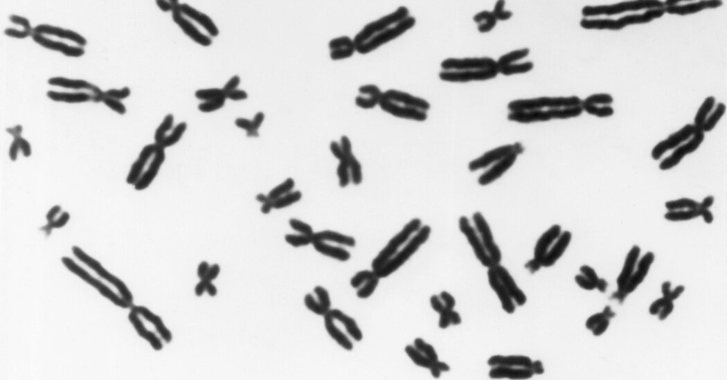
A new prenatal test developed by researchers at Columbia University Fertility Center can determine if a fetus or embryo has the right number of chromosomes at a fraction of the time and cost of currently available clinical genetic tests.
Currently available prenatal genetic tests cost thousands of dollars and take days to weeks to deliver results, adding to the emotional and financial stress of fertility treatment and pregnancy and impacting treatment options.
The new test, called STORK (Short-read Transpore Rapid Karyotyping), can be used in the doctor’s office at the point of care, delivers results in under two hours, and is about 10 times less expensive to process per sample than current tests.
Details about the test — which is awaiting authorization from New York State Department of Health before it can be offered to Columbia patients — and its performance were published in the New England Journal of Medicine.
“We are developing the most advanced technologies to solve some of the most ancient of afflictions — infertility and pregnancy loss,” says study leader Zev Williams, MD, PhD, the Wendy D. Havens Associate Professor of Women’s Health and chief of the division of Reproductive Endocrinology and Infertility at Columbia University Vagelos College of Physicians and Surgeons. “Our hope is that this test will help improve women’s health, lower costs, and improve access to treatment.”
The chromosomal abnormalities that this test can detect are, by far, the most common causes of miscarriage, structural anomalies, and developmental delays. Prenatal genetic testing is recommended for pregnant women who are age 35 or older, have a family history of genetic disorders, or have had one or more miscarriages. It is also used increasingly during in vitro fertilization (IVF) to test embryos prior to implantation to improve the chances of pregnancy and reduce the risk of miscarriage.

Read more →

Why heat makes us sleepy

On the hottest summer days, you may find yourself dozing off in the middle of the day. In some parts of the world, it’s a cultural norm to schedule “siestas” and shutter businesses during the warmest hours of the day. As it turns out, biology, not just culture, may be behind this.
Temperature affects the span of human behavior, from eating and activity levels to sleep-wake cycles. We may have a harder time sleeping in the summer and be slow to get out of bed on colder mornings. But the link between sensory neurons and neurons that control this cycle are not understood completely.
Northwestern University neurobiologists have found a few clues about what’s happening. In a new study, published today (Aug. 17) in the journal Current Biology, researchers found that fruit flies are pre-programmed to take a nap in the middle of the day. A follow-up to their 2020 Biology paper that identified a brain thermometer only active in cold weather, the new paper explores a similar “thermometer” circuit for hot temperatures.
“Changes in temperature have a strong effect on behavior in both humans and animals, and offer animals a cue that is time to adapt to the changing seasons,” said Marco Gallio, associate professor of neurobiology in the Weinberg College of Arts and Sciences. “The effect of temperature on sleep can be quite extreme, with some animals deciding to sleep off an entire season — think of a hibernating bear — but the specific brain circuits that mediate the interaction between temperature and sleep centers remain largely unmapped.”
Gallio led the study and said fruit flies are a particularly good model to study big questions like “why do we sleep,” and “what does sleep do for the brain” because they don’t attempt to disrupt instinct in the same way humans do when we pull all-nighters, for example. They also allow researchers to study the influence of external cues like light and temperature on cellular pathways.
Cells that stay on longer
The paper is the first to identify “absolute heat” receptors in fly head, which respond to temperatures above about 77 degrees Fahrenheit — the fly’s favorite temperature. As it turns out, the common laboratory fruit fly (Drosophila) has colonized nearly the entire planet by forming a close association with humans. Not surprisingly, its favorite temperature also matches that of many humans.

Read more →

Study combines lithophane, 3D printing to enable individuals to 'see' data regardless of level of eyesight

A research team led by Baylor University chemists has taken a groundbreaking step forward in eliminating the exclusion of individuals with blindness from chemistry education and experiences. In an article published today in Science Advances, the researchers detail how they used lithophane — an old-fashioned art form — and 3D printing to turn scientific data into tactile graphics that glow with video-like resolution, enabling universal visualization of the same piece of data by both blind and sighted individuals.
Although the lithophane is an ancient artistic medium, it has never been used — until now — to represent scientific data and imagery in a quantitative, controlled manner for tactile visualization and tactile integration.
The Baylor study — “Data for all: Tactile graphics that light up with picture-perfect resolution”” — compared how blind and sighted people interpreted lithophane data by touch or eyesight. The participant cohorts tested the five lithophane forms — gel electropherograms, micrographs, electronic and mass spectra, and textbook illustrations — interpreting all five lithophanes by tactile sensing or eyesight at 79% overall accuracy, according to the study.
The researchers focused on creating and testing lithophanes of data found in the chemical sciences because of the explicit and systematic exclusion of students with blindness from chemistry, which the researchers noted “can be viewed as a virtue by educators, parents, peers or self, on the basis of laboratory safety and the ‘visual’ nature of chemistry.”
“This research is an example of art making science more accessible and inclusive. Art is rescuing science from itself,” said Bryan Shaw, Ph.D., professor of chemistry and biochemistry, who leads the Shaw Research Group at Baylor and is corresponding author on the journal article. “The data and imagery of science — for example, the stunning images coming out from the new Webb telescope — are inaccessible to people who are blind. We show, however, that thin translucent tactile graphics, called lithophanes, can make all of this imagery accessible to everyone regardless of eyesight. As we like to say, ‘data for all.'”
New use for old art form
Likely created in China as early as the sixth century and popularized in Europe in the 1800s, lithophanes are thin engravings made from translucent materials (first porcelain and wax, now plastic) and initially appear opaque in ambient light. However, when back lit by any light source — from a ceiling light to sunlight — a lithophane glows like a digital image, with the scattering of light through the translucent material causing thinner regions to appear brighter and thicker regions to appear darker. Using free online software to convert a two-dimensional image to a 3D topograph, scientists in this study used 3D printing for the lithophanes.

Read more →

Researchers identify an Achilles' heel in neuroblastoma

Neuroblastoma, a childhood cancer that develops from neural cells on the adrenal glands, accounts for 15% of childhood cancer deaths. Almost half of children with high-risk neuroblastoma harbor extra copies of the gene MYCN (MYCN amplified), the primary driver of neuroblastoma and its resistance to therapy.
“Treating neuroblastoma by directly targeting MYCN has been challenging,” said Dr. Eveline Barbieri, corresponding author of a recent study published in the journal Nature Communications and assistant professor of pediatrics — hematology and oncology at Baylor College of Medicine and Texas Children’s Hospital. “In this study we investigated new strategies to improve the survival of children with MYCN amplified neuroblastoma by looking into metabolic vulnerabilities that we could exploit to overturn these tumors’ resistance to therapy.”
Barbieri and her colleagues used an unbiased, metabolomics analysis to compare the metabolic profiles of MYCN-amplified neuroblastomas to the profiles of non MYCN-amplified neuroblastomas. The results of their innovative approach showed that there were important differences between tumor cell utilization of specific nutrients for tumor growth in these two tumor groups.
“We found that MYCN amplification rewires a tumor’s lipid metabolism in a way that promotes the use and biosynthesis of fatty acids, a type of lipid cells can use as a source of energy,” Barbieri said. “Cells with extra copies of MYCN depend highly on fatty acids for their survival. We confirmed this both in MYCN-amplified cell lines and in MYCN- amplified patient tumor samples.”
Barbieri and her colleagues hypothesized that MYCN reroutes lipid metabolism so that fatty acids are readily available to cancer cells, thereby promoting tumor cell growth.
Looking into the mechanism
“When we investigated what prompted MYCN-amplified neuroblastomas to rely on fatty acids to grow, we discovered that MYCN directly upregulates or enhances the production of fatty acid transport protein 2 (FATP2), a molecule that mediates cellular uptake of fatty acids,” Barbieri said. “We then asked, what would happen if we interfered with FATP2 function in MYCN-amplified neuroblastomas?”

Read more →

New Method Improves Speed and Cost of Birth Defect Testing

If the technique is confirmed, women who have had miscarriages or those undergoing prenatal screening may no longer need to rely on centralized testing labs for results.After 10 years of effort, medical researchers at Columbia University have developed a very fast and cheap way to detect the extra or missing chromosomes that most often cause miscarriages or severe birth defects.The method, described Wednesday in the New England Journal of Medicine, takes less than two hours using a palm-size device and costs $200 per use. With current testing procedures, women can end up paying $1,000 to $2,000, often out of pocket.The technique, developed by Dr. Zev Williams, director of the Columbia University Fertility Center, and his colleagues, uses cells and tissues obtained from existing prenatal screening procedures of embryos and fetuses, or tissue obtained after miscarriages. Its key advantage is that the cells or tissue do not have to be sent to a testing lab — the analysis can be done in the same office that obtained the material, and results are ready in hours rather than days or weeks.Dr. Williams’s results still need to be confirmed by outside studies. But if the method were widely used, it could help more women who had lost pregnancies better understand what caused their miscarriages. For women who have embryos tested during in vitro fertilization, it would also avoid lengthy waits for results. The same would be true of women who have prenatal testing of fetuses with amniocentesis or chorionic villus sampling, known as C.V.S. And for women in states that have restricted abortion to within the first 15 or 18 weeks of a pregnancy, it could provide a bit more lead time in deciding whether to terminate a pregnancy.Current screening methods, sent to centralized labs, involve lengthy wait times for women. The longest delay in getting test results is for women who have had miscarriages. Those women may have to wait several weeks, because labs must first grow cells in order to have thousands to examine, said Dr. James Grifo, program director of the New York University Langone Fertility Center, who was not involved in the new study.Dr. Mark Hornstein, director of the division of reproductive endocrinology and infertility at Brigham and Women’s Hospital in Boston, said the cultured cells sometimes died before they could be tested.“It is very difficult to keep cells alive and not contaminated for three weeks,” Dr. Hornstein said. “Not infrequently, we get no growth. We wait three weeks, and then the lab tells us, ‘You have no cells.’”The new test avoids these delays by using a method called nanopore sequencing, which was developed to read the sequences of very long strands of DNA but has not been used to look for chromosomal anomalies.Dr. Williams and his colleagues modified the preparation of DNA samples to use nanopore sequencing with small DNA pieces. Each DNA segment should appear as two copies — one from each chromosome. If there are three copies, that means there is an extra chromosome; if there is one copy, that means a chromosome is missing.The new test uses a palm-size device for a method called nanopore sequencing, which was developed to read the sequences of very long strands of DNA. Columbia University Fertility CenterChromosome testing can provide important information for women and their doctors because extra or missing chromosomes are usually fatal to embryos. Fetuses that survive with them often have severe birth defects. The best-known example — Down syndrome — is “actually one of the mildest,” Dr. Williams said.That can be important for in vitro fertilization. The older a woman is, the more likely that an embryo will have chromosomal abnormalities. For 30-year-old women, the risk is 25 percent. It is 75 percent in women at age 42.Women who undergo I.V.F. and who opt to have their embryos tested before implantation usually have to pay for the testing themselves. The embryos are frozen to preserve them while testing goes on. The woman has to wait for the next month’s menstrual cycle for implantation.While test results using tissue from miscarriages take weeks, central labs get results from tests on embryos or fetuses in about three to five days. That is because cells from an embryo or fetus do not grow well in culture, Dr. Grifo explained, so the cells are tested sooner, but with a modest reduction in accuracy.But the new method yields results from those same tests on embryos or fetuses on the same day, within hours.The new test might also help alleviate a problem with prenatal testing in a handful of states that allow abortions only during the first 15 or 18 weeks of pregnancy.Amniocentesis is usually done between 15 and 20 weeks of pregnancy and C.V.S. between the 11th and 14th weeks. If a woman waits days for test results, it might be too late in some states to terminate her pregnancy.Dr. Williams and his colleagues tested their method by examining 218 samples, comparing their results to those from standard tests of the same samples.The results were identical for amniotic fluid and C.V.S. tissues. But in one of the 52 embryos tested, the standard test found an extra chromosome 21, meaning Down syndrome, while the new test did not.“We requested that the clinical lab retest that sample, but they didn’t have any material left to retest, so we can’t determine which result was correct,” Dr. Williams said.Tests of 10 miscarriage samples examined with the new method were different from those of the standard test, but it turned out the standard test was wrong.The group also showed that a group of technicians could quickly learn to do the test and get accurate results.“It’s a great start,” Dr. Grifo said. He added that he would like the accuracy of embryo testing to be 100 percent but said that he was confident that would happen.Dr. Hornstein, who was also not associated with the study, cautioned that the test was not yet ready for widespread use. There need to be independent studies that confirm that its results are accurate, and there must be evidence that it is “technically easy enough that any lab can do it,” he said.But he added that if those conditions are met, he expects the test to be adopted widely.Dr. Williams, who has applied for a patent, said he hoped to offer the test at Columbia soon. He submitted his data to New York State for approval.“Once it’s approved, we are ready to go,” he said.

Read more →

How Chewing Shaped Human Evolution

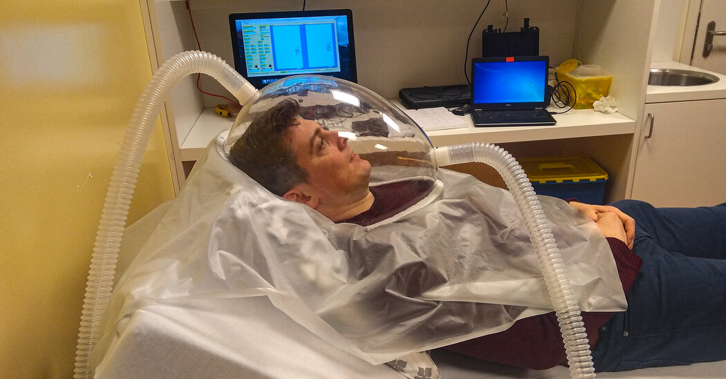
An experiment revealed that chomping on slightly tougher material requires markedly more energy. Spending less time on mastication may go hand in hand with human evolution.Humans spend about 35 minutes every day chewing. That adds up to more than a full week out of every year. But that’s nothing compared to the time spent masticating by our cousins: Chimps chew for 4.5 hours a day, and orangutans clock 6.6 hours.The differences between our chewing habits and those of our closest relatives offer insights into human evolution. A study published Wednesday in the journal Science Advances explores how much energy people use while chewing, and how that may have guided — or been guided by — our gradual transformation into modern humans.Chewing, in addition to keeping us from choking, makes the energy and nutrients in food accessible to the digestive system. But the very act of chewing requires us to expend energy. Adaptations to teeth, jaws and muscles all play a part in how efficiently humans chew.Adam van Casteren, an author of the new study and a research associate at the University of Manchester in England, says that scientists haven’t delved too deeply into the energetic costs of chewing partly because compared with other things we do, such as walking or running, it’s a thin slice of the energy-use pie. But even comparatively small advantages can play a big role in evolution, and he wanted to find out if that might be the case with chewing.To measure the energy that goes into chewing, Dr. van Casteren and his colleagues outfitted study participants with plastic hoods that look like “an astronaut’s helmet,” he said. The hoods were connected to tubes to measure oxygen and carbon dioxide from breathing. Because metabolic processes are fueled by oxygen and produce carbon dioxide, gas exchange can be a useful measure for how much energy something takes. The researchers then gave the subjects gum.The participants didn’t get the sugary kind, though; the gum bases they chewed were flavorless and odorless. Digestive systems respond to flavors and scents, so the researchers wanted to make sure they were only measuring the energy associated with chewing and not the energy of a stomach gearing up for a tasty meal.Measuring the chewing muscles with an ultrasound wand.Amanda HenryThe test subjects chewed two pieces of gum, one hard and one soft, for 15 minutes each. The results surprised researchers. The softer gum raised the participants’ metabolic rates about 10 percent higher than when they were resting; the harder gum caused a 15 percent increase.“I thought there wasn’t going to be as big a difference,” Dr. van Casteren said. “Very small changes in the material properties of the item you’re chewing can cause quite substantial increases in energy expenditure, and that opens up a whole universe of questions.”Because chewing tougher food — or in this case, tougher gum — takes significantly more energy, these findings suggest that the metabolic costs of chewing may have played an important role in our evolution. Making food easier to process through cooking, mashing food with tools and growing crops optimized for eating might have dialed down the evolutionary pressure for us to be super-chewers. Our evolving chewing needs may have even shaped what our faces look like.“One thing that we haven’t really been able to figure out is why the human skull is so funny-looking,” said Justin Ledogar, a biological anthropologist at East Tennessee State University, who was not involved with the study. Compared to our closest relatives, our facial skeletons are delicately built with jaws, teeth and chewing muscles that are all relatively small. “All this reflects a reduced reliance on forceful chewing,” he explained.But he added that our flatter faces and shorter jaws let us bite more efficiently. “It makes the whole process of feeding just metabolically less costly,” Dr. Ledogar said. Humans developed ways to chew smarter, not harder. Dr. van Casteren, who hopes to continue his research using actual foods, says he’s excited by the prospect of learning more about how humans evolved.“To know about the environmental and societal and dietary causes that led us to get here, it’s just infinitely interesting to me,” he said, because it enables humankind to “try and work out the foggy road ahead.”

Read more →

C.D.C. Investigates ‘Fast-Moving’ E. coli Outbreak

The agency said that no food had been identified yet as the source of the outbreak, which has sickened 14 people in Ohio and 15 in Michigan.Federal health officials said Wednesday that they were investigating an E. coli outbreak that has been linked to a growing number of illnesses in Ohio and Michigan.The Centers for Disease Control and Prevention said that no food had been identified yet as the source of the “fast-moving” outbreak, which has sickened 14 people in Ohio and 15 in Michigan. Nine people have been hospitalized. No deaths have been reported.The Michigan Department of Health and Human Services said on Tuesday that it had received reports of 98 cases of E. coli infections in August, compared with 20 cases during the same period last year. The investigation is in the early stages, the department said, and laboratory results have linked some of the cases to one another.“While reports of E. coli illness typically increase during the warmer summer months, this significant jump in cases is alarming,” Dr. Natasha Bagdasarian, Michigan’s chief medical executive, said in a statement.“This is a reminder to make sure to follow best practices when it comes to hand hygiene and food handling to prevent these kinds of food-borne illness,” she said.Anyone who experiences E. coli symptoms such as cramping, diarrhea or gastrointestinal distress should contact their health care provider, especially if such symptoms are severe, Dr. Bagdasarian said.Other common symptoms include vomiting and fever. Symptoms usually start three to four days after swallowing E. coli bacteria, and most people recover without treatment within five to seven days, according to the C.D.C.Some people may develop a type of kidney failure and require hospitalization, the C.D.C. said. The current outbreak has affected people from 6 to 91 years old.The Ohio Department of Health said that cases had been reported in Wood, Lucas, Mahoning, Clermont, Cuyahoga, Franklin, Lorain and Summit counties. Four people in Ohio have been hospitalized in connection with the outbreak, the department said.The C.D.C. urged people to prevent infection by washing their hands as well as their utensils and cooking surfaces, rinsing fruits and vegetables under running water, keeping raw meat, poultry and seafood away from other foods and cooking meat to a temperature high enough to kill germs.Heath officials also advise people to thaw foods in the refrigerator instead of on the counter. Other recommendations include not swallowing water in lakes, ponds, streams, swimming pools and backyard “kiddie” pools.E. coli is commonly found in the intestines, and infections can begin when someone ingests food contaminated with feces, according to the C.D.C.In 2019, an E. coli outbreak linked to romaine lettuce infected 167 people in 27 states, according to the C.D.C. No deaths were reported but 85 people were hospitalized, including 15 people who had kidney failure.Lab testing and data analysis showed the outbreak was caused by the same strain of E. coli that led to outbreaks linked to leafy greens in 2017 and to romaine lettuce in 2018.E. coli outbreaks last year were linked to packaged salads, baby spinach, cake mix and an unknown food source, according to the C.D.C.

Read more →

Raymond Damadian, Creator of the First M.R.I. Scanner, Dies at 86

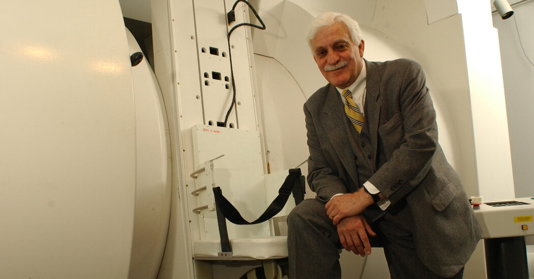
Incensed when two other men won the Nobel Prize in Medicine, he took out a newspaper ad that called his exclusion a “shameful wrong that must be righted.”Dr. Raymond Damadian, who built the first magnetic resonance imaging scanner, which revolutionized doctors’ ability to diagnose cancer and other illnesses — but who, to his dismay, saw the Nobel Prize for the science behind it go to two others — died on Aug. 3 at his home in Woodbury, N.Y. He was 86.The cause was cardiac arrest, said Daniel Culver, a spokesman for the Fonar Corporation, which Dr. Damadian founded in 1978.Since Dr. Damadian and his research assistants finished building the first M.R.I. scanner more than 40 years ago, it has become an essential piece of medical equipment, allowing doctors to peer inside the human body with more detail and greater resolution than X-rays and CT scans provide, without exposing patients to damaging radiation as many other technologies do.“We take it for granted now, but M.R.I. is absolutely spectacular,” Dr. Burton P. Drayer, the chairman of the radiology department at the Mount Sinai Health System in Manhattan, said in a phone interview. “M.R.I. is better at detecting cancers, particularly in the brain and spine.”But the question of who owned the idea, and the device, was a contentious one from the start.Two scientists whose research contributed to M.R.I. technology were later awarded a Nobel Prize, an honor that Dr. Damadian felt should have also gone to him. And no sooner had his machine come on the market than a handful of major corporations started producing their own versions, leading to years of court battles over patent rights.The vision of scanning the human body without radiation came to Dr. Damadian in the late 1960s, he said, when he was working on nuclear magnetic resonance spectroscopy — which, until then, had been used to identify the chemical makeup of the contents of a test tube — at Downstate Medical Center (now SUNY Downstate Health Sciences University) in Brooklyn.Working with rats, he discovered that when tissues were placed in a magnetic field and hit with a pulse of radio waves, cancerous ones emitted distinctly different radio signals than healthy ones.He published his findings in 1971 in the journal Science and was granted a patent three years later for an “apparatus and method for detecting cancer in tissue.” It took 18 months to build the first M.R.I., originally known as a nuclear magnetic resonance scanner, or N.M.R. Its first scan, on July 3, 1977, was of Lawrence Minkoff, one of Dr. Damadian’s assistants — a vivid and colorful image of his heart, lungs, aorta, cardiac chamber and chest wall.Dr. Damadian in his Brooklyn laboratory in 1977, the year his M.R.I. machine made the first human scan.via Fonar Corporation“Having birthed the original idea of the N.M.R. body scanner, we were intent on being the first to accomplish it,” Dr. Damadian said in the book “Gifted Mind: The Dr. Raymond Damadian Story, Inventor of the M.R.I.,” published in 2015, which he wrote with Jeff Kinley. “Failing to do so meant we might be denied the recognition for the original idea.”But the technology behind the M.R.I. had several fathers.Acknowledging that he was inspired by Dr. Damadian’s work, Paul C. Lauterbur of the State University of New York at Stony Brook had figured out how to translate the radio signals bounced off tissue into images. And Peter Mansfield of the University of Nottingham in England had developed mathematical techniques for analyzing the data, making the process more practical.Incorporating those advances, Dr. Damadian’s company, Fonar, based in Melville, N.Y., produced the first commercial scanner in 1980.But Fonar was soon confronted with competition from major corporations like General Electric, Johnson & Johnson, Siemens, Hitachi and Philips.Dr. Damadian sued them all for patent infringement. He lost his case against Johnson & Johnson when a federal judge in 1986 set aside a jury verdict in favor of Fonar,The judge, Dr. Damadian said, “did everything he could to sandbag us in court.”He won his biggest legal victory in 1997, when the U.S. Court of Appeals for the Second Circuit affirmed a lower court’s award of nearly $129 million in damages and interest from G.E. He also won smaller settlements from other manufacturers.“The court verdict gives us official recognition for origination,” Dr. Damadian told The New York Times after the G.E. decision.His pioneering work was also recognized when he received the National Medal of Technology from President Ronald Reagan in 1988 and was inducted into the National Inventors Hall of Fame in 1989. That same year he donated his first scanner, which he called Indomitable, to the Smithsonian Institution.But the recognition that he craved most — a Nobel Prize — eluded him.Dr. Damadian, left, with his 1977-model scanner and his colleagues Lawrence Minkoff, center, the subject of its first scan, and Michael Goldsmith.Raymond Vahan Damadian was born on March 16, 1936, in Manhattan, and grew up in Forest Hills, Queens. His father, Vahan, an immigrant from Turkey, was a newspaper photoengraver; his mother, Odette (Yazedjian) Damadian, was an accountant.Raymond studied violin for several years at Juilliard but diverted to science when he received a Ford Foundation scholarship to attend the University of Wisconsin, Madison. He majored in mathematics there and graduated with a bachelor’s degree in 1956. He received his medical degree four years later from the Albert Einstein College of Medicine in the Bronx and then became a fellow in biophysics at Harvard, where he became familiar with nuclear magnetic resonance technology.While working at Downstate and later at Fonar, Dr. Damadian was aware of Dr. Lauterbur, a chemist who was also working on M.R.I. imaging and with whom he shared the National Medal of Technology.In “Gifted Mind,” Dr. Damadian acknowledged that Dr. Lauterbur “realized that the N.M.R. signal differences in diseased and normal tissues I discovered could be used to construct a picture (image).”But in 2003, when Dr. Lauterbur and Dr. Mansfield won the Nobel Prize in Medicine for their contributions to the science of magnetic resonance imaging, Dr. Damadian was enraged.He wrote a letter to the American Medical Association, proclaiming that “some unconscionable scientific pilferer is trying to steal my entire life.”He then spent several hundred thousand dollars on an advertisement that ran in six major international newspapers. Headlined “The Shameful Wrong That Must Be Righted,” the ad claimed that the Nobel Prize committee had unfairly denied him the prize.At the bottom of the ad he provided a coupon, addressed to the Nobel Committee for Physiology or Medicine, to let readers tell the committee that “the TRUTH must have a place,” and that it should add him as the third recipient of the award.Dr. Hans Ringertz, chairman that year of the Swedish committee that awards the prize, had no comment on Dr. Damadian’s claims but told The Times that there was nothing to prevent Dr. Damadian from being nominated in the future.A year later, Dr. Damadian received one of the two annual Bower Awards given by the Franklin Institute, a science museum in Philadelphia. He was cited for his business leadership.“There is no controversy in this,” said Dr. Bradford A. Jameson, a professor of biochemistry at Drexel University who was the chairman of the committee that chose the winners. “If you look at the patents in this field, they’re his.”Dr. Damadian said then that he was no longer concerned with the Nobel dispute. But he told The Times, “If people want to reconsider history apart from the facts, there’s not much that I can do about that.”Dr. Damadian continued to innovate. He created open M.R.I. machines, which alleviate the claustrophobia patients can experience during scans when they are moved slowly through a tight tunnel, as well as mobile and stand-up scanners.In recent years, he was focused on research that included imaging cerebral spinal fluid as it flowed to the brain.Fonar made and installed about 500 M.R.I. scanners, but today it is focused on managing imaging centers in the United States and servicing existing scanners.Dr. Damadian is survived by his daughter, Keira Reinmund; his sons, Timothy, Fonar’s president and chief executive since 2016, and Jevan; nine grandchildren; three great-grandchildren; and a sister, Claudette Chan. His wife, Donna (Terry) Damadian, died in 2020.In 1982, as the industry he helped create was in its infancy, Dr. Damadian told Newsday that he had not lost his inventor’s zeal for what lay ahead.“In 1977, I knew my machine could be a reality,” he said. “But until then, it was like building a model airplane. Now I know it’s for real.”Kenneth Chang

Read more →

Professor working to heal homelessness, fix broken aid industry

While volunteering at an Indian orphanage in 1984, Dr. David Buck found his passion for helping those most in need during an encounter with the greatest humanitarian in human history — Mother Teresa. That unforgettable experience put him on a decades-long path to reshaping how communities care for the underserved.
“Mother Teresa asked us ‘Who among you wants to change the world?'” recalled Buck. “Internally I was screaming ‘Me, I do!'”
But Buck, now a professor and associate dean for community health at the University of Houston Tilman J. Fertitta Family College of Medicine, didn’t raise his hand.
“She said ‘For all of you who want to do that, there’s the door. Our task is much greater. Our task is to love one person at a time,'” he said. “So, I came back to the U.S. and knew I wanted to make a difference here at home.”
Buck recently reflected on his nearly 40 years of tackling the homelessness epidemic, repairing a fragmented aid industry, and challenging the status quo in medicine in a first-person account published in the Journal of Social Distress and Homelessness.
Early in his career, just after returning from that life-changing experience with Mother Teresa, Buck volunteered with Houston Catholic Worker House where he learned the biggest problem facing the homeless was access to health care. Not long after, he established two free health clinics and a dental clinic, but soon realized the root problem wasn’t just access. Instead, it was a lack of integration of all the services they needed.

Read more →

Cellular waste may supercharge immune cell function

The immune cells that protect us from infection and cancer seek out a wide array of fuel sources to power their function — including some long thought to be cellular waste products.
The findings, published today in Cell Metabolism, lay the foundation for future personalized dietary recommendations designed to supercharge the immune system and augment therapies for cancer and other diseases.
“Every process in the body is powered by metabolism, which in turn is fueled by the nutrients we consume through our diet,” said Russell Jones, Ph.D., chair of Van Andel Institute’s Department of Metabolism and Nutritional Programming and senior author of the study. “We found that immune cells are much more flexible in selecting the nutrient fuels they consume and, importantly, that they prefer some nutrients that were previously dismissed as waste. This understanding is crucial for optimizing T cell responses and developing new strategies for boosting our ability to fight off disease.”
T cells are the soldiers of the immune system and are tasked with combating bacteria, viruses and even cancer cells. They absorb nutrients from the foods we eat to generate the energy required to carry out their jobs.
The findings stem from a novel approach that could reshape how metabolism is studied. Historically, T cells are grown in the lab in dishes of nutrient-containing media. However, much of this media doesn’t fully reflect the rich array of nutrients found in the human body. To solve the problem, Jones and his colleagues developed media packed with a more diverse range of nutrients.
“Previously, we were giving immune cells a very basic diet — the equivalent of just eggs and toast,” Jones said. “We found that, when we offer them a full buffet, these cells actually prefer a wider array of ‘fuels’ than previously believed. This has major implications for how we tailor dietary recommendations as ways to promote health and combat disease.”
One example is lactate, a cellular waste product responsible for muscle aches after a long workout. When presented with glucose, a common sugar found in the body and in lab media, and lactate, the T cells preferentially used the lactate to power energy production — a decision that enhanced their function.
Lactate also is an important byproduct of cancer cells and facilitates cancer’s ability to invade other tissues and evade attack by the immune system. Some research suggests that too much lactate may be bad for T cells. The work from Jones’ group indicates that, at lower levels, lactate may actually enhance T cell function.
The findings also suggest that T cell function and survival is strongly influenced by the nutrients available in their environment. Going forward, Jones and his colleagues aim to delve deeper into the intricate relationships between metabolism and the immune system in search of new insights to further illuminate how these crucial systems collaborate.
Authors include Irem Kaymak, Ph.D., Katarzyna M. Luda, Ph.D., Lauren R. Duimstra, Eric H. Ma, Ph.D., Joseph Longo, Ph.D., Michael S. Dahabieh, Ph.D., Brandon M. Oswald, McLane J. Watson, Ph.D., Susan M. Kitchen-Goosen, Lisa M. DeCamp, Shelby E. Compton, Zhen Fu, Ph.D., Kelsey S. Williams, Ph.D., and Ryan D. Sheldon, Ph.D., of VAI; Brandon Faubert, Ph.D., of University of Chicago; and Ralph J. DeBerardinis, Ph.D., of UT Southwestern Medical Center.
Research reported in this publication was supported by Van Andel Institute (Jones), and an Allen Distinguished Investigator Award, a Paul G. Allen Frontiers Group advised grant of the Paul G. Allen Family Foundation (Jones).
Jones is supported by the National Institute of Allergy and Infectious Diseases of the National Institutes of Health under award no. R01AI165722. Support for authors on this project include a postdoctoral fellowship award from Fonds de la Recherche du Québec-Santé (FRQS) (Dahabieh); a VAI Metabolism and Nutrition (MeNu) Program Pathway-to-Independence Award (Longo); National Cancer Institute award no. T32CA251066-01A1) (Watson) and award no. R35CA2202901 (DeBerardinis); and the Howard Hughes Medical Institute Investigator Program (DeBerardinis).

Read more →