Supercomputer creates the most realistic virtual brain ever

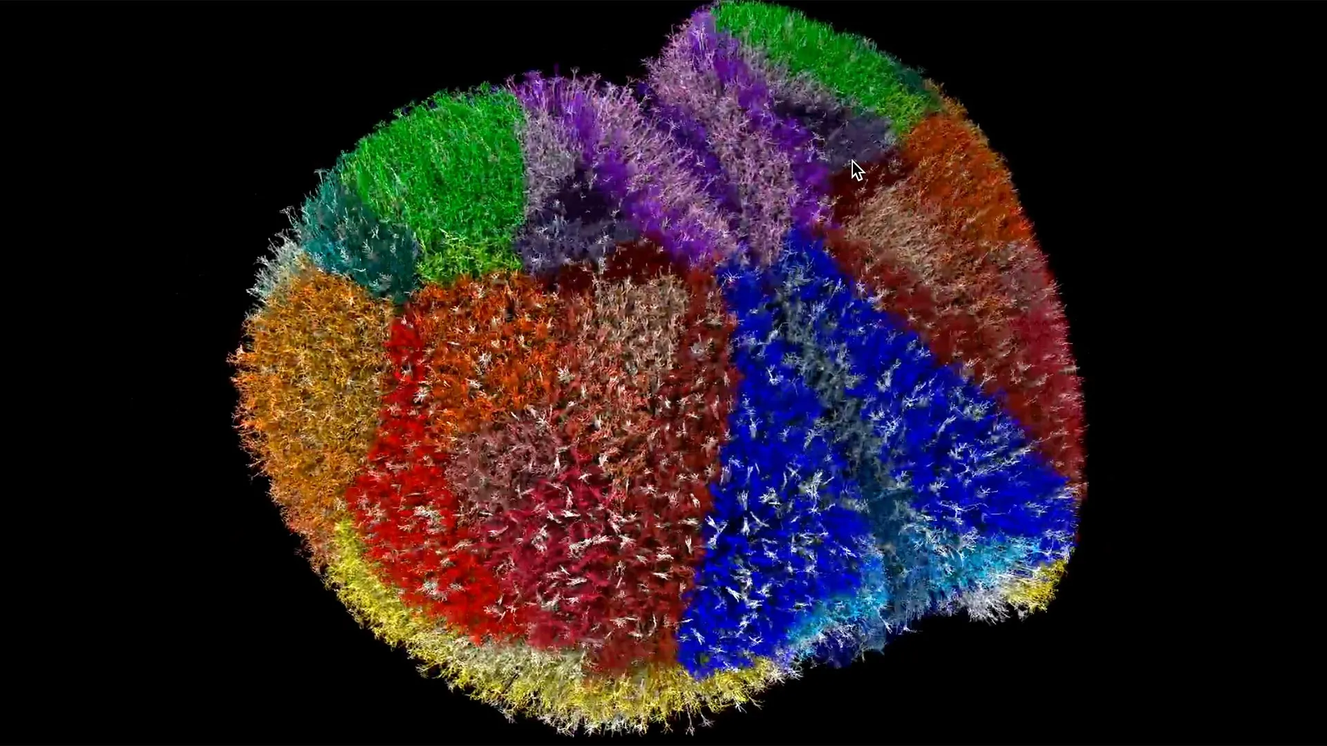
Using the strength of one of the fastest supercomputers on the planet, scientists have created one of the most comprehensive and biologically realistic animal brain simulations ever developed. This digital reconstruction of the entire mouse cortex gives researchers a new way to explore brain function by recreating conditions such as Alzheimer’s or epilepsy inside a virtual environment. It allows them to track how damage moves through neural circuits and to investigate processes involved in cognition and consciousness. The simulation includes nearly ten million neurons, 26 billion synapses, and 86 connected brain regions, capturing both structure and activity at high resolution.
This major accomplishment was enabled by Supercomputer Fugaku, Japan’s premier high-performance system capable of performing quadrillions of calculations per second. Scientists from the Allen Institute and Tadashi Yamazaki, Ph.D., at Japan’s University of Electro-Communications, partnered with three additional Japanese organizations to lead this work. The full findings will be detailed in a paper scheduled for release at SC25, the top global supercomputing conference taking place in mid-November.
A New Way to Explore Disease and Brain Function
Researchers can use this virtual cortex to study how neurological disorders form, how brain waves contribute to attention, and how seizures move through neural networks. In the past, these kinds of questions required real brain tissue and could only be addressed through individual experiments. With this model, scientists can test many ideas in a digital space. These simulations may offer early clues about how brain disorders begin before symptoms appear and provide a safe way to evaluate potential therapies.
“This shows the door is open. We can run these kinds of brain simulations effectively with enough computing power,” said Anton Arkhipov, Ph.D., an investigator at the Allen Institute who worked on the project. “It’s a technical milestone giving us confidence that much larger models are not only possible, but achievable with precision and scale.”
This collaborative effort brings together deep neuroscience knowledge and the processing power of a world-class machine. The Allen Institute contributed the biological foundation of the virtual brain using data from the Allen Cell Types Database and the Allen Connectivity Atlas, while Fugaku handled the massive computations needed to generate the model.
How Researchers Created the Whole Cortex Simulation
Fugaku, developed by RIKEN and Fujitsu, ranks among the fastest computers ever built and can process more than 400 quadrillion operations every second. To grasp the scale of that number, counting to it at one count per second would take over 12.7 billion years (approximately the age of the universe: 13.8 billion years). The system’s name, “Fugaku,” refers to Mount Fuji and reflects the machine’s far-reaching capability and towering performance.

“Fugaku is used for research in a wide range of computational science fields, such as astronomy, meteorology, and drug discovery, contributing to the resolution of many societal problems,” said Yamazaki. “On this occasion, we utilized Fugaku for a neural circuit simulation.”
The supercomputer is assembled from many small processing units called nodes. These nodes are organized into units, shelves, and racks, forming a system of 158,976 total nodes that can handle enormous amounts of data and calculations.
From Biological Data to a Living Digital Cortex
Using the Allen Institute’s Brain Modeling ToolKit, the team converted biological data into a functioning digital reconstruction of the cortex. To simulate living neuronal behavior, a tool called Neulite transformed mathematical equations into virtual neurons capable of spiking, signaling, and communicating as real neurons do.
Watching the simulation is similar to observing live brain activity. The model reproduces fine details of neuron structure, synapse activity, and electrical signaling across cell membranes. “It’s a technical feat, but it’s only the first step,” said Yamazaki. “God is in the details, so in the biophysically detailed models, I believe.”
“Our long-term goal is to build whole-brain models, eventually even human models, using all the biological details our Institute is uncovering,” said Arkhipov. “We’re now moving from modeling single brain areas to simulating the entire brain of the mouse.” With computational systems this powerful, the possibility of a complete, biologically accurate brain model is moving from concept to reality. Scientists are entering a new era in which understanding the brain also means being able to construct one.
This cutting-edge research was made possible by an international team including Laura Green, Ph.D.; Beatriz Herrera, Ph.D.; Kael Dai, B.Sc.; Rin Kuriyama, M.Sc.; and Kaaya Akira, Ph.D.

Read more →

Scientists discover metformin may block key exercise benefits

A widely used medication for diabetes prevention may unexpectedly interfere with one of the most reliable ways to lower disease risk: regular physical activity.
A Rutgers-led team reported in The Journal of Clinical Endocrinology & Metabolism that metformin appeared to weaken several important benefits normally gained from exercise. These include improvements in blood vessel function, overall fitness and the body’s ability to control blood sugar.
Since 2006, medical guidance has encouraged people with elevated blood sugar to combine metformin with consistent exercise. The expectation was that two well-established treatments would reinforce one another. According to the Rutgers researchers, the evidence suggests a different outcome.
“Most health care providers assume one plus one equals two,” said Steven Malin, a professor in the Department of Kinesiology and Health in the School of Arts and Sciences and the lead author of the study. “The problem is that most evidence shows metformin blunts exercise benefits.”
How the Study Tested Metformin’s Impact
To explore this issue, Malin and his colleagues enrolled 72 adults considered at risk for metabolic syndrome, which is a cluster of conditions known to raise the likelihood of both diabetes and heart disease. Participants were separated into four groups: high-intensity exercise with placebo, high-intensity exercise with metformin, low-intensity exercise with placebo and low-intensity exercise with metformin.
Over a 16-week training period, the research team measured how well participants’ blood vessels responded to insulin. This response helps vessels widen and deliver oxygen, hormones and nutrients after eating.

The results showed that exercise alone strengthened vascular insulin sensitivity. Blood vessels became more responsive to insulin, allowing greater blood flow to the muscles. This is important because insulin-assisted dilation helps move glucose from the bloodstream into body tissues, lowering blood sugar after meals.
Metformin Significantly Reduced Expected Gains
When metformin was added, these improvements became noticeably smaller. The medication also appeared to limit gains in aerobic capacity and lessen positive changes in inflammation and fasting blood glucose.
“Blood vessel function improved with exercise training, regardless of intensity,” Malin said. “Metformin blunted that observation, suggesting one type of exercise intensity is not better either with the drug for blood vessel health.”
These findings are concerning because exercise is intended to support healthy blood sugar levels and improve physical capability. If metformin reduces these effects, patients relying on both may not receive the degree of protection they expect.
“If you exercise and take metformin and your blood glucose does not go down, that’s a problem,” Malin said. “People taking metformin also didn’t gain fitness. That means their physical function isn’t getting better and that could have long-term health risk.”
Real-Life Consequences for Daily Function and Well-Being

Malin noted that exercise-driven fitness improvements influence everyday activities such as climbing stairs, playing with children or maintaining an active social life. If these gains are diminished, overall quality of life can suffer.
The researchers emphasized that the findings should not prompt anyone to stop taking metformin or abandon exercise. Instead, the study highlights the need for physicians to closely consider how these interventions interact and to monitor patient progress. Malin hopes that future studies will help identify approaches that preserve the positive effects of both.
Why Metformin May Interfere With Exercise Adaptation
The reason metformin dampens exercise benefits is still being investigated. Malin explained that metformin partly works by inhibiting specific mitochondrial processes. This action reduces oxidative stress and helps regulate blood sugar. However, the same interference may block the cellular changes typically triggered by exercise, including better mitochondrial efficiency and improved aerobic performance. In essence, the mechanism that makes metformin effective may also hinder the body’s ability to fully respond to physical training.
Earlier studies have suggested a similar pattern, but this trial is one of the first to look closely at vascular insulin sensitivity, a key factor in both glucose control and cardiovascular health. By demonstrating that metformin can limit changes in both major arteries and small capillaries across different exercise intensities, the researchers highlight how complex these combined treatments can be.
Implications for Diabetes Prevention and Clinical Guidelines
Malin pointed out that nearly 35 million people in the United States live with type 2 diabetes, and prevention strategies often depend on a mix of lifestyle changes and medication. If these approaches do not work together as expected, long-term risks may increase.
“We need to figure out how to best recommend exercise with metformin,” Malin said. “We also need to consider how other medications interact with exercise to develop better guidelines for doctors to help people lower chronic disease risk.”
Other Rutgers researchers involved in the study include: Sue Shapses, professor in the Department of Nutritional Sciences at the School of Environmental and Biological Sciences; Andrew Gow, professor of pharmacology and toxicology at the Ernest Mario School of Pharmacy; Ankit Shah, assistant professor in the Department of Medicine at Robert Wood Johnson Medical School; Tristan Ragland, a former post-doctoral fellow in Department of Kinesiology and Health; Emily Heiston, project scientist and clinical coordinator in the Applied Metabolism and Physiology Laboratory; and Daniel Battillo, a former doctoral student in the Department of Kinesiology and Health.

Read more →

A silent kidney crisis is spreading faster than anyone expected

A growing number of people worldwide are now believed to have reduced kidney function, according to a new analysis. The number of affected individuals increased from 378 million in 1990 to 788 million in 2023. As populations have expanded and grown older, the condition has reached a point where it is now listed among the top 10 causes of death across the globe.
Researchers from NYU Langone Health, the University of Glasgow, and the Institute for Health Metrics and Evaluation (IHME) at the University of Washington led the investigation. Their work focused on chronic kidney disease, a condition in which the kidneys gradually lose the ability to clear waste and extra fluid from the bloodstream. Early stages often produce no noticeable symptoms, while advanced disease may require dialysis, kidney replacement therapy, or a transplant.
Global Burden and Mortality Trends
The study estimates that roughly 14% of adults worldwide live with chronic kidney disease. In addition, approximately 1.5 million people died from the condition in 2023. When changes in population age structures are taken into account, this represents more than a 6% increase in deaths since 1993.
“Our work shows that chronic kidney disease is common, deadly, and getting worse as a major public health issue,” said study co-senior author Josef Coresh, MD, PhD, director of NYU Langone’s Optimal Aging Institute. “These findings support efforts to recognize the condition alongside cancer, heart disease, and mental health concerns as a major priority for policymakers around the world.”
Growing Health Priority for Global Agencies
In May, the World Health Organization added chronic kidney disease to its list of health priorities aimed at reducing early deaths from noncontagious diseases by one-third before 2030. Coresh, who is also the Terry and Mel Karmazin Professor of Population Health at the NYU Grossman School of Medicine, notes that identifying current patterns in the disease is essential for developing effective strategies.

The new report was published online Nov. 7 in The Lancet and, according to the authors, represents the most detailed assessment of chronic kidney disease in nearly ten years. It is also being presented at the American Society of Nephrology’s annual Kidney Week conference.
How the Study Was Conducted
The research was part of the Global Burden of Disease (GBD) 2023 study, considered the most wide-ranging effort to track health-related loss in countries over long periods of time. Findings from this initiative frequently inform public health policy and guide future research.
To generate the new estimates, the team examined 2,230 scientific papers and national health datasets from 133 countries. Their work centered not only on diagnoses and deaths, but also on the level of disability associated with chronic kidney disease.
Links to Heart Disease and Leading Risk Factors
Another key conclusion was that impaired kidney function is a major contributor to heart disease. The study reports that it accounted for about 12% of global cardiovascular deaths. In 2023, chronic kidney disease ranked as the 12th leading cause of disability-related reductions in quality of life. High blood sugar, high blood pressure, and high body mass index (a measure of obesity) were identified as the most significant risk factors for developing the condition.

Most individuals in the study were in the early stages of chronic kidney disease. Coresh emphasizes that this is a crucial window for intervention, as timely treatment and lifestyle changes can prevent the need for dialysis or transplantation later on.
Access to Treatment Remains Uneven Worldwide
Coresh also points out that in parts of sub-Saharan Africa, Southeast Asia, Latin America, and other lower-income regions, many people do not receive dialysis or transplants. These therapies are less accessible and often too expensive for widespread use in those areas.
“Chronic kidney disease is underdiagnosed and undertreated,” said study co-lead author Morgan Grams, MD, PhD. “Our report underscores the need for more urine testing to catch it early and the need to ensure that patients can afford and access therapy once they are diagnosed.”
New Therapies but Slow Global Progress
Grams, the Susan and Morris Mark Professor of Medicine at the NYU Grossman School of Medicine, notes that several new medications introduced over the past five years can slow disease progression and reduce the likelihood of heart attack, stroke, and heart failure. Even so, she explains that it will take time before the benefits of these treatments are reflected worldwide.
She also warns that because chronic kidney disease is often not tested for, its true global prevalence may be even higher than the study suggests.
Study Support and Research Team
The project was funded by National Institutes of Health grant R01DK100446, the Gates Foundation, and the National Kidney Foundation.
Coresh serves as a scientific adviser and equity holder in Healthy.io, a health technology company that provides remote clinical testing and related services. He also works as a consultant for SomaLogic. These roles are disclosed and managed according to NYU Langone Health policies.
Along with Coresh and Grams, Patrick Mark, PhD, at the University of Glasgow, and Lauryn Stafford, MS, at IHME at the University of Washington in Seattle, were co-lead authors.
Additional co-senior authors included Jennifer Lees, PhD, at the University of Glasgow, and Theo Vos, PhD, and Liane Ong, PhD, at IHME at the University of Washington in Seattle.

Read more →

Catherine’s first speech in two years calls for dignity for carers

The Princess of Wales has called for more dignity for carers and a greater respect for values such as “tenderness” in business, in her first public speech in two years.At an event in the City of London, Catherine called on 80 top business leaders to play their part in ensuring employees can have healthy family lives, including caring responsibilities.”I believe in restoring the dignity to the quiet, often invisible work of caring, of loving well, as we look to build a happier, healthier society,” the princess told the Future Workforce Summit.The event was part of Catherine’s campaign to raise awareness about the importance of children’s early years and building healthy family relationships.This was Catherine’s first speech since November 2023 and the first since her cancer diagnosis, which was revealed in March 2024.Since then, she has delivered recorded messages on social media, including about her gradual recovery after chemotherapy, but this has been the first speech since then to have been delivered in person.It marks another step in her return to public life and also reflects her style, with the event beginning with a poem read by Alex Wharton, former children’s laureate for Wales.Her speech, confidently delivered, reflected her deeply-held, personal message about the need for a greater emphasis on kindness and recognising that “love is the first and most essential bond”.”The love we feel in our earliest years fundamentally shapes who we become and how we thrive as adults,” said the princess.She told business leaders about the need for families to be able to provide a supportive home for their children, as part of what she called the “weave of love”.The speech was delivered high up in a city skyscraper, with London stretched out below like a model village.Catherine called on business leaders to re-think their workplaces and create a different kind of model for their priorities, in which companies were “valuing time and tenderness just as much as productivity and success”.The princess, who set up the Centre for Early Childhood before her illness, called for a more rounded way of measuring success.”As business leaders you will face the daily challenge of finding the balance between profitability and having a positive impact. But the two are not, and should not be incompatible,” she said.The event also heard from Robert Waldinger, a psychiatrist at Harvard Medical School, who said research showed that people who had been in nurturing relationships in early childhood were still likely to be more emotionally secure in their old age.Former England football manager Sir Gareth Southgate, addressing this business gathering, said that when he was trying to motivate players, he had found that youngsters from difficult family backgrounds might not have the same level of trust as those who had grown up in closer family relationships.He said that coaches carried out personality profiles on the players to understand the “way they were wired”.He talked about his own experiences of responding to moments of pressure, using a combination of “sleep, good nutrition and limiting alcohol” and breathing exercises to control stress.Sir Gareth also remembered his own recovery after the “public humiliation” of missing a penalty in the Euro 96 semi-final when he said “half the world was watching”.He said that it meant “slowly taking small steps to recover your confidence” and he talked of the importance of people, in any type of workplace, being able to “feel comfortable in your own skin”.This gathering was part of the Business Taskforce set up by the princess to try to get business support for her campaign to recognise the importance of early childhood.Delegates at the event heard from leaders of companies supporting the project including Aviva, NatWest Group, Iceland, Ikea, Lego and Deloitte.

Read more →

Eleven countries added to methanol poisoning warning list

The UK Foreign Office has added a further 11 countries to its list warning of the risks of methanol poisoning from tainted alcoholic drinks.The updated travel guidance includes Bangladesh, India, Iran, Jordan, Libya, Malawi, Malaysia, Morocco, Nepal, Papua New Guinea, and Rwanda following “a global increase in the number of reported cases” of methanol poisoning.Last month, Ecuador, Japan, Kenya, Mexico, Nigeria, Peru, Russia and Uganda were added to the existing list, following a number of high-profile incidents including the death of six tourists in Laos in 2024.Travellers are being warned that even small amounts of methanol can cause blindness or death within 12 to 48 hours.Methanol is a toxic industrial alcohol used in antifreeze, windscreen washer fluid and paint thinner. It is not meant for human consumption.While it is naturally produced during the manufacture of alcoholic drinks, commercial firms reduce it to low, safe levels for human consumption through the distillation process.But in some countries, it is illegally mixed into alcoholic drinks to cut costs, and because it is tasteless and odourless, it is impossible to detect.The Foreign Office is running a campaign to advise on reducing risks and recognising symptoms of methanol poisoning.Foreign Office minister Hamish Falconer said all travellers should know the signs of methanol poisoning.”If you’re drinking spirits overseas, stick to trusted places and avoid homemade alcohol or free shots,” Falconer said.”If something feels off, like a hangover that’s way worse than normal or vision problems – get medical help fast.”The full list of countries listed as posing a risk of methanol poisoning are: Brazil, Bangladesh, Cambodia, Costa Rica, Ecuador, Fiji, India, Indonesia, Iran, Jordan, Kenya, Laos, Libya, Malawi, Malaysia, Mexico, Morocco, Nepal, Nigeria, Papua New Guinea, Peru, Russia, Rwanda, Thailand, Turkey, Uganda, and Vietnam.

Read more →

How do cold weather health alerts work?

50 minutes agoShareSaveJennifer ClarkeBBC NewsShareSaveGetty ImagesAmber cold health alerts have been issued for the North East and West of England, and Yorkshire and the Humber. A yellow cold alert is in place for the rest of England.They will remain in place until 08:00 GMT on Saturday 22 November.How does the weather health alert system work?The weather alert service is run by the UK Health Security Agency (UKHSA) and the Met Office.The alerts warn the public when high or low temperatures pose a potential risk to health. Cold health alerts are issued between 1 November and 30 March, and heat health alerts are published between 1 June and 30 September.As well as warning the public, the system sends guidance directly to NHS England, the government and healthcare professionals during periods of adverse weather.Alerts are categorised according to severity and include:headline weather conditions expected in the coming daysdetails of how weather conditions will affect each regionlinks to additional information, advice and guidanceThe system was designed to help reduce illness and deaths during periods of extreme hot and cold weather.What do the alert levels mean?The level of alert is based on Met Office forecasts and data.There are four levels ranging from green (least severe) to red (most severe):GreenGreen is the normal level, when advice is given on how people should prepare to respond if temperatures rise or fall.YellowYellow alerts are issued during periods of hot or cold weather that are only likely to affect those who are particularly vulnerable, for example the elderly, or those with existing health conditions.AmberAmber alerts are issued in situations that could potentially put the whole population at risk. The NHS may see increased demand on GPs and ambulances, for example.Travel disruption is also likely.RedA red alert is the most severe.It is issued in situations when hot or cold weather would be a significant risk to life, even for the healthy population, and could lead to failures of critical national infrastructure, such as power outages or roads and rail lines being closed.What does cold weather do to the body?Flu and certain other diseases that cause colds and sore throats spread more easily in the winter months. Pneumonia, when there is inflammation in the lungs because of an infection, is more common after cold weather.Cases of norovirus – the winter vomiting bug – tend to rise too.Prof Damian Bailey, from the University of South Wales, made a programme for BBC Radio 4’s Inside Health about the effects of cold on the body.Presenter James Gallagher spent 30 minutes in a room where the air temperature dropped from 21C to 10C.During the experiment, the blood flow to his brain fell, his blood pressure and heart rate increased, the temperature of his extremities – arms, legs and head – dropped, and he took significantly longer to complete a series of puzzles.Getty ImagesProf Bailey said 18C is the body’s tipping point – below that and the body has to work hard to maintain its core temperature. That is one reason why heart attacks and strokes are more common in the winter months.When it is not possible to keep rooms heated to 18C, he recommends wearing gloves, warm socks and a woolly hat, eating a higher carbohydrate diet and generating more body heat by moving around.

Read more →

Blocking one enzyme may break the link between alcohol and liver disease

Scientists have identified an unexpected link between the way the body processes sugar and the development of alcohol addiction. This connection also points to a promising therapeutic target for treating alcohol-associated liver disease (ALD) and alcohol use disorder (AUD).
A study published in Nature Metabolism by researchers at the University of Colorado Anschutz reports that alcohol activates a metabolic pathway that causes the body to produce fructose internally. Fructose is the same sugar found in many sweetened foods and drinks. This internal production relies on an enzyme called ketohexokinase (KHK), which appears to strengthen alcohol-seeking behavior while also contributing to liver damage.
Blocking KHK Reduces Drinking and Liver Injury
Experiments in mice showed that animals lacking KHK had a noticeably lower interest in alcohol. They consumed less in voluntary drinking tests, performed differently in reward-based experiments, and showed reduced activity in brain regions associated with addictive behavior.
The study also found that liver injury caused by alcohol did not develop when KHK was disrupted through genetic methods or medication. These mice had less fat buildup, less inflammation, and less scarring in their livers. The findings suggest that limiting fructose metabolism could slow or even prevent the progression of alcohol-related liver disease.
Breaking the Cycle Between Sugar and Alcohol
“Our findings show that alcohol doesn’t just damage the liver directly, it hijacks the body’s sugar metabolism in a way that enhances drinking behavior and worsens liver injury,” said Miguel A. Lanaspa, DVM, PhD, associate research professor at CU Anschutz and senior author. “By targeting fructose metabolism, we may be able to break this cycle and develop new treatments for both alcohol addiction and liver disease.”
Because alcohol-associated liver disease and metabolic dysfunction-associated steatotic liver disease (MASLD) rely on similar fructose-driven processes, the researchers suggest that treatments aimed at blocking fructose metabolism could help people with liver disease related to either alcohol or diet.

A Shared Metabolic Pathway for Liver Damage
“This discovery highlights an unexpected intersection between sugar and alcohol metabolism,” said Richard Johnson, MD, professor at CU Anschutz and study co-author. “It opens exciting possibilities for developing treatments that target a common pathway underlying both metabolic and alcohol-related liver diseases.”
The results offer a promising new direction for tackling alcohol addiction and liver disease, two conditions with limited effective treatment options.

Read more →

Amazon scorpion venom shows stunning power against breast cancer

The venom of an Amazonian scorpion species may contain a compound capable of helping treat breast cancer, a disease that remains one of the top causes of death among women.
Researchers at the University of São Paulo’s Ribeirão Preto School of Pharmaceutical Sciences (FCFRP-USP) in Brazil have pinpointed a molecule in the toxin of Brotheas amazonicus that appears to attack breast cancer cells in a way similar to a widely used chemotherapy medication.
These early findings were generated through a collaboration with scientists from the National Institute for Amazonian Research (INPA) and the Amazonas State University (UEA), and were presented during FAPESP Week France in the Occitanie region of southern France.
“Through bioprospecting, we were able to identify a molecule in the species of this Amazonian scorpion that is similar to that found in the venoms of other scorpions and that acts against breast cancer cells,” said Eliane Candiani Arantes, a professor at FCFRP-USP and the coordinator of the project.
Turning Venom Components Into Biopharmaceutical Tools
Teams at FCFRP-USP and partner institutions have long worked to clone and express bioactive molecules, including proteins from rattlesnake and scorpion venom. These efforts take place within projects supported by FAPESP and connected to the Center for Translational Science and Development of Biopharmaceuticals (CTS), housed at the Center for the Study of Venoms and Venomous Animals (CEVAP) at São Paulo State University (UNESP) in Botucatu.
One result of this research is CEVAP’s patented fibrin sealant, described as a “biological glue.” It is produced from serinoproteinase enzymes extracted from snake venom (including Bothrops neuwiedi pauloensis and Crotalus durissus terrificus) combined with cryoprecipitate enriched with fibrinogen from buffalo, cattle, or sheep.

When applied, these components form a fibrin structure resembling the body’s natural clotting and tissue repair processes. The sealant has been investigated for use in nerve repair, bone healing, and restoring movement following spinal cord injury. It is currently undergoing phase three clinical trials, which represent the final evaluation stage required before approval of a new therapy.
Advancing Fibrin Sealant Technology Through Genetic Expression
Recently, researchers cloned and expressed another rattlesnake serine protease known as cholinein-1. Its amino acid sequence differs from gyroxine, a toxin taken directly from rattlesnake venom and used in fibrin sealant production.
“Our idea now is to obtain this serine protease through heterologous expression [in a fragment or complete gene from a host organism that doesn’t have it naturally] in Pichia pastoris,” Arantes explained.
Using this same yeast species, first isolated in France in 1950, the researchers also plan to produce an endothelial growth factor called CdtVEGF. This molecule was originally identified in the venom of Crotalus durissus terrificus.
“This growth factor favors the formation of new vessels. If we combine it with colinein-1, we can create an improved fibrin sealant compared to the one being developed at CEVAP, with the possibility of expanding the industrial scale, since it can be obtained through heterologous expression,” she said.

Through similar genetic expression approaches, the team identified two neurotoxins in scorpion venom with immunosuppressive effects. Working with collaborators at INPA and UEA, they also found a molecule named BamazScplp1 in the venom of Brotheas amazonicus that appears to have anti-tumor potential.
Laboratory tests showed that the peptide’s impact on breast cancer cells was comparable to paclitaxel, a commonly prescribed chemotherapy treatment. It primarily triggers necrosis, a form of cell death previously associated with molecules from other scorpion species.
“We also intend to obtain these molecules through heterologous expression,” said Arantes.
Developing New Cancer Therapies With Radioisotopes
In Campinas, in the state of São Paulo, researchers at a Research, Innovation and Dissemination Center (RIDC) funded by FAPESP — the Cancer Theranostics Innovation Center (CancerThera) — are pursuing a different therapeutic strategy. Their goal is to combine diagnosis and targeted treatment in a single approach.
This method originated in Germany and involves attaching various radioisotopes to molecules that target specific tumors. These tagged molecules can then be used in imaging and treatment.
“Depending on the type of radiation emitted by the isotope we attach to the molecule — whether positron or gamma — we can produce images of it using the tomography equipment available at CancerThera. When we document that an isotope captures too much of a particular molecule, we can replace it with another that emits more intense radiation locally and thus treat tumors,” explained Celso Darío Ramos, a professor at the School of Medical Sciences at the State University of Campinas (FCM-UNICAMP) and one of CancerThera’s lead researchers.
One group at the center focuses on identifying molecules that accumulate in different cancers, while the clinical team evaluates how known compounds might be repurposed.
“We’ve been studying known molecules from hematological cancers, primarily multiple myeloma, as well as other unknown molecules from head and neck cancer, liver cancer, sarcomas, lung cancer, colorectal cancer, and gastric cancer, among others. In addition, we’ve also been studying thyroid cancer, which has been treated with radioactive material, radioactive iodine, for many years, but some patients are resistant. That’s why we’re trying to identify another treatment possibility, with a different radioactive material, for these patients,” Ramos told Agência FAPESP.
A Personalized Cancer Vaccine Built From Dendritic Cells
Another experimental strategy is under development at the Biomedical Sciences Institute at the University of São Paulo (ICB-USP), where researchers are exploring an immunotherapy based on dendritic cells.
These cells are important components of the immune system, and their functioning is often compromised in cancer patients, explained José Alexandre Marzagão Barbuto, a professor at ICB-USP and the project coordinator.
“A few years ago it was discovered that it’s possible to take monocytes from the blood cells of cancer patients and turn them into dendritic cells in the laboratory. But the dendritic cells produced in this way are often diverted to induce tolerance.”
To address this issue, the team created dendritic cells from healthy donors and fused them with cancer cells from patients, producing a personalized vaccine designed to activate the immune system against the individual’s own tumor.
Results from studies involving various cancers, including more recent tests with glioblastoma patients, suggest that this strategy can be effective when the immune response it generates is properly controlled.
“The immune system interprets this vaccine, based on dendritic cells from a healthy donor fused with the patient’s tumor cells, as a transplant and reacts violently,” said Barbuto. “We did the first studies on patients with melanoma and kidney cancer, and the results were very good, and others with glioblastoma. Now we’re hoping to carry out a phase three clinical study.”
Using AI to Improve MRI Predictions for Brain Cancer
Researchers at the Cancer University Institute of Toulouse (IUCT-Oncopole) in France are also contributing to the understanding of glioblastoma. Their work investigates whether artificial intelligence applied to magnetic resonance imaging can reliably indicate whether chemotherapy patients have a DNA modification associated with treatment outcomes and survival.
The modification, known as “MGMT promoter region methylation,” influences how the MGMT protein is produced and regulated.
“MGMT methylation status is an important prognostic factor, but it requires biopsies that aren’t necessarily representative of the entire tumor and can vary in recurrence,” said Elizabeth Moyal, a researcher at IUCT-Oncopole and coordinator of the project.
Partnering with computer scientist Ahmed Berjaoui from IRT Saint-Exupéry, the team adopted AI techniques originally designed for aerospace applications to help resolve these challenges.
“We’ve developed a model capable of predicting survival with high accuracy, ranging from 80% to 90%, and which surpasses other existing techniques,” said Berjaoui.

Read more →

Hundreds of hospice beds unused amid financial crisis

Some 380 hospice beds out of around 2,000 lie empty in England because of financial pressures, say bosses.Hospice UK has told BBC News this is up from 300 a year ago and illustrates the severe challenges facing the sector.Beds are left empty to save money – since staffing and caring is costly – and so are unavailable to patients.Hospices are run by charities, raising between two-thirds and three-quarters of their income from donations and private fund raising. They depend on the rest from the NHS, and managers say this funding has not kept pace with costs, such as employer national insurance.Hospice leaders say their organisations are “on the brink of a financial crisis”.A Department of Health and Social Care spokesperson said the government had already invested £100 million to improve hospice facilities and had committed £80 million for children’s and young people’s hospices over three years.”We recognise there is more to do and we are exploring how we can improve the access, quality and sustainability of all-age palliative care and end of life care in line with the 10-Year Health Plan,” a spokesman added. Hospice UK says five of its members have announced “cost reductions” or cutbacks since early October. In some cases job losses are being made.One of them is Ashgate Hospice in Derbyshire which has warned staff that 52 are at risk of redundancy. Bed numbers are also being reduced – from 15 to six – and the proposals would mean 600 fewer patients being cared for each year. The hospice has blamed energy bills and rising staff salaries with NHS funding not matching the increases.Meanwhile, Arthur Rank Hospice in Cambridge says a cut in NHS funding will mean inpatient beds being reduced from 21 to 12 – what it described as “a devastating decision”. Garden House Hospice Care in Hertfordshire has announced what it calls “the most serious financial challenge in its history” and has launched a consultation process which may lead to more than 20 redundancies.Charlie King, director of external affairs at Hospice UK, said: “The financial situation facing hospices is untenable, with even more beds out of use this year than last year. “We know many hospices have waiting lists and demand for end of life care is rising, so it’s not a case of lack of demand. Hospices desperately want to reach everyone who needs them, but financial pressure is holding them back.”Mr King argued that an overhaul of hospice funding was needed because ministers were pushing for more care to be shifted from hospitals into the community. He added that with assisted dying potentially on the horizon, well-funded end of life care would be a vital safeguard.Ministers unveiled an emergency funding plan this year with £100 million available for hospices in England. But the money was specifically for capital spending on improving buildings and facilities rather than for day to day running costs. Funding for future years for adult hospices has yet been announced though the government has come up with an £80 million three year plan for children’s hospices.

Read more →

Life-changing surgery means boy gets first sleepover

5 hours agoShareSaveBeth Cruse,West of England and James Diamond,West of England, BathShareSaveBBCThe mum of a seven-year-old boy with a rare genetic condition says life-changing surgery means he is finally able to have his very first sleepover.William, from Bath in Somerset, has Treacher Collins syndrome which is a condition that affects the growth of a child’s skull and facial bones.In May, he underwent an eight-hour surgery where doctors rebuilt his jaw using bone grafts from his ribs – allowing him to breathe, eat and swallow independently.”Now he’s looking at his eighth birthday with his world opening up,” said William’s mum Kate.Kate told BBC Radio Bristol that William has a “severe” case of Treacher Collins syndrome, which disrupted the development of his bones and caused his airways to be blocked.”From birth he struggled to breathe and was given a tracheostomy at two-weeks-old,” she said.A tracheostomy is a procedure where a hole is made at the front of the neck and a tube is inserted through the opening.”It is taken very seriously, he had to be with someone at all times who knew what to do in the event of an emergency,” Kate added.Surgeons at Bristol Children’s Hospital have successfully reversed the procedure, removing William’s breathing tube and taking a small part of bone from his ribs to create new jaw joints.”A frame was put into his lower jaw and we were turning these screws to try and artificially pull it forwards,” Kate said.The surgery means William no longer needs to be constantly supervised by an adult who knows what to do if he stops breathing. “He’s desperate to go down water slides and he’s lined up his little besties to have sleepovers and to go on play dates,” she said.”On his first sleepover I think I won’t sleep a wink that whole night, it will be enormous but for all the right reasons.”More on this storyRelated Internet Links

Read more →