Study of ancient skulls sheds light on human interbreeding with Neanderthals

Research has established that there are traces of Neandertal DNA in the genome of modern humans. Now an exploratory study that assessed the facial structure of prehistoric skulls is offering new insights, and supports the hypothesis that much of this interbreeding took place in the Near East — the region ranging from North Africa to Iraq.
“Ancient DNA caused a revolution in how we think about human evolution,” says Steven Churchill, co-author of the study and a professor of evolutionary anthropology at Duke University. “We often think of evolution as branches on a tree, and researchers have spent a lot of time trying to trace back the path that led to us, Homo sapiens. But we’re now beginning to understand that it isn’t a tree — it’s more like a series of streams that converge and diverge at multiple points.”
“Our work here gives us a deeper understanding of where those streams came together,” says Ann Ross, corresponding author of the study and a professor of biological sciences at North Carolina State University.
“The picture is really complicated,” Churchill says. “We know there was interbreeding. Modern Asian populations seem to have more Neandertal DNA than modern European populations, which is weird — because Neandertals lived in what is now Europe. That has suggested that Neandertals interbred with what are now modern humans as our prehistoric ancestors left Africa, but before spreading to Asia. Our goal with this study was to see what additional light we could shed on this by assessing the facial structure of prehistoric humans and Neandertals.”
“By evaluating facial morphology, we can trace how populations moved and interacted over time,” Ross explains. “And the evidence shows us that the Near East was an important crossroads, both geographically and in the context of human evolution.”
For this study, the researchers collected data on craniofacial morphology from the published literature. This ultimately resulted in a data set including 13 Neandertals, 233 prehistoric Homo sapiens, and 83 modern humans.
The researchers focused on standard craniofacial measurements, which are reproducible, and used those measurements to assess the size and shape of key facial structures. This then allowed the researchers to do an in-depth analysis to determine whether a given human population was likely to have interbred with Neandertal populations, as well as the extent of that likely interbreeding.
“Neandertals had big faces,” Churchill says. “But size alone doesn’t establish any genetic link between a human population and Neandertal populations. Our work here involved a more robust analysis of the facial structures.”
The researchers also accounted for environmental variables that are associated with changes in human facial characteristics, to determine the likelihood that connections they established between Neandertal and human populations were the result of interbreeding rather than other factors.
“We found that the facial characteristics we focused on were not strongly influenced by climate, which made it easier to identify likely genetic influences,” Ross says. “We also found that facial shape was a more useful variable for tracking the influence of Neandertal interbreeding in human populations over time. Neandertals were just bigger than humans. Over time, the size of human faces became smaller, generations after they had bred with Neandertals. But the actual shape of some facial features retained evidence of interbreeding with Neandertals.”
“This was an exploratory study,” Churchill says. “And, honestly, I wasn’t sure this approach would actually work — we have a relatively small sample size, and we didn’t have as much data on facial structures as we would have liked. But, ultimately, the results we got are really compelling.
“To build on this, we’d like to incorporate measurements from more human populations, such as the Natufians, who lived more than 11,000 years ago on the Mediterranean in what is now Israel, Jordan and Syria.”
Story Source:
Materials provided by North Carolina State University. Original written by Matt Shipman. Note: Content may be edited for style and length.
Read more →

Researchers unfolded elegant equations to explain the enigma of expanding origami

Most materials — from rubber bands to steel beams — thin out as they are stretched, but engineers can use origami’s interlocking ridges and precise folds to reverse this tendency and build devices that grow wider as they are pulled apart.
Researchers increasingly use this kind of technique, drawn from the ancient art of origami, to design spacecraft components, medical robots and antenna arrays. However, much of the work has progressed via instinct and trial and error. Now, researchers from Princeton Engineering and Georgia Tech have developed a general formula that analyzes how structures can be configured to thin, remain unaffected, or thicken as they are stretched, pushed or bent.
Kon-Well Wang, a professor of mechanical engineering at the University of Michigan who was not involved in the research, called the work “elegant and extremely intriguing.”
Wang said the paper “creates new tools and paths for the technical community to harness and pursue that will further elevate the functionalities of advanced origami and metamaterials. The impact is tremendous.”
In a paper published Aug. 3 in the Proceedings of the National Academy of Sciences, Paulino and his colleagues lay out their general rule for the way a broad class of origami responds to stress. The rule applies to origami formed from parallelograms (such as a square, rhombus or rectangle) made of thin material. In their article, the researchers use origami to explore how structures respond to certain kinds of mechanical stress — for example, how a rectangular sponge swells in a bowtie shape when squeezed in the middle of its long sides. Of particular interest was how materials behave when stretched, like a stick of chewing gum that thins as it is pulled at both ends. The ratio of compression along one axis with stretching along the other is called the Poisson ratio.
“Most materials have a positive Poisson ratio. If, for example, you pick up a rubber band and stretch it, it will become thinner and thinner before it breaks,” said Glaucio Paulino, the Margareta Engman Augustine Professor of Engineering at Princeton. “Cork has a zero Poisson ratio, and that is the only reason you can put the cork back in a wine bottle. Otherwise, you would break the bottle.”
The researchers were able to write a set of equations to predict how origami-inspired structures will behave under this kind of stress. They then used the equations to create origami structures with a negative Poisson ratio — origami structures that grew wide instead of narrower when their ends were pulled, or structures that snapped into dome shapes when bent instead of sagging into a saddle shape.

Read more →

Optimization of human small intestinal organoids

Researchers from the Organoid group (formerly the Clevers group) have improved human small intestinal organoids — miniature versions of the small intestine. This will help them to better study the functioning of the small intestine during health and disease. Specifically, the researchers managed to develop organoids that contain mature Paneth cells, which were not present in the previous human small intestinal organoids. The results of the study were published on the 23rd of August in Cell Stem Cell.
The development of organoids has meant a lot for research on the functioning of human organs and tissues. This has to do with the fact that the mini-organs give a good representation of human biology. However, some of the currently available human organoids, such as the small intestinal organoids, do not yet resemble the original organ fully. Researchers from the Organoid group therefore developed a new, optimized version of the miniature small intestines: organoids that include all cell types of the human small intestine.
Missing Paneth cells
The small intestine contains a wide variety of cells, such as enterocytes, stem cells and Paneth cells. Together, these cells form a barrier between the side where the food passes and the side where the blood vessels and immune cells are. The stem cells in the small intestines continuously create all mature cell types, thereby keeping the barrier in good shape. In particular, Paneth cells are important for preventing infections. They do so by producing antimicrobial peptides that act against harmful bacteria. If active Paneth cells are absent, the small intestine will be more prone to infections. This is a problem in several diseases, among which inflammatory bowel disease (IBD). The previous organoids lacked Paneth cells and did therefore not fully represent healthy human small intestines, while the original mouse organoids described by Toshi Sato in Nature (2009) were complete. This indicated that there was room for improvement of human organoids.
Surprising finding
To induce the formation of Paneth cells in human small intestinal organoids, the researchers studied the effect of various molecules. They discovered that the molecule Interleukin-22 (IL-22) increased the numbers and activity of Paneth cells. The effect of IL-22 on Paneth cells is surprising. Researcher Gui-Wei He explains why: “At the moment, people believe that IL-22 can promote stem cell function. Our study actually showed that IL-22 does not do this, but rather stimulates the activation of Paneth cells.” The discovered function of IL-22 was therefore used to increase the numbers of active Paneth cells in human small intestinal organoids. This led to the development of organoids that mimic healthy small intestines.
Future directions
Now that optimized human small intestinal organoids are available, researchers can expand on what they study. For instance, researchers can use the organoids to make mutations in the DNA of the cells. In this way, they can determine how mutations — occurring in diseases such as IBD — affect the function of the small intestine.
Story Source:
Materials provided by Hubrecht Institute. Note: Content may be edited for style and length.

Read more →

People with similar faces likely have similar DNA

A collection of photos of genetically unrelated lookalikes, along with DNA analysis, revealed that strong facial similarity is associated with shared genetic variants. The work appears August 23rd in the journal Cell Reports.
“Our study provides a rare insight into human likeness by showing that people with extreme lookalike faces share common genotypes, whereas they are discordant at the epigenome and microbiome levels,” says senior author Manel Esteller of the Josep Carreras Leukaemia Research Institute in Barcelona, Spain. “Genomics clusters them together, and the rest sets them apart.”
The number of people identified online as virtual twins or doubles who are genetically unrelated has increased due to the expansion of the World Wide Web and the possibility of exchanging pictures of humans across the planet. In the new study, Esteller and his team set out to characterize, on a molecular level, random human beings that objectively share facial features.
To do so, they recruited human doubles from the photographic work of François Brunelle, a Canadian artist who has been obtaining worldwide pictures of lookalikes since 1999. They obtained headshot pictures of 32 lookalike couples. The researchers determined an objective measure of likeness for the pairs using three different facial recognition algorithms.
In addition, the participants completed a comprehensive biometric and lifestyle questionnaire and provided saliva DNA for multiomics analysis. “This unique set of samples has allowed us to study how genomics, epigenomics, and microbiomics can contribute to human resemblance,” Esteller says.
Overall, the results revealed that these individuals share similar genotypes, but differ in their DNA methylation and microbiome landscapes. Half of the lookalike pairs were clustered together by all three algorithms. Genetic analysis revealed that 9 of these 16 pairs clustered together, based on 19,277 common single-nucleotide polymorphisms.
Moreover, physical traits such as weight and height, as well as behavioral traits such as smoking and education, were correlated in lookalike pairs. Taken together, the results suggest that shared genetic variation not only relates to similar physical appearance, but may also influence common habits and behavior.
“We provided a unique insight into the molecular characteristics that potentially influence the construction of the human face,” Esteller says. “We suggest that these same determinants correlate with both physical and behavioral attributes that constitute human beings.”
A few study limitations include the small sample size, the use of 2D black-and-white images, and the predominance of European participants. Despite these caveats, the findings may provide a molecular basis for future applications in various fields such as biomedicine, evolution, and forensics.
“These results will have future implications in forensic medicine — reconstructing the criminal’s face from DNA — and in genetic diagnosis — the photo of the patient’s face will already give you clues as to which genome he or she has,” Esteller says. “Through collaborative efforts, the ultimate challenge would be to predict the human face structure based on the individual’s multiomics landscape.”
This work was funded by the governments of Catalonia and Spain, as well as the Cellex Foundation.
Story Source:
Materials provided by Cell Press. Note: Content may be edited for style and length.

Read more →

Identifying melanoma patients with high-risk disease

The use of additional adjuvant therapy beyond initial treatment has greatly improved outcomes and reduced the risk of disease recurrence for high-risk patients with melanoma. While there is a consensus regarding the use of adjuvant therapy in many high-risk patients, the use of adjuvant therapy in patients with early stage 3A disease is unclear. In a new study published in the Journal of Clinical Oncology, Moffitt Cancer Center physicians, along with a team of international researchers from eight other cancer centers, report on their identification of high-risk patients with stage 3A disease and microscopic lymph node metastases who would benefit from adjuvant therapy.
Physicians choose therapies for patients with melanoma according to the stage and characteristics of the primary tumor, the presence or absence of metastatic disease that has spread to other sites and other patient characteristics. Stage 3 disease represents patients who have metastatic spread to the local/regional lymph nodes and is commonly treated with either neoadjuvant therapy or upfront surgery followed by adjuvant therapy plus or minus further surgery for high-risk patients or the patients who had neoadjuvant therapy. This stage is heterogeneous and includes stage 3A that has metastases that can be seen only with a microscope, up to stage 3D that involves bulky regional lymph node metastases. Currently, the indications for when to use adjuvant therapy in patients with stage 3 melanoma are more routinely used with higher risk, stage 3B to 3D tumors; however, it is unclear when to recommend adjuvant therapy for patients with stage 3A disease.
A team of researchers from North America, Australia and Europe conducted a study to determine which patients with stage 3A disease were at high risk for poor outcomes and could benefit from adjuvant therapy. They included 3,607 patients in their study who had early-stage primary melanoma and underwent a biopsy of their lymph node closest to their primary tumor, known as the sentinel lymph node. Of these patients, 3,199 were determined to have thinner and earlier stage primary melanoma (stage 1B) tumors that did not have microscopic metastases detected in their sentinel lymph node, while the remaining 408 patients were classified as stage 3A due to the presence of sentinel lymph node microscopic metastases.
The researchers analyzed the survival patterns of the patients with stage 3A disease. They determined that the number of lymph nodes affected with metastases did not have an impact on survival outcomes, but the size of the microscopic metastases did. Patients who had metastases less than 0.3 millimeters had a significantly better survival than patients who had metastases greater than or equal to 0.3 millimeters. The five-year disease-specific survival rate was 94.1% for patients who had metastases less than 0.3 millimeters and 80.3% for patients with metastases greater than or equal to 0.3 millimeters. Similar differences in survival were observed between the groups for overall disease-free survival and distant metastasis-free survival. Furthermore, the researchers determined that the low-risk stage 3A group of patients had similar survival outcomes to patients with stage 1A disease.
These combined observations demonstrate that patients who have stage 3A disease with metastases of greater than or equal to 0.3 millimeters are at a higher risk of disease progression and worse outcomes, while patients with stage 3A disease and metastases of less than 0.3 millimeters have better outcomes similar to those seen in patients with stage 1A disease. As a result, these patient subgroups may benefit from different treatment strategies.
“The data suggest that early stage 3A patients with micrometastases of maximum tumor dimension less than 0.3 millimeters could be considered for observation and might not benefit from adjuvant therapy, whereas patients with micrometastases of greater than or equal to a 0.3 millimeter maximum tumor dimension might derive the most benefit from adjuvant therapy when looking at subgroups of stage 3A patients,” said Jonathan Zager, M.D., principal investigator of this study and senior member of Moffitt’s Department of Cutaneous Oncology.
Zager added these recommendations differ somewhat from current treatment guidelines and could change patient management guidelines in the future with more data from larger retrospective reviews with more centers or from clinical trials investigating this observation. The researchers hope their observations will lead to better clarity regarding the need for adjuvant therapy in populations of stage 3A patients, improve patient outcomes among high-risk patients and reduce the need for unnecessary treatments among low-risk patients.
This study was supported by the National Health and Medical Research Council of Australia (APP1141295, APP1093017).
Story Source:
Materials provided by H. Lee Moffitt Cancer Center & Research Institute. Note: Content may be edited for style and length.

Read more →

Your Doppelgänger Is Out There and You Probably Share DNA With Them

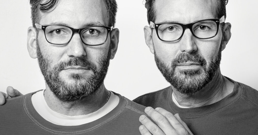
Charlie Chasen and Michael Malone met in Atlanta in 1997, when Mr. Malone served as a guest singer in Mr. Chasen’s band. They quickly became friends, but they didn’t notice what other people around them did: The two men could pass for twins.Mr. Malone and Mr. Chasen are doppelgängers. They look strikingly similar, but they are not related. Their immediate ancestors aren’t even from the same parts of the world; Mr. Chasen’s forebears hailed from Lithuania and Scotland, while Mr. Malone’s parents are from the Dominican Republic and the Bahamas.The two friends, along with hundreds of other unrelated look-alikes, participated in a photography project by François Brunelle, a Canadian artist. The picture series, “I’m not a look-alike!,” was inspired by Mr. Brunelle’s discovery of his own look-alike, the English actor Rowan Atkinson.The project has been a hit on social media and other parts of the internet, but it’s also drawn the attention of scientists who study genetic relationships. Dr. Manel Esteller, a researcher at the Josep Carreras Leukaemia Research Institute in Barcelona, Spain, had previously studied the physical differences between identical twins, and he wanted to examine the reverse: people who look alike but aren’t related. “What’s the explanation for these people?” he wondered.In a study published Tuesday in the journal Cell Reports, Dr. Esteller and his team recruited 32 pairs of look-alikes from Mr. Brunelle’s photographs to take DNA tests and complete questionnaires about their lifestyles. The researchers used facial recognition software to quantify the similarities between the participants’ faces. Sixteen of those 32 pairs achieved similar overall scores to identical twins analyzed by the same software. The researchers then compared the DNA of these 16 pairs of doppelgängers to see if their DNA was as similar as their faces.Elisa Berst and Corinne Barois, Paris, 2010.Ana Maria Sánchez and Katherine Romero, Bogotá, Colombia, 2014.Pedro López Soto and Albert Pueyo Kaotico, Barcelona, 2015.Stella Cappiello and Nunzia Girardi, Bari, Italy, 2015.Dr. Esteller found that the 16 pairs who were “true” look-alikes shared significantly more of their genes than the other 16 pairs that the software deemed less similar. “These people really look alike because they share important parts of the genome, or the DNA sequence,” he said. That people who look more alike have more genes in common “would seem like common sense, but never had been shown,” he added.However, DNA alone doesn’t tell the whole story of our makeup. Our lived experiences, and those of our ancestors, influence which of our genes are switched on or off — what scientists call our epigenomes. And our microbiome, our microscopic co-pilot made up of bacteria, fungi and viruses, is further influenced by our environment. Dr. Esteller found that while the doppelgängers’ genomes were similar, their epigenomes and microbiomes were different. “Genetics put them together, and epigenetics and microbiome pulls them apart,” he said.This discrepancy tells us that the pairs’ similar appearances have more to do with their DNA than with the environments they grew up in. That surprised Dr. Esteller, who had expected to see a bigger environmental influence.Because the doppelgängers’ appearances are more attributable to shared genes than shared life experiences, that means that, to some extent, their similarities are just the luck of the draw, spurred on by population growth. There are, after all, only so many ways to build a face.“Now there are so many people in the world that the system is repeating itself,” Dr. Esteller said. It’s not unreasonable to assume that you, too, might have a look-alike out there.Anna-Maria Tenta and Helena Joas, Munich, 2013.Garrett Levenbrook and Roniel Tessler, New York, 2013.Karen Chu and Ashlee Wong, Culver City, Calif., 2013.Jeanne Bédard and Jessica Gagnon, Montréal, 2015.Dr. Esteller is hopeful that the study’s findings will help doctors diagnose illness in the future — if people have similar enough genes to look alike, they might share predilections for diseases too.“There seems to be something pretty strong in terms of genetics that is making two individuals who look alike also having genome-wide similar profiles,” said Olivier Elemento, the director of the Englander Institute for Precision Medicine at Weill Cornell Medicine in New York, who was not involved with the study. Discrepancies between DNA’s predictions and people’s actual appearances might alert doctors to problems, he said.Dr. Esteller also suggested that there could be links between facial features and behavioral patterns, and that the study’s findings might one day aid forensic science by providing a glimpse of the faces of criminal suspects known only from DNA samples. However, Daphne Martschenko, a postdoctoral researcher at the Stanford Center for Biomedical Ethics who was not involved with the study, urged caution in applying its findings to forensics.“We’ve already seen plenty of examples of how existing facial algorithms have been used to reinforce existing racial bias in things like housing and job hiring and criminal profiling,” Dr. Martschenko said, adding that the study “raises a lot of important ethical considerations.”Despite the potential pitfalls of linking people’s appearances with their DNA or their behavior, Mr. Malone and Mr. Chasen said the look-alike project, and the knowledge that we all might have a secret twin out there, was a means of bringing people together. The two have remained friends for 25 years; when Mr. Chasen got married last week, Mr. Malone was the first person he called. While not everyone with similar DNA shares such a bond, Mr. Malone said that he saw Mr. Brunelle’s photography project as “another way to connect all of us in the human race.”

Read more →

Significant boost in rates of type 2 diabetes among children during COVID-19 pandemic

In a multi-site study of medical records, researchers at Johns Hopkins Children’s Center and across the United States say they have documented a steep rise in type 2 diabetes among children during the COVID-19 pandemic.
In a report on the findings, published Aug. 17 in The Journal of Pediatrics, the investigators note it is unclear whether the virus infection itself was a factor in the rise, and they point to the switch to virtual learning and shutdown of sports and school activities as “environmental factors” that likely increased risk.
Before the pandemic, type 2 diabetes was increasing among children around the world, and because rates of childhood diabetes are known to rise and fall over time, the investigators launched a nationwide review of medical records to assess the impact of the pandemic, according to Sheela N. Magge, M.D., M.S.C.E., director of the Division of Pediatric Endocrinology at the Children’s Center.
Magge, an associate professor of pediatrics at the Johns Hopkins University School of Medicine and co-first author of the paper, says reduced physical activity and weight gain are well-known risk factors for type 2 diabetes. “During the COVID-19 lockdown, children were removed from normal day-to-day routines like going to school, playing sports and other hobbies,” Magge says. “Not only were they less physically active, they were confined to their homes and spent a lot more time watching TV, playing video games, or with other electronic devices.”
Type 2 diabetes is a chronic disorder that affects the body’s ability to regulate, use and process sugar. Without treatment and control, it can cause heart disease, nerve and kidney damage, impaired vision and other irreversible injury to organs.
Although generally associated with adults, an estimated one-third of American youth are considered at risk because of being overweight and obese. Magge adds that previous research from other institutions has shown that children diagnosed with diabetes appear to get complications faster than adults.

Read more →

Air pollution is associated with heart attacks in non-smokers

Research presented at ESC Congress 2022 supports a causal relationship between air pollution and heart attacks since smokers, who already inhale smoke, were unaffected by dirty air.
Study author Dr. Insa de Buhr-Stockburger of Berlin Brandenburg Myocardial Infarction Registry (B2HIR), Germany said: “The correlation between air pollution and heart attacks in our study was absent in smokers. This may indicate that bad air can actually cause heart attacks since smokers, who are continuously self-intoxicating with air pollutants, seem less affected by additional external pollutants.”
This study investigated the associations of nitric oxide, particulate matter with a diameter less than 10 µm (PM10), and weather with the incidence of myocardial infarction in Berlin. Nitric oxide originates from combustion at high temperatures, in particular from diesel vehicles. Combustion is also a source of PM10, along with abrasion from brakes and tyres, and dust.
The study included 17,873 patients with a myocardial infarction between 2008 and 2014 enrolled in the B2HIR.2 Daily numbers of acute myocardial infarction were extracted from the B2HIR database along with baseline patient characteristics including sex, age, smoking status, and diabetes. Daily PM10 and nitric oxide concentrations throughout the city were obtained from the Senate of Berlin. Information on sunshine duration, minimum and maximum temperature, and precipitation were retrieved from the Berlin Tempelhof weather station and merged with the data on myocardial infarction incidence and air pollution.
The researchers analysed the associations between the incidence of acute myocardial and average pollutant concentrations on the same day, previous day, and an average of the three preceding days among all patients and according to baseline characteristics. Associations between the incidence of acute myocardial and weather parameters were also analysed.
Regarding pollution, myocardial infarction was significantly more common on days with high nitric oxide concentrations, with a 1% higher incidence for every 10 µg/m3 increase. Myocardial infarction was also more common when there was a high average PM10 concentration over the three preceding days, with a 4% higher incidence for every 10 µg/m3 increase. The incidence of myocardial infarction in smokers was unaffected by nitric oxide and PM10 concentrations.
Regarding weather, the incidence of myocardial infarction was significantly related to the maximum temperature, with a 6% lower incidence for every 10°C rise in temperature. No associations with sunshine duration or precipitation were detected.
Dr. de Buhr-Stockburger said: “The study indicates that dirty air is a risk factor for acute myocardial infarction and more efforts are needed to lower pollution from traffic and combustion. Causation cannot be established by an observational study. It is plausible that air pollution is a contributing cause of myocardial infarction, given that nitric oxide and PM10 promote inflammation, atherosclerosis is partly caused by inflammatory processes, and no associations were found in smokers.”
Story Source:
Materials provided by European Society of Cardiology. Note: Content may be edited for style and length.

Read more →

Parents adopt unhealthy food routines for family wellbeing in place of unaffordable activities

New study study suggests a key reason parents on a low-income buy unhealthy foods for their families is to compensate for non-food related activities which support social wellbeing, but that they are unable to afford.
The study from the Centre for Food Policy at City, University of London sheds light on the food buying habits of low-income parents across England. It looked at how these families’ food practices may be influenced by their ‘food environment’, i.e. where people can buy and eat food outside of the home, as well as advertising and promotions they come across, but also the wider socioeconomic factors in their lives that may be affecting their decision making.
The findings support the well-established view that a food environment where unhealthy foods are ubiquitous, cheap and heavily marketed, drives parents to feed their families on them. However, they further suggest that when parents are unable to afford social activities with their children, like visiting a ‘soft play’ centre or holidays even a short distance away, they are additionally driven to compensate with family ‘treats’ taking the form of unhealthy food routines.
Examples of such routines identified in the study include family visits to fast-food outlets like the local ‘chippy’ (fish and chips shop), kebab shop, or (famously branded) burger restaurant, or even food related events at home such as family snacks time in front of a movie or board game.
The study involved 60 parents on low incomes as participants, recruited equally from deprived neighbourhoods across three regions of England: Great Yarmouth, Stoke-on-Trent and the London Borough of Lewisham. Participants were aged over 18, a parent of a child in school of nursey and the primary shopper in the family. Reflecting the highly gendered nature of food work, 56 participants were women.
All participants took part in semi-structured interviews relating to practices of purchasing, preparing and consuming foods in the family, and the roles of different family members, including children, in enacting those practices. Fifty-eight of the participants took part in a photo elicitation exercise over a week where they took photos of things that made it harder or easier for them to buy the food they wanted for their families. Twenty-two of the participants also took part in a ‘shop-along’ interview where they guided the interviewing researcher around the shops of their choice, and what they bought.

Read more →

Surprising culprit worsens stroke, TBI damage

In the aftermath of a stroke or TBI, a group of amino acids that typically support brain function contribute significantly to the brain destruction that can follow both these injuries, scientists report.
The new study provides for the first time the surprising evidence that four common nonexcitatory amino acids that usually make proteins which are essential to brain function, instead cause irreversible, destructive swelling of both the astrocytes that support neurons and the neurons themselves, says Dr. Sergei Kirov, neuroscientist in the Department of Neuroscience and Regenerative Medicine at the Medical College of Georgia.
“There are many ways to kill neurons. This is one that people have not thought about,” says Kirov, corresponding author of the study published in the journal GLIA.
A stroke resulting from a ruptured or blocked blood vessel in the brain as well as traumatic brain injury, or TBI, both disrupt the uniquely, super-tightly woven endothelial cells that line blood vessels in the brain, which help ensure that nothing escapes from our blood that might hurt our brain. It’s called the blood brain barrier and following these types of significant brain events, the protective barrier can become leaky and its components can escape.
Plasma, the fluid part of the blood, is an early escapee into the already stressed nearby brain areas, and with it comes these amino acids, which make proteins, a fundamental component of our cells and muscle. Normally small molecules like oxygen are the ones that can pass through the blood brain barrier, and larger molecules like these amino acids, have a tightly regulated system that ensures that the right amount of the right factor gets delivered directly to the cells that need them.
Transporters inside the membranes of the endothelial cells enable select items, including amino acids as well as glucose, to be delivered and may carry some other molecule out on the return trip.

Read more →