Synthetic mouse embryo develops beating heart
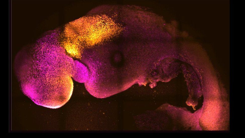
Published13 hours agoSharecloseShare pageCopy linkAbout sharingImage source, Amadei and HandfordScientists in Cambridge have created synthetic mouse embryos in a lab, without using eggs or sperm, which show evidence of a brain and beating heart. The mouse embryos, developed using stem cells, only lasted for eight days.But the research team say it could improve understanding of the earliest stages of organ development – and why some pregnancies fail.Other scientists caution that while the technique is promising there are still many hurdles to overcome.The researchers from the University of Cambridge and the California Institute of Technology (Caltech) are the latest to publish their results in the journal Nature.Researchers from Israel also published similar findings recently.The Cambridge team has been studying the early stages of pregnancy for the past decade but so much of it is hidden from view in the womb.By mimicking natural processes in a laboratory, they found a way to get three types of stem cells from mice to interact and grow into embryo-like structures.The synthetic mouse embryos only lasted for eight days, due to defects – but they reached the point where a brain began to develop.Professor Magdalena Zernicka-Goetz, professor of mammalian development and stem cell biology at Cambridge and professor of biology at Caltech, said it was “a dream come true” and could offer a glimpse into how organs are formed.Image source, Amadei and Handford”This period of human life is so mysterious, so to be able to see how it happens in a dish – to have access to these individual stem cells, to understand why so many pregnancies fail and how we might be able to prevent that from happening – is quite special,” she said.The advance could also mean less reliance on animals for research and a useful way to test new drugs.’Very early stage’However, Prof Alfonso Martinez Arias, from the Universitat Pompeu Fabra in Barcelona, said: “This is an advance but at a very early stage of development, a rare event which, while superficially looking like an embryo, bears defects which should not be overlooked.”The researchers now plan to work on keeping the synthetic embryos developing for a day or two longer, which is difficult to do without creating a synthetic placenta.Eventually, their ambition is to develop similar embryos from human stem cells – but this is still a long way off, and ethically much more complicated.At present, UK law permits human embryos to be studied in the laboratory only up to the fourteenth day of development, but there are no rules around synthetic embryos.Prof Robin Lovell-Badge, from the Francis Crick Institute, said that should change.”Given the similarity with real embryos, it follows that consideration also needs to be given as to whether and how such integrated stem cell-based embryo models should be regulated,” he said. He added that it was important not to think of the embryo-like models “as being the real thing – even if they are getting close”.”If these had been derived from human stem cells, and it is accepted that these should never be transplanted into a uterus, we will never know if they are equivalent.”More on this storyHuman cells grown in monkey embryos spark debate15 April 2021Artificial ’embryos’ created in the lab3 March 2017Human-pig ‘chimera embryos’ detailed26 January 2017Embryo study shows ‘life’s first steps’4 May 2016

Read more →

Neurofeedback shows promise for addressing 'chemo brain'

Restoring normal functioning in the brains of cancer patients through neurofeedback could potentially alleviate the mental fogginess that many report after treatment, according to a new pilot study from UCLA researchers.
The study is one of the first to indicate that neurofeedback, or electroencephalogram (EEG) biofeedback, could help address cognitive deficits of cancer patients experiencing “chemo brain,” a myriad of symptoms that could include problems with memory, concentration and organization, as well as other symptoms like trouble sleeping and emotional difficulties. Previous research has found that neurofeedback, in which brain waves are trained to operate in optimal frequency patterns, has helped improve cognitive function in patients with cognitive impairments like attention-deficit/hyperactivity disorder, stroke and seizures, as well as helped regulate brain activity in patients with substance use and post-traumatic stress disorders.
“The history of neurofeedback shows that it’s helpful for a whole range of disorders and symptoms. This study was an opportunity for seeing whether neurofeedback is something that could be helpful with chemo brain,” said Stephen Sideroff, a professor at UCLA’s Department of Psychiatry & Biobehavioral Sciences who led the study and has used neurofeedback training with patients for over 20 years.
The study was published this month in the peer-reviewed Journal of Complementary and Integrative Medicine.
The study by Sideroff and UCLA colleagues David Wellisch and Valerie Yarema included nine female breast cancer patients between the ages of 21 and 65 who had completed chemotherapy at least one year earlier and complained of debilitating symptoms of chemo brain, which brought significant disruptions to their work and personal lives. A clinical nurse practitioner conducted a brief mental status interview with each patient to confirm that they had persistent difficulties with concentration, memory, organization and confusion. The patients selected for the study did not have a current breast cancer diagnosis, a present or recent diagnosis of a major depressive disorder or other mental illness, or used cognitive-altering medications that might confound study results.
Before the neurofeedback training sessions began, the study participants received neurocognitive and psychological tests, as well as a quantitative EEG to measure brain wave frequencies that could be compared to normative data. The pre-training quantitative EEGs shows that each study participant had abnormal brain waive activity compared to healthy adult brains.

Read more →

Distress leads to higher COVID vaccine rates, less adherence to distancing guidelines, study finds

People who were more distressed — showing signs of anxiety or depression — during the COVID-19 pandemic were less likely to follow some best practice recommendations from the Centers for Disease Control and Prevention, according to a new study by Washington University in St. Louis researchers.
They found, however, that those same people were more likely than their non-distressed peers to get vaccinated. The authors refer to this as differential distress: when people act safely in one aspect while disregarding safety in another, both in response to the same psychological distress. This creates a conundrum for those trying to determine how best to communicate risks and best practices to the public.
The research comes from the Department of Psychological & Brain Sciences in Arts & Sciences. It was led by professors Leonard Green and Joel Myerson. The team included professors Michael Strube and Sandra Hale and Bridget Bernstein, a research technician.
Their study of 810 people revealed that distress was less likely to affect older people either way, despite their higher risk for severe outcomes if infected with SARS-CoV-2. The findings, published July 27 in the journal Frontiers in Psychology, suggest that fear messaging, which is intended to scare people and can increase their levels of distress, may not be the most effective way to encourage people to change behaviors.
“These findings do not point to a straightforward public health messaging campaign,” Myerson said. “Instead, officials may have to consider more finely tailored messages for different populations in order to achieve best outcomes: more attention to CDC recommendations as well as more people getting vaccinated.”
This is the second study from this team to analyze the ways people changed behaviors during the pandemic. The first study, published in November in the journal PLoS One, looked at social distancing and hygiene behaviors across a range of demographics. The results suggested that distress was closely tied to the way people responded to recommendations about social distancing. People who were more distressed were less likely to observe social distancing recommendations, perhaps as a way to maintain social connections that can ease anxiety and depression.
In the latest work, researchers again asked people about their adherence to the latest CDC recommendations, including newer recommendations outlining when to wear a mask and suggesting that people avoid spending lots of time inside with others. The results showed similar correlations to the previous study among age, distress and behavior changes.
In terms of public health and effective messaging, one of the most pressing issues to arise after publication of the first study was the introduction of vaccines — and the myriad ways people felt about them. Looking at four categories — fully vaccinated; partially vaccinated; unvaccinated but likely to get one; unvaccinated and unlikely to get one — several findings stood out: People who had been fully vaccinated were more likely than those who were partially vaccinated to have close interactions with others following their shots. Relative to those who said they were unlikely to get vaccinated, those who said they were likely to do so thought their chance of infection was higher. Depending on the person’s age, they responded differently to the same level of stress. Overall, for example, the higher level of distress someone had, the less likely they were to social distance, but the more likely they were to get vaccinated. Both of these correlations became weaker, however, as people aged.Fear messaging that tries to scare people into following guidelines tends to be useful only for a one-time event, Green said. “Ostensibly, getting vaccinated should count as such an event.” But as breakthrough cases increase and boosters add up, vaccinations are no longer one and done; they are instead a series of events, spread out over more than a year.
Although fear-based messaging may encourage younger people to get vaccinated, it also diminishes their resolve to stick to mitigation behaviors like social distancing. Without doing both, the risk of breakthrough infections could continue to rise.
And, the research shows, messaging becomes less effective as people age — and become more susceptible to severe illness if they’re infected.
“Part of the solution to the problem of differential distress may be to avoid the distress altogether,” Green said, by forgoing the fear campaign. Instead, a gentler approach may be warranted. “Our previous work suggests that what really motivates many people to change behaviors for the better is considering how their actions can benefit, or harm, other people.”

Read more →

Study uncovers differences in saliva bacteria of students with recent suicidal thoughts

A new University of Florida study has found that bacteria in the saliva of college students who reported recent thoughts of suicide differed in significant ways from those found in students who had not experienced recent suicidal thinking.
While there is a growing body of research on mental health and the human microbiome, this is the first study to look at bacterial differences in the saliva of those with and without recent suicidal thoughts, also called suicidal ideation. Recent suicidal ideation was defined as thoughts of suicide within the two weeks before the saliva sample was taken.
Controlling for the influence of other factors known to impact mental health, such as diet and sleep, the researchers found that students with recent suicidal thoughts had higher levels of bacteria associated with periodontal disease and other inflammatory health conditions.
They also found that these students had lower levels of Alloprevotella rava, a bacterium known to produce a compound that promotes brain health. These students also shared a genetic variation that the researchers found may influence the presence of Alloprevotella rava in the mouth.
“These results are exciting because they tell us which bacteria we need to look at more closely. Our question now is, what are these bacteria doing biologically that affects mental health?” said Angelica Ahrens, first author of the study and a postdoctoral researcher in the UF/IFAS microbiology and cell science department. Ahrens led the study as part of her doctoral program in the UF/IFAS College of Agricultural and Life Sciences.
“Eventually, we hope this line of research could help predict suicidal ideation based on a person’s microbiome and could inform pro- or prebiotic treatments for those at risk,” said Ahrens.
The study analyzed saliva collected from nearly 500 undergraduate students taking classes in the microbiology and cell science department at UF. These students also completed the Patient Health Questionnaire-9, which is used to screen for depression symptoms and asks respondents to share if they have had thoughts of suicide within the last two weeks. Those who reported recent suicidal ideation were referred to on-campus mental health services.
“Mental health and suicide are serious issues on college campuses, and our students were very interested in being a part of research that can help address this problem. We are continuing to collect data for follow-up studies and hope more students and universities will become involved,” said Eric Triplett, chair of the microbiology and cell science department and senior author of the study.
Depression and suicidal ideation are relatively common among college-aged adults: A 2020 study by the CDC found that up to a quarter of people between ages 18 to 24 had seriously thought about suicide within the last month.
For this initial study, students came to the lab to provide a saliva sample, but today participants can opt to send in their saliva sample by mail using a collection kit developed by the researchers.
“This at-home method is very convenient for students and also helps us build a more diverse dataset and test different variables. For example, we would like to look at the saliva microbiome of people who have been diagnosed with depression and are taking antidepressants,” Ahrens said.
“While various treatments and lifestyle changes can help, there is still much to be learned about how the human microbiome affects mental health and could be harnessed to improve it,” Ahrens said.
Story Source:
Materials provided by University of Florida. Original written by Samantha Murray. Note: Content may be edited for style and length.

Read more →

How do molecular motors convert chemical energy in to mechanical work?

Molecular motors are complex devices composed of many different parts that consume energy to perform various cellular activities. In short, molecular machines transform energy into useful work. Understanding the mechanistical aspects underlying these motors begins with generating a detailed description of their overall architecture and atomic organisation. However, to uncover the core mechanisms energizing these motors it is essential to decode all of the molecular dynamics in atomic detail.
Now, the research team of Thomas C. Marlovits from the Centre for Structural Systems Biology CSSB at DESY and the University Medical Center Hamburg-Eppendorf (UKE) in Hamburg reveals the architecture, complete functional cycle and the mechanism of such a molecular motor: They report in the journal Nature, how a ‘RuvAB branch migration complex’ converts chemical energy into mechanical work to perform recombination and repair of DNA. DNA recombination is one of the most fundamental biological processes in living organisms. It is the process by which chromosomes “swap” DNA either to generate genetic diversity, by creating new offspring, or to maintain genetic integrity, by repairing breaks in existing chromosomes. During DNA recombination, four DNA arms separate from their double-helix formations and join together at an intersection known as a Holliday junction. Here the DNA arms exchange strands in a process called active branch migration.
The essential energy needed for this branch migration to occur comes from a molecular machinery that scientists have tagged as the RuvAB branch migration complex. This complex assembles around the Holliday junction and is made of two motors labelled RuvB AAA+ ATPases, that fuel the reaction, and a RuvA stator. The research team has now provided an intricate blueprint that explains how the RuvB AAA+ motors work under the regulation of the RuvA protein to perform synchronized DNA movement.
The active branch migrations energized by the RuvB AAA+ motor molecule are very fast and highly dynamic. To determine the individual steps of this process, the scientists used time-resolved cryo electron microscopy to observe the motor’s machinery in slow motion. “We basically provided the RuvB AAA+ motor with a slower burning fuel which allowed us to capture the biochemical reactions as they occur,” explains Marlovits.
The scientist captured over ten million images of the motor machinery interacting with the Holliday junction. Jiri Wald (CSSB, UKE and part of the Vienna BioCenter PhD Program), the paper’s first author, combed through the immense amount of data and carefully classified the subtle changes occurring in each image. Using the high-performance computing facility at DESY, the scientists were then able to put all the puzzle pieces together to generate a high-resolution movie detailing how the RuvAB complex functions on the molecular scale.
“We were able to visualize seven distinct states of the motor and demonstrate how the interconnected elements work together in a cyclical manner,” explains Wald. “We also demonstrated that the RuvB motor converts energy into a lever motion which generates the force that drives branch migration. We were amazed by the discovery that the motors use a basic lever mechanism to move the DNA substrate. Overall, the sequential mechanism, coordination and force generation manner of the RuvAB motor share conceptual similarities with combustion engines.”
AAA+ motors are often used in other biological systems, such as protein transport, therefore this detailed model of the RuvB AAA+ motor can be used as a blueprint for similar molecular motors. “We understand how the motor works and now we can put this motor into another system with some minor adaptations,” explains Marlovits. “We are essentially presenting core principles for AAA+ motors.”
The Marlovits group’s future work will explore ways to interfere with the function of AAA+ motors. This could provide the basis for the development of a new generation of drugs, which would disrupt the mechanisms of such a motor in pathogens and thus halt the spread of infection. “We are excited to explore the possibilities that exist now that we have a blueprint of the RuvB AAA+ motor,” notes Wald.
Scientists from CSSB, UKE, the Institute of Molecular Biotechnology, the Research Institute of Molecular Pathology, both in Vienna, Austria, and DESY contributed to this research.
CSSB is a joint initiative of ten research partners from Northern Germany, including three universities and six research institutes that devotes itself to infection biology research.
DESY is one of the world’s leading particle accelerator centres and investigates the structure and function of matter — from the interaction of tiny elementary particles and the behaviour of novel nanomaterials and vital biomolecules to the great mysteries of the universe. The particle accelerators and detectors that DESY develops and builds at its locations in Hamburg and Zeuthen are unique research tools. They generate the most intense X-ray radiation in the world, accelerate particles to record energies and open up new windows onto the universe. DESY is a member of the Helmholtz Association, Germany’s largest scientific association, and receives its funding from the German Federal Ministry of Education and Research (BMBF) (90 per cent) and the German federal states of Hamburg and Brandenburg (10 per cent).

Read more →

Cells: Strong at the right place and time

Researchers from TU Delft and NWO institute AMOLF discovered how certain molecular bonds make living cells both flexible, in order to move, as well as strong, in order to withstand forces. Paradoxically, it turns out that these force-sensitive catch bonds are weak and inactive most of the time, but travel to specific places where and when cells become damaged. This discovery will be published in Nature Materials on 25 August.
Molecular catch bond proteins can be found in many different tissues, both within and between cells. These bonds fall apart regularly, as most biological bonds do, but they have a peculiar property: if you pull hard at a catch bond, it actually starts binding tighter. Researchers discovered that this ability strengthens the material in specific places where the bond experiences stress. The discovery is a breakthrough, 20 years after the first finding of such bonds. Also, this marks the first time that the researchers have witnessed catch bonds working together within biological materials.
Both flexible and strong
Former AMOLF researcher Yuval Mulla explains: “We usually define how strong something is by one of two ways: a material can either deform well — stretch very far without breaking, such as rubber — or the material can bear much force, for instance a brick; although it’s strong, it can only stretch a little before it will break. Studying the nature of catch bonds, we found that these molecular bonds were able to do both: be flexible and strong, even though their molecular bindings are weak. And then we considered: might catch bonds explain why living cells combine the stretchiness of rubber with the strength of a brick?”
To test these ideas, the researchers measured the mechanical properties of cytoskeletal networks that they reconstituted in the lab, collaborating with the Biophysics group to pull at single bonds. They found that many of the bonds are just floating around, binding briefly only to let go again. However, when the researchers deformed the networks, they found that many bonds travel to particularly damaged sites to bind. Mulla: “Because the catch bonds accumulate at weak spots when and where they are needed to make the network very strong.”
Relation to diseases
The study included a mutant version of the same protein, one which is known to occur with a genetic disease that leads to kidney failure. Unlike a regular catch bond, the researchers found that this mutant version was always active. This increased bond strength makes it difficult for the mutant to move around, but, paradoxically, also makes the networks weaker as the bonds do not accumulate where needed, says group leader Gijsje Koenderink: “By understanding the mutant protein better, in the future we might also understand the process of kidney failure. In addition, we hope to understand how catch bonds play a role in how invasive cancer cells are.”
Material perspective on life
The research group of professor Gijsje Koenderink at Delft University of Technology is primarily interested in material properties of living matter. A central theme in her group is the fact that living cells and tissues need to be dynamic and flexible, but also strong: “This property is different from any synthetic materials we know,” says Koenderink. “Our ambition is to learn new design principles from living materials to make synthetic materials that can be both flexible and strong at the same time. In fact, we are currently working together with chemists and biophysicists like Sander Tans at AMOLF to try and make such synthetic catch bonds.”
Story Source:
Materials provided by Delft University of Technology. Note: Content may be edited for style and length.

Read more →

Artificial intelligence model can detect Parkinson's from breathing patterns, researchers show

Parkinson’s disease is notoriously difficult to diagnose as it relies primarily on the appearance of motor symptoms such as tremors, stiffness, and slowness, but these symptoms often appear several years after the disease onset. Now, Dina Katabi, the Thuan (1990) and Nicole Pham Professor in the Department of Electrical Engineering and Computer Science (EECS) at MIT and principal investigator at MIT Jameel Clinic, and her team have developed an artificial intelligence model that can detect Parkinson’s just from reading a person’s breathing patterns.
The tool in question is a neural network, a series of connected algorithms that mimic the way a human brain works, capable of assessing whether someone has Parkinson’s from their nocturnal breathing — i.e., breathing patterns that occur while sleeping. The neural network, which was trained by MIT PhD student Yuzhe Yang and postdoc Yuan Yuan, is also able to discern the severity of someone’s Parkinson’s disease and track the progression of their disease over time.
Yang is first author on a new paper describing the work, published today in Nature Medicine. Katabi, who is also an affiliate of the MIT Computer Science and Artificial Intelligence Laboratory and director of the Center for Wireless Networks and Mobile Computing, is the senior author. They are joined by Yuan and 12 colleagues from Rutgers University, the University of Rochester Medical Center, the Mayo Clinic, Massachusetts General Hospital, and the Boston University College of Health and Rehabilition.
Over the years, researchers have investigated the potential of detecting Parkinson’s using cerebrospinal fluid and neuroimaging, but such methods are invasive, costly, and require access to specialized medical centers, making them unsuitable for frequent testing that could otherwise provide early diagnosis or continuous tracking of disease progression.
The MIT researchers demonstrated that the artificial intelligence assessment of Parkinson’s can be done every night at home while the person is asleep and without touching their body. To do so, the team developed a device with the appearance of a home Wi-Fi router, but instead of providing internet access, the device emits radio signals, analyzes their reflections off the surrounding environment, and extracts the subject’s breathing patterns without any bodily contact. The breathing signal is then fed to the neural network to assess Parkinson’s in a passive manner, and there is zero effort needed from the patient and caregiver.
“A relationship between Parkinson’s and breathing was noted as early as 1817, in the work of Dr. James Parkinson. This motivated us to consider the potential of detecting the disease from one’s breathing without looking at movements,” Katabi says. “Some medical studies have shown that respiratory symptoms manifest years before motor symptoms, meaning that breathing attributes could be promising for risk assessment prior to Parkinson’s diagnosis.”
The fastest-growing neurological disease in the world, Parkinson’s is the second-most common neurological disorder, after Alzheimer’s disease. In the United States alone, it afflicts over 1 million people and has an annual economic burden of $51.9 billion. The research team’s algorithm was tested on 7,687 individuals, including 757 Parkinson’s patients.
Katabi notes that the study has important implications for Parkinson’s drug development and clinical care. “In terms of drug development, the results can enable clinical trials with a significantly shorter duration and fewer participants, ultimately accelerating the development of new therapies. In terms of clinical care, the approach can help in the assessment of Parkinson’s patients in traditionally underserved communities, including those who live in rural areas and those with difficulty leaving home due to limited mobility or cognitive impairment,” she says.
“We’ve had no therapeutic breakthroughs this century, suggesting that our current approaches to evaluating new treatments is suboptimal,” says Ray Dorsey, a professor of neurology at the University of Rochester and Parkinson’s specialist who co-authored the paper. Dorsey adds that the study is likely one of the largest sleep studies ever conducted on Parkinson’s. “We have very limited information about manifestations of the disease in their natural environment and [Katabi’s] device allows you to get objective, real-world assessments of how people are doing at home. The analogy I like to draw [of current Parkinson’s assessments] is a street lamp at night, and what we see from the street lamp is a very small segment … [Katabi’s] entirely contactless sensor helps us illuminate the darkness.”
This research was performed in collaboration with the University of Rochester, Mayo Clinic, and Massachusetts General Hospital, and is sponsored by the National Institutes of Health, with partial support by the National Science Foundation and the Michael J. Fox Foundation.
Story Source:
Materials provided by Massachusetts Institute of Technology. Original written by Alex Ouyang, Abdul Latif Jameel Clinic for Machine Learning in Health. Note: Content may be edited for style and length.

Read more →

These neurons have food on the brain

A gooey slice of pizza. A pile of crispy French fries. Ice cream dripping down a cone on a hot summer day. When you look at any of these foods, a specialized part of your visual cortex lights up, according to a new study from MIT neuroscientists.
This newly discovered population of food-responsive neurons is located in the ventral visual stream, alongside populations that respond specifically to faces, bodies, places, and words. The unexpected finding may reflect the special significance of food in human culture, the researchers say.
“Food is central to human social interactions and cultural practices. It’s not just sustenance,” says Nancy Kanwisher, the Walter A. Rosenblith Professor of Cognitive Neuroscience and a member of MIT’s McGovern Institute for Brain Research and Center for Brains, Minds, and Machines. “Food is core to so many elements of our cultural identity, religious practice, and social interactions, and many other things that humans do.”
The findings, based on an analysis of a large public database of human brain responses to a set of 10,000 images, raise many additional questions about how and why this neural population develops. In future studies, the researchers hope to explore how people’s responses to certain foods might differ depending on their likes and dislikes, or their familiarity with certain types of food.
MIT postdoc Meenakshi Khosla is the lead author of the paper, along with MIT research scientist N. Apurva Ratan Murty. The study appears in the journal Current Biology.
Visual categories
More than 20 years ago, while studying the ventral visual stream, the part of the brain that recognizes objects, Kanwisher discovered cortical regions that respond selectively to faces. Later, she and other scientists discovered other regions that respond selectively to places, bodies, or words. Most of those areas were discovered when researchers specifically set out to look for them. However, that hypothesis-driven approach can limit what you end up finding, Kanwisher says.

Read more →

'Dangerous' and 'extremely dangerous' heat stress to become more common by 2100

Record-breaking heat waves have occurred recently from Delhi to the Pacific Northwest, and the number of these deadly events is expected to increase. New research from the University of Washington and Harvard University gives a range of heat impacts worldwide by the end of this century, depending on future emissions of greenhouse gases.
The study was published Aug. 25 in the open-access journal Communications Earth & Environment.
“The record-breaking heat events of recent summers will become much more common in places like North America and Europe,” said lead author Lucas Vargas Zeppetello, who did the research as a doctoral student at the UW and is now a postdoctoral researcher at Harvard. “For many places close to the equator, by 2100 more than half the year will be a challenge to work outside, even if we begin to curb emissions.”
“Our study shows a broad range of possible scenarios for 2100,” he added. “This shows that the emissions choices we make now still matter for creating a habitable future.”
The study looks at a combination of air temperature and humidity known as the “heat index” that measures impact on the human body. A “dangerous” heat index is defined by the National Weather Service as 103 F (39.4 C). An “extremely dangerous” heat index is 124 F (51 C), deemed unsafe to humans for any amount of time.
“These standards were first created for people working indoors in places like boiler rooms — they were not thought of as conditions that would happen in outdoor, ambient environments. But we are seeing them now,” Vargas Zeppetello said.

Read more →

'Synthetic' embryo with brain and beating heart grown from multiple stem cells

Researchers from the University of Cambridge have created model embryos from mouse stem cells that form a brain, a beating heart, and the foundations of all the other organs of the body — a new avenue for recreating the first stages of life.
The team, led by Professor Magdalena Zernicka-Goetz, developed the embryo model without eggs or sperm, and instead used stem cells — the body’s master cells, which can develop into almost any cell type in the body.
The researchers mimicked natural processes in the lab by guiding the three types of stem cells found in early mammalian development to the point where they start interacting. By inducing the expression of a particular set of genes and establishing a unique environment for their interactions, the researchers were able to get the stem cells to ‘talk’ to each other.
The stem cells self-organised into structures that progressed through the successive developmental stages until they had beating hearts and the foundations of the brain, as well as the yolk sac where the embryo develops and gets nutrients from in its first weeks. Unlike other synthetic embryos, the Cambridge-developed models reached the point where the entire brain, including the anterior portion, began to develop. This is a further point in development than has been achieved in any other stem cell-derived model.
The team say their results, the result of more than a decade of research that progressively led to more and more complex embryo-like structures and reported in the journal Nature, could help researchers understand why some embryos fail while others go on to develop into a healthy pregnancy. Additionally, the results could be used to guide repair and development of synthetic human organs for transplantation.
“Our mouse embryo model not only develops a brain, but also a beating heart, all the components that go on to make up the body,” said Zernicka-Goetz, Professor in Mammalian Development and Stem Cell Biology in Cambridge’s Department of Physiology, Development and Neuroscience. “It’s just unbelievable that we’ve got this far. This has been the dream of our community for years, and major focus of our work for a decade and finally we’ve done it.”
For a human embryo to develop successfully, there needs to be a ‘dialogue’ between the tissues that will become the embryo, and the tissues that will connect the embryo to the mother. In the first week after fertilisation, three types of stem cells develop: one will eventually become the tissues of the body, and the other two support the embryo’s development. One of these extraembryonic stem cell types will become the placenta, which connects the fetus to the mother and provides oxygen and nutrients; and the second is the yolk sac, where the embryo grows and where it gets its nutrients from in early development.

Read more →