Drug effects of ketamine in mice can depend on the sex of the human experimenter, study finds

Many researchers who work with mice can tell you that mice behave differently depending on who is handling them. Anecdotal reports and some existing scientific reports indicate that mice tend to be more fearful and uptight around men, and relaxed and comfortable around women. Whether this behavior actually affects research results though, remains a sort of the elephant in the room that not many people seem to want to address.
Now, researchers at the University of Maryland School of Medicine (UMSOM) have shown that mice respond more to the antidepressant effects of the drug ketamine when administered by men and not by women. The group demonstrated that the response of mice detected in a specific region of their brain from handling by a man is essential for ketamine’s effect to work. Then, the researchers identified the mechanism behind this response.
The researchers say that while the influence of the sex of the scientist administering ketamine is not directly relevant to the human response to ketamine, the brain mechanism underlying their findings could help determine why some people do not respond to ketamine anti-depressant therapy and suggest ways to potentially make this therapy work better for those patients who do not respond well.
The findings were published on August 30 in Nature Neuroscience.
“Our findings in mice suggests that activating a specific stress circuit in the brain may be a way to improve ketamine treatment. Our thought is that you may be able to provide a more robust antidepressant effect if you combine the ketamine with activation of this brain region, either a drug that spurs this process in the brain or even some sort of specific stressor,” said Todd Gould, MD, Professor of Psychiatry at UMSOM.
Dr. Gould’s team anecdotally noticed that ketamine’s antidepressant-like effects only seemed to work consistently when male researchers administered the treatment to mice. The team reached out to other labs studying mouse responses to ketamine, who reported the same issues, but no one had yet systematically documented the phenomena and investigated the cause. At the time, most of Dr. Gould’s team was women and so figuring out why the experiments did not work when women performed them was essential to the team getting workable data, so they could move forward with project.

Read more →

Wearables take 'logical' step toward onboard control

For all the talk about embedding computers in clothing, here’s an interesting option. Make the clothing the computer, and do it without electricity.
Mechanical engineers at Rice University’s George R. Brown School of Engineering are trying the concept on for size with a set of textile-based pneumatic computers capable of digital logic, onboard memory and user interaction.
The lab’s “fluidic digital logic” takes advantage of how air flows through a series of “kinked” channels to form bits, the 1s and 0s in computer memories.
The idea is to have such textile-based logic gates support pneumatic actuators, potentially in conjunction with an energy harvesting system developed by the Preston lab, to help people with functional limitations with their day-to-day tasks.
The research backed by a recent National Science Foundation CAREER Award appears in the Proceedings of the National Academy of Sciences.
Preston said the lab’s logic-enabled textiles can be mass produced using existing clothes-manufacturing processes and are resilient enough to withstand everyday use. The researchers claimed the embedded gates are both comfortable and tough enough to drive a truck over without damaging them. (And they proved it.)
“The idea of using fluids to construct digital logic circuits is not new,” he said. “And in fact, in the last decade, people have been moving towards implementing fluidic logic in soft materials, things like elastomers. But so far, no one had taken the step to implement it in sheet-based materials, a feat which required redesigning the entire approach from first principles.”

Read more →

Researchers visualize alpha-synuclein pathology in living patients with a neurodegenerative disorder

Multiple system atrophy (MSA) is a neurodegenerative disorder characterized by the aggregation of α-synuclein in the brain. Now, scientists from Japan, in collaboration with three pharmaceutical companies, have developed a radioligand that facilitates the imaging of α-synuclein aggregates in patients with MSA. Their findings have the potential to completely change the scenario of diagnosing neurodegenerative diseases.
α-synuclein is a neuronal protein involved in functions like vesicle trafficking and neurotransmitter release. It is typically found in abundance in a healthy brain. However, the aggregation of α-synuclein has been closely linked to several neurodegenerative disorders, including Parkinson’s disease, multiple system atrophy (or MSA), and Lewy body dementia.
MSA is a movement disorder that also affects the autonomic nervous system, which controls essential functions such as movement, breathing, and digestion. Thus, the imaging of α-synuclein aggregates in vivo (or directly in a living organism), could be a potential diagnostic confirmation of MSA. However, the road to the live imaging of α-synuclein has been marred with obstacles, including the lack of sensitive imaging agents.
Now, a collaborative effort by researchers from the National Institutes for Quantum Science and Technology, including Dr. Makoto Higuchi and Dr. Kiwamu Matsuoka from the Quantum Life and Medical Science Directorate, Institute for Quantum Medical Science has completely changed the scenario with three pharmaceutical companies — Eisai Co., Ltd., Ono Pharmaceutical Co., Ltd., and Takeda Pharmaceutical Company Limited. They have successfully visualized α-synuclein aggregates in the brains of patients. To achieve this feat, the team developed a radioligand, 18F-SPAL-T-06, to be used as a probe for positron emission tomography (PET). “The pre-competitive collaboration between a research institute and three pharmaceutical companies enabled us to develop the radioligand, 18F-SPAL-T-06, for the in vivo imaging of α-synuclein aggregates,” says Dr. Higuchi, crediting teamwork for their success. The team’s findings have been published in the journal Movement Disorders.
Prior to the clinical assessments, in vitro studies on the binding properties of 18F-SPAL-T-06 had been conducted on the postmortem brain tissue of patients with MSA and healthy individuals, showing promising results. For the first-in-human imaging studies, the researchers enrolled three patients who were clinically diagnosed with MSA and one 72-year-old healthy control (HC). Among the three patients with MSA, two were identified as having MSA with predominant Parkinsonism (MSA-P) and one with MSA with predominant cerebellar ataxia (MSA-C). PET scans with 18F-SPAL-T-06 was performed on all the patients and specific binding was estimated by the radioligand retention in the tissue. “Remarkably, we observed enhanced 18F-SPAL-T-06 retention in the putamen, pons, and cerebellar white matter and peduncles
of the patients with MSA-P and MSA-C, in sharp contrast to minimal radio signals in the corresponding areas in the brain of the HC,” explains Dr. Higuchi.
The researchers also found that 18F-SPAL-T-06 has a high affinity for MSA-type α-synuclein aggregates and that it does not cross-react with other off-target components, indicating its high specificity and consequent potential use as a probe for MSA diagnosis.
With respect to the long-term applications of their work, Dr. Higuchi and Dr. Matsuoka share: “We are encouraged by our findings, and investigations into the visualization of α-synuclein aggregates in other α-synucleinopathies are currently underway.”

Read more →

Understanding the expanded role of clinical ethicists

The COVID-19 pandemic brought many troubling ethical issues to the frontlines of clinical care, creating significant distress for clinicians, patients, and families. Behind the scenes, clinical ethicists managed those issues to support front-line workers and were integral to hospital operations.
Until now, little has been published about the expanded role clinical ethicists played during this time and their role in supporting hospital operations across the country. A new study from the University of Pennsylvania School of Nursing (Penn Nursing) reports how clinical ethicists provided consultation and guidance in situations of uncertainty, distress, or disagreement, often with imperfect information.
“Although the clinical ethicists’ role has been studied under usual circumstances, very little is understood about their experience during the pandemic, the ethical challenges they faced, and how they addressed their own ethical challenges or received support,” says the study’s lead investigator, Connie M. Ulrich, PhD, RN, FAAN, Lillian S. Brunner Chair in Medical and Surgical Nursing, Professor of Nursing and Professor of Medical Ethics and Health Policy at Penn Nursing.
The results of the study have been published in the journal AJOB Empirical Bioethics. The article “Ethical Challenges Experienced by Clinical Ethicists During COVID-19” is available online.
“By understanding the experiences of clinical ethicists during the COVID-19 pandemic and the ethical challenges they faced, we can better understand how their expanded role links to both clinical and organizational outcomes,” says Dr. Ulrich. “Moreover, it is important to know whether this role has now moved beyond traditional conceptions and the resulting future educational needs.”
Co-authors of the article include Janet A. Deatrick, PhD, RN, FAAN, Professor Emerita of Nursing; Jesse Wool, MBE, BSN; and Liming Huang, all of Penn Nursing; Nancy Berlinger of the Hastings Center; and Christine Grady of the National Institutes of Health.
Story Source:
Materials provided by University of Pennsylvania School of Nursing. Note: Content may be edited for style and length.

Read more →

Brain bubbles: Researchers describe the dynamics of cavitation in soft porous material

A tiny bubble popping within a liquid seems more fanciful than traumatic. But millions of popping vapor bubbles can cause significant damage to rigid structures like boat propellers or bridge supports. Can you imagine the damage such bubbles could do to soft human tissues like the brain? During head impacts and concussions, vapor bubbles form and violently collapse, creating damage to human tissue. Purdue University fluid mechanics researchers are now one step closer to understanding these phenomena.
“When a bubble collapses inside a liquid, it generates pressure shock waves,” said Hector Gomez, professor of mechanical engineering and principal investigator. “The process of forming a vapor cavity and its collapse is what we call cavitation.”
“Cavitation has been studied since the 1800s,” said Pavlos Vlachos, the St. Vincent Health Professor of Healthcare Engineering and director of the Regenstrief Center for Healthcare Engineering. “It’s a very complex field of study because it involves non-equilibrium thermodynamics, continuum mechanics, and many other factors on a scale of micrometers and microseconds. After hundreds of years of research, we are only just now starting to understand these phenomena.”
Even less is known about bubbles that collapse in soft porous materials, such as the brain or other body tissues. That’s significant, because understanding how those bubbles behave could lead to a better understanding of concussions — or even be used to deliver targeted medications inside the body.
In new research published in the Proceedings of the National Academies (PNAS) Nexus, Gomez, Vlachos, and collaborators presented the development of a mathematical model to describe the dynamics of these cavitation bubbles in a deformable porous medium.
Cavitation occurs throughout the human body — for example, cracking your knuckles is the sound of bubbles popping in your joints’ synovial fluid. When the fluids inside the body are subjected to pressure waves — such as when football players endure head impacts — bubbles could form in the fluid surrounding the brain. And just like the bubbles that damage boat propellers, bubbles bursting near the brain could damage its soft tissue.

Read more →

Beyond neurons: How cells called astrocytes contribute to brain disorders

Neurons often get most of the credit for keeping our brains sharp and functioning — as well as most of the blame when it comes to brain diseases. But star-shaped cells called astrocytes, another abundant cell in the human brain, may bear the brunt of the responsibility for exacerbating the symptoms of some neurodevelopmental disorders. Salk Institute scientists have now identified a molecule produced by astrocytes that interferes with normal neuron development in Rett, fragile X and Down syndromes.
As the team reports in Nature Neuroscience on August 30, 2022, blocking the molecule reduces the signs of disease in mice brains.
“These findings are part of a new push to look at how all the cells in the brain, not just neurons, interact in neurodevelopmental disorders,” says Associate Professor Nicola Allen, who led the new study. “This opens the door to potential therapeutics to treat these disorders by targeting astrocytes.”
In recent years, scientists have discovered that astrocytes play key roles in brain development and disease. Isolated neurons, for instance, don’t form connections and communicate unless astrocytes are present. If astrocytes affected by disease are mixed with healthy neurons, the neurons begin showing signs of disease. Similarly, if neurons affected by neurodevelopmental disorders are exposed to healthy astrocytes, their function improves.
However, researchers haven’t been able to pin down what molecules from astrocytes are responsible.
In the new study, Allen and colleagues isolated astrocytes and neurons from the developing brains of mice with genetic mutations causing Rett, fragile X or Down syndrome or from healthy animals. Then they determined the levels of 1,235 different proteins produced by each set of astrocytes. They found hundreds of proteins present at higher or lower levels in each disease, with 120 proteins in common between all three diseases — 88 at higher-than-usual levels, and 32 at lower-than-usual levels.

Read more →

How COVID-19 may have reversed gains in maternal and child mortality

Declines in essential health care utilization during the COVID-19 pandemic in low- and lower-middle-income countries devastatingly impact women and children’s health, according to a new study publishing August 30 in the open-access journal PLOS Medicine by Tashrik Ahmed of the World Bank, US, and colleagues. In some of the world’s poorest countries, the projected corresponding increases in child and maternal mortality can erase years of progress and cause thousands of preventable deaths.
Pandemics can affect health service utilization through numerous pathways. These include limitations in infrastructure, health workforce and supply chains, as well as altered patient behavior that can be attributed to changes in public transportation, mobility restrictions and fear of contracting illness. Early studies of the COVID-19 pandemic predicted that these service disruptions presented a threat to delivery of non-COVID healthcare services.
In the new study, researchers used data on service utilization from 18 countries in Africa and the Middle East to estimate the percent change in health services delivered between March 2020 and June 2021, compared to pre-pandemic levels. Across the countries, they found an average decline in outpatient consultation of 13.1%, and average declines of 2.6% to 4.6% for maternal and child services. The largest service disruptions occurred at the pandemic’s start and during months with strict mobility restrictions. Using a mathematical model, the group projected corresponding increases of 3.6% in child mortality and 1.5% in maternal mortality.
“This work demonstrates how the COVID-19 pandemic has reversed years of progress in the health of women and children, especially those in the most vulnerable communities,” says Dr. Ahmed. “As countries tackle multiple crises that continue to restrict service delivery and utilization, these findings can help them promote effective policies to strengthen health systems and recover with greater resilience.”
Story Source:
Materials provided by PLOS. Note: Content may be edited for style and length.

Read more →

Study reveals fentanyl's effects on the brain

Fentanyl is used to supplement sedation and to relieve severe pain during and after surgery, but it’s also one of the deadliest drugs of the opioid epidemic. In research conducted by investigators at Massachusetts General Hospital (MGH) and published in PNAS Nexus, tests of the brain’s electrical activity revealed fentanyl’s effects over time and indicated that the drug stops people’s breathing before other noticeable changes and before they lose consciousness.
In the study, electroencephalogram (EEG) tests were run for 25 patients undergoing general anesthesia for surgeries lasting 2 hours or more. The researchers discovered that certain EEG patterns were associated with respiration, sedation, and loss of consciousness.
“We found that fentanyl produces a specific EEG signature distinct from other anesthetic drugs, which could make it possible to monitor its effects to enable safer, more precise, and personalized opioid administration,” says senior author Patrick L. Purdon, PhD, the Nathaniel M. Sims Endowed Chair in Anesthesia Innovation and Bioengineering at MGH. “For example, think of patients with COVID-19 who are sedated in the ICU or patients undergoing surgery — currently there is no way to know if opioids are working in these unconscious patients.”
The EEG tests by Purdon and his colleagues also revealed that fentanyl begins to impair breathing about 4 minutes before there is any change in alertness and at 1,700-times lower drug concentrations than those that cause sedation. “This explains why fentanyl is so deadly: it stops people’s breathing before they even realize it,” says Purdon.
The findings make it clear that no amount of fentanyl would be safe outside of a clinical setting with trained specialists. As fentanyl exposure is likely to remain a persistent risk during illicit use, the rapid respiratory depression the researchers observed supports the need for increased availability of medical observation or supervision units, naloxone, and other tools to reduce the risk of death among individuals with substance use disorder.
Additional co-authors include Gustavo A. Balanza, Kishore M. Bharadwaj, Andrew C. Mullen, Amanda M. Beck, Erin C. Work, Francis J. McGovern, Timothy T. Houle, and Eric, T. Pierce.
This work is supported by funds from the National Institutes of Health through a National Institute on Drug Abuse grant.
Story Source:
Materials provided by Massachusetts General Hospital. Note: Content may be edited for style and length.

Read more →

Researchers uncover where and why proteins malfunction in Parkinson's disease

Scientists at the Francis Crick Institute, UCL and the University of Edinburgh have uncovered how a build-up of harmful protein starts to happen within neurons in Parkinson’s disease, ultimately causing nerve cell death. By looking at how, where and why this build-up happens, the work provides unique insight into a key biological process driving Parkinson’s.
Parkinson’s is a progressive neurodegenerative disease that causes tremors, slowing of movements, stiffness and can progress to cause severe cognitive problems. It affects around 145,000 people in the UK, with this number expected to increase as more people live longer.
Parkinson’s is caused by a loss of neurons in specific parts of the brain. In affected nerve cells, a protein called alpha-synuclein misfolds and clumps together into harmful structures. The mechanisms behind this are not yet fully understood.
In their paper, published in Nature Neuroscience today (August 30), the researchers developed a new sensitive approach to study what happens to alpha-synuclein during the earliest stages of disease.
Using neurons derived from cells donated by people with inherited forms of Parkinson’s, as well as from healthy individuals, the team were able to visualise where, why, and how this protein starts to misfold and clump inside nerve cells.
The interdisciplinary team of neurologists, chemists and structural biologists found that alpha-synuclein contacts the membranes, or linings, of structures within nerve cells. When it contacts the membrane of the mitochondria, part of the cell responsible for generating energy, this triggers the misfolding and clumping of alpha-synuclein.

Read more →

Paxlovid Cuts Covid Deaths Among Older People, Israeli Study Finds

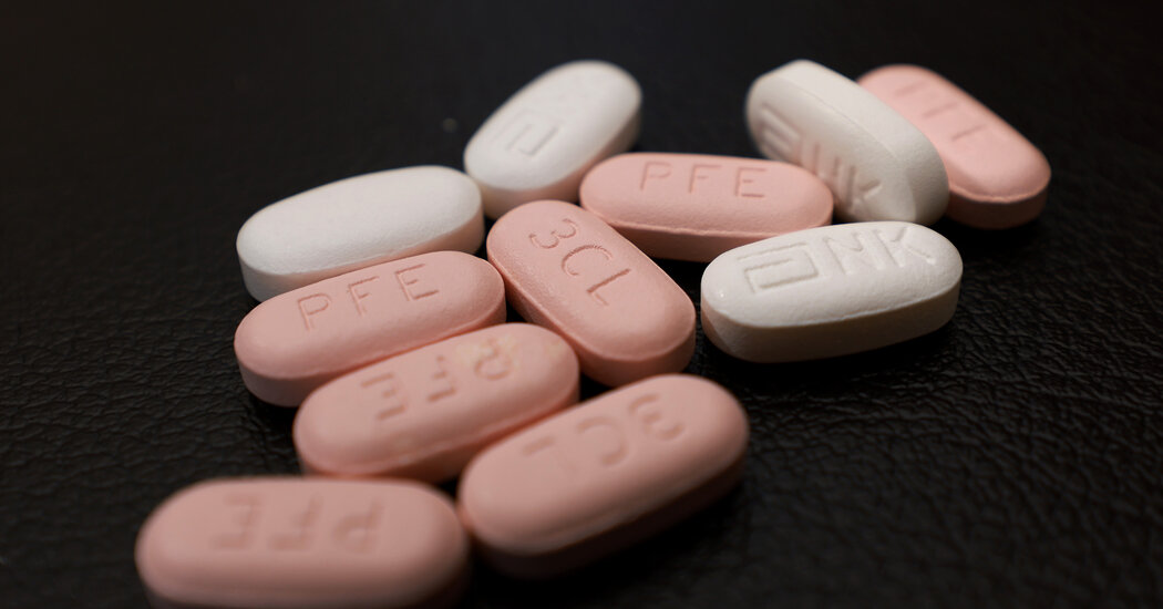
Among patients under 65, however, the drug made little difference in hospitalization or death rates.Paxlovid, the Covid-19 treatment made by Pfizer, reduced hospitalizations and deaths in older patients during the Omicron surge in Israel earlier this year, but made no difference for patients under 65 at high risk for severe disease, new research has found.The study is one of the first published examinations of the real-life effectiveness of Paxlovid against the Omicron variant, now the dominant version of the coronavirus. Pfizer’s trials of Paxlovid were conducted during a surge of the Delta variant last year, and included only unvaccinated individuals.There have been lingering questions about how effective the medication is against the Omicron variant, and among patients who are vaccinated or have some immunity from a prior bout of Covid. The drug has been available to Americans since December.The new study did not address another pressing mystery: how often patients experience “rebound” cases of Covid after taking the drug. Jill Biden, the first lady, exited a second isolation period on Monday after her infection returned following a course of Paxlovid.On Friday, Dr. Ashish Jha, the White House Covid-19 response coordinator, said on Twitter that while there was confusion over who should take Paxlovid, the data still indicated that it should be administered to anyone aged 50 and older soon after they develop Covid symptoms, as well as to anyone with health conditions leaving them vulnerable to severe illness.Although the Israeli study found that the drug had no benefit for adults aged 40 to 64 with underlying health problems, other research has suggested that it can improve outcomes. One study in Hong Kong, not yet peer-reviewed or published in a journal, reported benefits among patients aged 50 to 64.Researchers at Massachusetts General Brigham health system reported that Paxlovid significantly reduced hospitalizations among patients aged 50 to 64, with a pronounced effect among unvaccinated individuals and those with obesity. Dr. Jha said on Twitter that there was no reason to think the drug’s benefits would accrue only to older or more vulnerable populations. He noted that there were few side effects (the most notable is a metallic taste in the mouth), and that there was no shortage of Paxlovid in the United States.“Of course a drug that stops virus replication in a 70 yr old will do the same in 60 yr old,” Dr. Jha wrote. Nearly 200,000 Americans aged 50 to 64 have died of Covid, he noted.Pfizer’s own studies found that Paxlovid reduced the risk of hospitalizations and deaths by 88 percent in unvaccinated individuals at high risk for severe Covid, so long as the drug was taken within the first five days of symptom onset.The new research, published on Wednesday in The New England Journal of Medicine, showed the drug to be effective mostly in older Covid patients.“The big story is that it works, and saves quite a few lives and hospitalizations,” said Dr. Ronen Arbel, the study’s first author and a health outcomes expert at Clalit Health Services in Tel Aviv. “It’s very important that it’s helpful for older patients.”Other authors included Yael Wolff Sagy, Dr. Doron Netzer and Ariel Hammerman, all affiliated with Clalit Health Services, a large health care provider in Israel. The researchers reviewed medical records of almost 110,000 members of Clalit who tested positive for Covid from January through March, when the Omicron variant was dominant.The patients were at least 40 years old and were considered to be at high risk for severe disease. Most had either been vaccinated, previously infected with Covid, or both. The mean age of patients was 60, and more than half were women.About 4,000 were treated with Paxlovid, and the medication was highly effective when administered to individuals 65 and over, the researchers found.Among the 42,821 patients who were 65 and older, 766 patients who did not get Paxlovid were hospitalized for Covid, while only 11 patients who got Paxlovid were hospitalized, for a relative reduction in risk of 73 percent.Deaths were significantly reduced in the older patients treated with Paxlovid. Only two of 2,484 treated patients died, compared with 158 of the 40,337 untreated patients, a risk reduction of 79 percent.The drug had little effect on younger adults, however, making no significant dent in deaths or hospitalizations, which were as low in this group as among the older treated patients.Dr. Arbel said he and his colleagues had hoped to examine the rebound phenomenon, but the symptom reporting data was not reliable enough to do so.“I know it’s a big story in the United States, but I’m not sure anyone died or was hospitalized” after a rebound, he said. “It’s almost not relevant.”Although the drug does not appear to have much impact on younger and midlife adults, he said some physicians may still choose to prescribe it to patients under age 65.“For a 62-year-old unvaccinated person with complications, there may be reason to give the drug,” he said.

Read more →