Human Brain Cells Grow in Rats, and Feel What the Rats Feel
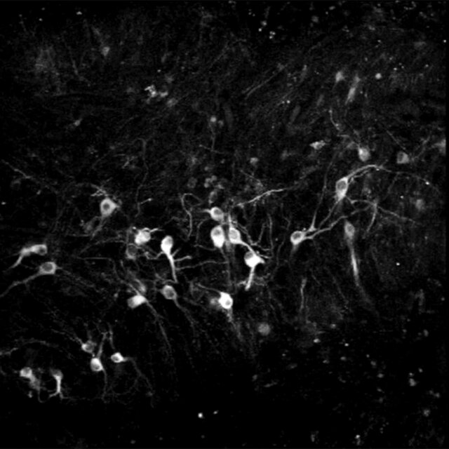
Human brain “organoids” wired themselves into rats’ nervous systems, influencing the animals’ sensations and behaviors.Scientists at Standford observed spontaneous activity of human neurons that had been transplanted into a rat. Pasca Lab, Stanford UniversityScientists have successfully transplanted clusters of human neurons into the brains of newborn rats, a striking feat of biological engineering that may provide more realistic models for neurological conditions such as autism and serve as a way to restore injured brains.In a study published on Wednesday, researchers from Stanford reported that the clumps of human cells, known as “organoids,” grew into millions of new neurons and wired themselves into their new nervous systems. Once the organoids had plugged into the brains of the rats, the animals could receive sensory signals from their whiskers and help generate command signals to guide their movements.Dr. Sergiu Pasca, the neuroscientist who led the research, said that he and his colleagues were now using the transplanted neurons to learn about the biology underlying autism, schizophrenia and other developmental disorders.“If we really want to tackle the biology of these conditions, we’re going to need more complex models of the human brain,” Dr. Pasca said.In 2009, after training in medicine in Romania, Dr. Pasca joined Stanford as a postdoctoral researcher to learn how to create human neurons in a dish. He and his colleagues took skin cells from volunteers and bathed them in chemicals that caused them to change character. Now they were more like embryo cells, which can become any tissue in the body.With the addition of more chemicals, the researchers coaxed the cells to develop into neurons. They could then observe pulses of voltage shoot down the length of the neurons as they lay in a dish.Dr. Pasca and his colleagues carried out the same experiment again, this time using skin cells from people with Timothy syndrome, a rare form of autism caused by a single mutation that leads to serious heart problems as well as impaired language and social skills.Growing Timothy syndrome neurons in a dish, Dr. Pasca could see a number of differences between them and typical neurons. They produced extra amounts of signaling chemicals such as dopamine, for example.But examining single cells could reveal only a limited number of clues about the condition. Dr. Pasca suspected that he could learn more by studying thousands of neurons joined together in circuits called brain organoids.A new chemical recipe allowed Dr. Pasca to mimic the condition inside the developing brain. Bathed in this broth, skin cells turned into progenitor brain cells, which in turn became tangles of neurons found in the brain’s outer layers, called the cortex.In a later study, he and his colleagues connected three organoids: one made of cortex, another of spinal cord and a third of muscle cells. Stimulating the cortex organoid caused the muscle cells to contract.A slice of rat brain showing the human cortical organoid in bright green.Pasca Lab, Stanford UniversityBut organoids are far from being miniature brains. For one thing, their neurons remain stunted. For another, they are not as electrically active as ordinary neurons in a living brain. “It’s clear that there are a number of limitations to these models,” Dr. Pasca said.Scientists began putting organoids in living brains, theorizing that a petri dish limited an organoid’s development. In 2018, the neuroscientist Fred Gage and his colleagues at the Salk Institute for Biological Studies transplanted human brain organoids into the brains of adult mice. The human neurons continued to mature as the mouse brain suppled them with blood vessels.Since then, Dr. Gage and other researchers have implanted organoids into the back of brain, where mice perceive signals from their eyes. When the animals saw pulsing flashes of white light, the human-organoid neurons responded in much the same way the mouse’s own cells did, according to a study published online in June that has not yet been peer-reviewed.Dr. Pasca and his team were also working on organoid transplants, but they chose to put them into young rodents rather than adults. A day or two after a rat was born, the scientists injected an organoid the size of a poppy seed into a region of the brain called the somatosensory cortex, which processes touch, pain and other signals from across the body. In rats, the region is especially sensitive to signals from their whiskers.The human neurons multiplied in the rat brain until they numbered about three million, making up about a third of the cortex on one side of the rat brain. Each cell in the organoid grew six times longer than it would have in a petri dish. The cells also became about as active as neurons in human brains.Even more strikingly, the human organoids spontaneously wired themselves into the rat brain. They connected not just to nearby neurons, but to distant ones as well.Those connections made the human neurons sensitive to the rat’s senses. When the researchers blew puffs of air over the rat’s whiskers, its human organoid crackled in response.Dr. Pasca and his colleagues also ran experiments to see how the organoids affected the behavior of the rats, using a water fountain in their chambers.After 15 days of training, the rats learned they could get a drink from the fountain when their organoid was stimulated. The human organoids were apparently sending messages to the reward-seeking regions of the rats’ brains.These species-blending experiments raise provocative ethical questions. Before starting the work, Dr. Pasca consulted with experts at the Center for Law and the Biosciences at Stanford, who urged him to pay special attention to the animals’ pain and well-being.“You’re not just worried about how many mice are in a cage, or how well they’re fed,” said Henry Greely, a Stanford law professor. “This is a new kind of thing. You don’t know what you might see.”Dr. Sergiu Pasca, a neuroscientist at Stanford University, led the research.Timothy ArchibaltDr. Pasca’s team found no evidence that the rats experienced pain, became prone to seizures or suffered a loss of memory or control of their movements. “It turns out that the rats tolerate the human graft really well,” Dr. Pasca said.Giorgia Quadrato, a neurobiologist at the University of Southern California who was not involved in the new study, noted that the human organoids did not make the rats more human. On learning tests, for example, they scored no better than other rats.“They are rats, and they stay rats,” Dr. Quadrato said. “This should be reassuring from an ethical perspective.”But that might not hold true if scientists were to put human organoids in a close relative of humans, like a monkey or a chimpanzee. “It would be a good opportunity to set guidelines to operate in the right ethical framework in the future,” she said.Dr. Pasca said that the similarity between primates and humans might allow the organoids to grow more and take on a bigger role in the animal’s mental processes. “It’s not something that we would do, or would encourage doing,” he said.Instead, he is using the implanted organoids to study neurological disorders. In one experiment, Dr. Pasca’s team implanted an organoid from a patient with Timothy syndrome on one side of a rat’s brain and implanted another organoid without the mutation on the other side.Both organoids grew in the rats. But the Timothy syndrome neurons developed twice as many branches for receiving incoming signals, called dendrites. What’s more, the dendrites were shorter.Dr. Pasca hopes that he will be able to observe differences in the way rats behave when they carry brain organoids from people with autism and other neurological conditions. Such experiments could help reveal how certain mutations alter the way the brain works.Dr. Isaac Chen, a neurosurgeon and organoid researcher at the University of Pennsylvania who was not involved in the research, saw another possibility in the new study: the repair of injuries to human brains.Dr. Chen envisioned growing brain organoids from the skin of a patient with a damaged cortex. Once injected into the brain of the patient, the organoid might grow and wire up with healthy neurons.“This idea is definitely out there,” he said. “It’s just a matter of, How do we take advantage of it, and take it to the next level?”

Read more →

Simple test could lead to more personalized treatment of rare ovarian cancer

A global study into mucinous ovarian cancer could help oncologists recommend the best treatment for women who are diagnosed early with the condition.
By looking down a microscope for two different ‘patterns of invasion’ — the way that cancer cells invade ovarian tissue — oncologists can better predict which patients may have better or worse prognoses and can target treatment accordingly. The finding was reported in a paper published today in Clinical Cancer Research, a journal of the American Association for Cancer Research.
“Mucinous ovarian cancer is a rare type of ovarian cancer. It actually has more in common with gastrointestinal cancers, and can be hard to diagnose and hard to treat once it has spread beyond the ovaries,” says lead author Nicki Meagher, who has just completed her PhD in the Molecular Oncology group, UNSW School of Clinical Medicine.
She says that observing which of the two types of invasion patterns that the cancer cells form could help specialists decide on treatment strategies.
“We’ve shown for the first time that women who have early-stage disease — meaning they have tumours that haven’t spread beyond the ovary — have much poorer survival chances in the first two years from diagnosis if they have what we call an infiltrative pattern of invasion.
“Knowing this in the early stage of the disease means we can identify patients who could benefit from additional chemotherapy following surgery to remove their ovaries.”
The two patterns of invasion are defined by the way the cancer cells organise themselves when viewed under a microscope. The infiltrative pattern of invasion associated with poorer health outcomes shows cancer cells spreading in an uneven, haphazard way through the ovarian tissue. The other pattern is known as expansile, where cells expand through tissue in a more orderly manner, and is associated with better prognoses.

Read more →

Toward a fully edible sensor showing if frozen food has previously thawed

When you’re standing in the frozen food aisle, it’s nearly impossible to know whether that Salisbury steak has thawed and refrozen — a process with potentially harmful consequences. So, researchers reporting in ACS Sensors have designed a food-grade device from edible materials, including table salt, red cabbage and beeswax, that lets you know. The proof-of-concept sensor provides a color readout when it’s warmed above a specific temperature, which is tunable from -58 F to 32 F.
Keeping food cold while it’s transported and stored is essential to retaining its flavor and quality, reducing the risk of food poisoning and minimizing waste. While researchers have developed devices that alert manufacturers when cold items are exposed to unwanted temperatures, they only indicate changes above freezing. To create a sensor for frozen products, one solution could be to use materials with electrical properties that are altered upon melting. It would also be ideal if such changes could produce a signal, such as a visible color change. In addition, an edible electronic device, which uses only food and consumable components, would be the safest way to monitor food. So, Ivan Ilic, Mario Caironi and colleagues set out to develop the first fully edible, self-powered temperature sensor with a visible color indicator for use with frozen products.
The researchers started by building a device that generated an electrical current as it defrosted, connecting magnesium and gold electrodes through an electrolyte solution held in a plastic container. They tested the device with solutions of frozen edible electrolytes, including table salt and calcium-containing salts, and naturally electrolyte-rich foods, including a grape, melon and apple. As the solutions defrosted, they conducted current between -58 F and 32 F, which the researchers say could be fine-tuned, based on the amount and identity of the salt. Next, this device was connected to a color-changing system, containing tin and gold electrodes and red cabbage juice, that produced an irreversible shift from reddish purple to blue when current was applied.
In the final step, the team put all of the parts together in a block of beeswax that held the temperature-activated and indicator solutions in separate chambers, and demonstrated that the self-powered device could be used for frozen food monitoring. The researchers say that their proof-of-concept sensor paves the way for edible materials to be used in inexpensive, safe technologies that alert customers to a frozen product’s storage history.
The authors acknowledge funding from the European Research Council, the European Union’s Horizon 2020 program and the Sustainability Activity of Istituto Italiano di Tecnologia.
Story Source:
Materials provided by American Chemical Society. Note: Content may be edited for style and length.

Read more →

Powerful enzyme that tamps down inflammation holds promise for protecting the eyes in diabetes, premature birth

An enzyme under study to treat certain cancers is also showing promise in reducing the significant vision damage that can result from diabetes and premature birth, scientists report.
Inflammation is considered a hallmark of cancer. It’s pervasive as well in both of these potentially blinding eye conditions, in which inadequate oxygen to the eyes prompts growth of new blood vessels to better deliver oxygen, but which instead often obstruct the vision pathway and become leaky, which causes swelling, further hindering vision.
Scientists at the Medical College of Georgia report in newly published studies in the journals Cell Death and Disease and Cells, increasing evidence that making more of the enzyme arginase 1, or A1, available helps alleviate these unhealthy responses and interrupt a natural body response that promotes destructive ongoing, high levels of inflammation in both diabetic retinopathy and retinopathy of prematurity.
Key to the process is making less of the amino acid L-arginine available. With diabetes, for example, high blood sugar and lipid levels as well as oxidative stress increase expression of inducible nitric oxide synthase, or iNOS, which uses the L-arginine to help produce even more inflammation and promote disease progression. The way it’s supposed to work is iNOS goes up in response to an infection then high-expressing A1 cells move in to turn iNOS off and inflammation down.
That’s because A1 competes with iNOS for L-arginine. They theorized that more A1, which actually breaks down L-arginine into two products, would make less L-arginine available to “feed this (unhealthy) iNOS explosion,” and help tamp down the vicious cycle of inflammation and related damage, says Dr. William Caldwell, pharmacologist and chair emeritus of the MCG Department of Pharmacology and Toxicology.
“If you reduce L-arginine levels, iNOS cannot work. This will make things better,” Caldwell says.

Read more →

Are smartwatch health apps to detect atrial fibrillation smart enough?

Extended cardiac monitoring in patients and the use of implantable cardiovascular electronic devices can increase detection of atrial fibrillation (AF), but the devices have limitations including short battery life and lack of immediate feedback. Can new smartphone tools that can record an electrocardiogram (ECG) strip and make an automated diagnosis overcome these limitations and facilitate timely diagnosis? The largest study to date, in the Canadian Journal of Cardiology, published by Elsevier, finds that the use of these devices is challenging in patients with abnormal ECGs. Better algorithms and machine learning may help these tools provide more accurate diagnoses, investigators say.
“Earlier studies have validated the accuracy of the Apple Watch for the diagnosis of AF in a limited number of patients with similar clinical profiles,” explained lead investigator Marc Strik, MD, PhD, LIRYC institute, Bordeaux University Hospital, Bordeaux, France. “We tested the accuracy of the Apple Watch ECG app to detect AF in patients with a variety of coexisting ECG abnormalities.”
The study included 734 consecutive hospitalized patients. Each patient underwent a 12-lead ECG, immediately followed by a 30-second Apple Watch recording. The smartwatch’s automated single-lead ECG AF detections were classified as “no signs of atrial fibrillation,” “atrial fibrillation,” or “inconclusive reading.” Smartwatch recordings were given to an electrophysiologist who conducted a blinded interpretation, assigning each tracing a diagnosis of “AF,” “absence of AF,” or “diagnosis unclear.” A second blinded electrophysiologist interpreted 100 randomly selected traces to determine the extent to which the observers agreed.
In approximately one in every five patients, the smartwatch ECG failed to produce an automatic diagnosis. The risk of having a false positive automated AF detection was higher for patients with premature atrial and ventricular contractions (PACs/PVCs), sinus node dysfunction, and second- or third-degree atrioventricular-block. For patients in AF, the risk of having a false negative tracing (missed AF) was higher for patients with ventricular conduction abnormalities (interventricular conduction delay) or rhythms controlled by an implanted pacemaker.
The cardiac electrophysiologists had a high level of agreement for differentiation between AF and non-AF. The smartphone app correctly identified 78% of the patients who were in AF and 81% who were not in AF. The electrophysiologists identified 97% of the patients who were in AF and 89% who were not.
Patients with PVCs were three times more likely to have false positive AF diagnoses from the smartwatch ECG, and the identification of patients with atrial tachycardia (AT) and atrial flutter (AFL) was very poor.
“These observations are not surprising, as smartwatch automated detection algorithms are based solely on cycle variability,” Dr. Strik noted, explaining that PVCs cause short and long cycles, which increase cycle variability. “Ideally, an algorithm would better discriminate between PVCs and AF. Any algorithm limited to the analysis of cycle variability will have poor accuracy in detecting AT/AFL. Machine learning approaches may increase smartwatch AF detection accuracy in these patients.”
In an accompanying editorial, Andrés F. Miranda-Arboleda, MD, and Adrian Baranchuk, MD, Division of Cardiology, Kingston Health Science Center, Kingston, ON, Canada, observed that this is the first “real-world” study focusing on the use of the Apple Watch as a diagnostic tool for AF.
“It is of remarkable importance because it allowed us to learn the performance of the Apple Watch in the diagnosis of AF is significantly affected by the presence of underlying ECG abnormalities. In a certain manner, the smartwatch algorithms for the detection of AF in patients with cardiovascular conditions are not yet smart enough. But they may soon be,” Dr. Miranda-Arboleda and Dr. Baranchuk said.
“With the growing use of smartwatches in medicine, it is important to know which medical conditions and ECG abnormalities could impact and alter the detection of AF by the smartwatch in order to optimize the care of our patients,” Dr Strik said. “Smartwatch detection of AF has great potential, but it is more challenging in patients with pre-existing cardiac disease.”
Story Source:
Materials provided by Elsevier. Note: Content may be edited for style and length.

Read more →

How poliovirus takes over cells from within

For the first time, researchers at Umeå University, Sweden, can now show how the dreaded poliovirus behaves when it takes over an infected cell and tricks the cell into producing new virus particles. Polio was thought to be almost eradicated, but infection has now been rediscovered in London and New York.
“We now have a completely different understanding of how the virus acts and thus better opportunities for research to perhaps find new ways to curb the virus’ progress in the future,” says Lars-Anders Carlson at the Department of Medical Chemistry and Biophysics at Umeå University.
The dreaded poliovirus belongs to the same large family, enteroviruses, as several common colds. It has been known for some time that enteroviruses drastically rearrange the inside of infected, but it has not been known exactly how, simply because technology has not allowed us to see so deeply into the cells. Thanks to the advanced cryo-electron microscope in Umeå, researchers have for the first time been able to take three-dimensional images of how the poliovirus forms and takes over human cells.
“We were surprised to see how the virus transforms processes in the cell that are otherwise used to destroy viruses to produce new viruses instead,” says Lars-Anders Carlson.
The researchers were able to identify the site in the cell where the poliovirus forms new virus particles, by seeing sites with half-assembled viruses. Surprisingly, this “virus factory” in the cell turned out to be surfaces in the cell that resembled an otherwise normal process in the cell, autophagy. Autophagy is a relatively recently discovered process in cells that was subject of the 2016 Nobel Prize. Normally, autophagy serves to break down particles that the cell wants to get rid of, such as virus particles. But the poliovirus manages to reprogram this defence mechanism against viruses to produce more virus instead.
The researchers found that certain proteins are particularly important. The VSP34 protein is used by the virus to build new virus particles. When the researchers inhibited VSP34, they could see that the virus could barely assemble whole viruses, but mostly only half virus particles. Another important protein is called ULK1, which slows down the production of viruses. The researchers could see that the amount of virus exploded when this protein was inhibited. This confirms the theory that the poliovirus breaks down this “brake.”
Once the virus has multiplied in the cell, the particles must be released to infect new cells. This is done by releasing the particles in small packets, called vesicles. Here, the researchers also made a surprising discovery; a careful sorting of what is packed into the vesicles takes place. Only viruses that are correctly formed and carry the genetic material of the virus are placed in the vesicles, while empty virus particles are not allowed in. In this way, the virus may spread more efficiently.
“The new knowledge we are contributing about the role of autophagy in virus formation may provide new insights for the development of future antivirals that could complement vaccines. We have good reason to believe that our findings are valid for the large group of viruses to which poliovirus belongs, enteroviruses. There is no vaccine against most enteroviruses, but an antiviral that acts on the autophagy system could be effective against many of them. However, there is still a long way to go,’ says Lars-Anders Carlson.
Polio is rightly a dreaded disease that can cause paralysis and death. The poliovirus starts in the intestines but can then attack the spinal cord. There is still no cure for the disease, but the only way to prevent it is to be vaccinated. In much of the world, vaccination campaigns have been so successful that the disease is considered virtually eliminated. However, polio has persisted in some countries in Asia, Africa and the Middle East. The poliovirus is mainly transmitted through feces. In 2022, the virus was again detected in sewage in New York and London. In addition, New York has had the first new case in ten years of a person getting paralysed due to polio infection.
The reappearance of polio in developed countries may be partly due to a decline in vaccination rates, as the disease was considered almost eradicated, and also due to increasing resistance to vaccination.
The study was a collaboration with researchers at the National Institutes of Health, USA, and Monash University, Australia. It is published in the scientific journal Nature Communications.
Story Source:
Materials provided by Umea University. Note: Content may be edited for style and length.

Read more →

Study explores links between people taking multiple medications and dementia diagnosis

People with dementia are likely to have taken more than three medications for other health conditions in the five years directly before their diagnosis, according to new research.
The study is the first to provide an in-depth exploration of the links between evolving polypharmacy — which involves a patient being prescribed more than one drug at any given time — and a dementia diagnosis.
Published in the Aging and Disease journal, it is based on an analysis of the records of more than 33,000 dementia patients in Wales between 1990 to 2015.
Experts in e-health used machine learning techniques to identify potentially damaging patterns in a patient’s medicine usage, and how these patterns evolve in the run-up to diagnosis.
They found that in the 20 years leading up to them being diagnosed, the proportion of patients taking three or more medications rose from 5.5% (for the period 16 to 20 years prior to diagnosis) to 82.16% among those less than five years from a diagnosis.
Researchers also found that as the development towards dementia progressed, the patterns of polypharmacy shifted from being clearly distinct to being more closely associated with particular medical conditions.
And of those closest to their diagnosis, almost two-thirds (66.55%) were found to be taking multiple medicines for a combination of respiratory or urinary infections, arthropathies and rheumatism, and cardio-vascular disease. A further 22% of patients were found to be taking medicines for infections, arthropathies and rheumatism, cardio-metabolic disease and depression.
The study was supported by the Health Data Research UK and conducted by an international team of researchers from the University of Plymouth, Aptuit (an Evotec company), Swansea University Medical School, and the University of Oxford.
Shangming Zhou, Professor of e-Health at the University of Plymouth, led the study. He said: “Given the rise in dementia cases internationally, the need to understand how patterns of polypharmacy evolve before and after a dementia diagnosis are important for devising a safe treatment programme for each patient. Our aim in this study was to help doctors find ways to prescribe multiple items of dementia medication safely and without reducing their effectiveness. The use of machine learning has been vital in helping us understand how these patterns develop, and our hope is we can now use this knowledge to treat patients.”
It has previously been established that when multiple types of preventative medication are being prescribed, the benefits of the drugs may be reduced and the chances of harm from drug interaction and side effects increased.
Those requiring hospital treatment who are taking multiple medications are also known to have a higher likelihood of re-admission within three months after being discharged.
With the number of people with dementia in the UK projected to rise to 1.6 million by 2040, researchers hope this new study will inform safe prescribing practices and encourage doctors to prescribe medicine combinations developed with a view to minimising cognitive impairments.
Story Source:
Materials provided by University of Plymouth. Original written by Alan Williams. Note: Content may be edited for style and length.

Read more →

Therapeutic games and brain stimulation mitigates cognitive decline in older adults

Older people may be able to boost their working memory with a new approach that couples online therapeutic games with a non-invasive brain stimulation technique.
Working memory is critical for people to function well in everyday life. This volatile form of memory holds and manipulates a finite amount of information over a short time interval, enabling people to interact with their environment in an effective and efficient manner. Working memory typically declines with age, with the decline in its capacity causing daily difficulties in people with Parkinson’s disease, dementia, and stroke.
Scientists and clinicians from the University of Birmingham, UK, Dalhousie University in Nova Scotia, Canada, and the University of Trento, Italy have devised a new technology to mitigate this decline.
The investigators refer to the technology as cognitive needs and skills training, or COGNISANT, and research published in Frontiers in Ageing Neuroscience shows it can provide particular benefit for older people who have low working memory capacity (WMC).
The online therapeutic exercises, developed to improve working memory, attention and vigilance, are packaged in the type of engaging interface that will be familiar to online game or App users. Brain stimulation was administered via a mobile wireless device that delivers a small (2milliAmpère) transcranial direct current stimulation (tDCS) during training.
The study involved healthy people aged 55 to 76 years old, who were split into two groups. Both groups did the online games for 20 minutes day, over a five-day period. While one group also received tDCS, the other group wore the tDCS device, which resembles a swimming cap, but did not receive tDCS.

Read more →

Humor and body image linked

Women who use positive humour to describe themselves have higher levels of ‘body appreciation’ than those who use self-deprecating humour, according to new research from the University of Surrey.
Researchers also found that along with having a negative view of their body, those who used self-defeating humour were also more likely to have poor eating habits.
Dr Fabio Fasoli, Lecturer in Social Psychology at the University of Surrey, said:
“Having a negative body image can affect all areas of a person’s life and lead to depression and social anxiety. Often people don’t talk openly about how they view their bodies, but how a person speaks about themselves through humour can provide valuable insights into such feelings.”
In the first study of its kind, researchers from the University of Surrey explored the relationship between different styles of humour, body perception and eating behaviours in women. Findings from this study can be used by clinical psychologists and eating disorder therapists to better understand their patients and promote the use of positive humour to improve their body image.
Researchers surveyed 216 people, analysing their humour styles and perceptions of their bodies. The team from Surrey found that women who used self-defeating humour, putting themselves down in an aggressive fashion, were more likely to be highly critical of their bodies and possess a strong desire to be slimmer. Those who used such humour were also found to be emotional eaters, often making themselves feel worse about their bodies and putting them at risk of obesity and resulting illnesses.
In addition, it was identified that those who used self-enhancing humour, making yourself the target of the humour in a good-natured way, reported more body appreciation, and were associated with body kindness. Those who used this type of humour were less likely to be emotional eat than their peers.
The Surrey team found that women who use both forms of humour reported a positive body image and had better eating habits than those who primarily engaged in self-defeating humour.
Jane Ogden, Professor of Health Psychology at the University of Surrey, said:
“People using themselves as the butt of jokes is often a technique to get approval from others, but it can also signify that something more worrying is going on in that person’s life. This type of humour can indicate that a person is struggling with their body image and self-esteem, which can have a long-lasting effect on their life.”
Story Source:
Materials provided by University of Surrey. Note: Content may be edited for style and length.

Read more →

One in 20 suffer long-term Covid effects, study finds

Published17 hours agoSharecloseShare pageCopy linkAbout sharingImage source, Getty ImagesOne in 20 people suffer long-term effects after Covid, a study has found.The long Covid in Scotland study, led by the University of Glasgow, found that effects were more likely to occur after severe infections requiring hospitalisation.The research showed older women from deprived communities were most at risk.The study, set up in May 2021, found those who were vaccinated before becoming unwell appeared to be protected from some long-term symptoms.The most reported symptoms included breathlessness, chest pain, palpitations, and confusion or “brain fog”. The latest headlines from ScotlandSymptoms could range from mild to moderate – but having long-term effects might not necessarily result in a long Covid diagnosis.Long Covid was more likely in those with pre-existing physical and mental health problems, such as a respiratory disease or depression.’Elephant on my chest’Image source, Jayne GemmellJayne Gemmell, 55, from Cairneyhill in Fife, contracted Covid in March 2020 and is still living with the effects. She already had ME and fibromyalgia but was able to work full-time.Jayne said the day she fell ill with Covid she felt extremely tired, although she didn’t consider it significant.”The second day I just felt like I had an elephant sitting on my chest,” she said. “I just had this overwhelming fatigue and overwhelming discomfort and tightness and heaviness. “I took some time off work expecting that I might need a few days or a week and actually never back to work.”Jayne says she has been left with worsened chronic fatigue and pain. Medics also believe she has developed asthma.She said she was “very grateful” to the NHS and Chest Heart & Stroke Scotland who have supported her rehabilitation. The Glasgow University research found that 6% of people felt they had not recovered at all, while 42% reported feeling only partially recovered between six and 18 months following infection.Jill Pell, professor of Public Health who led the study, said: “While most people recover quickly and completely after infection with Covid, some people develop a wide variety of long-term problems. “Therefore, understanding long Covid is essential to inform health and social care support.”What are the symptoms of long Covid?Mum puts hopes in experimental Long Covid trialThe study also found that those with asymptomatic infection had no long-term impact.It used 33,281 people who had developed laboratory-confirmed Covid infections and 62,957 never-infected individuals from the general population.’You feel like a forgotten tribe’Jane Ormerod from Aberdeen, first caught Covid in March 2020. It began with a bad headache and a sore throat. She said: “I didn’t have a cough at that time. I had a high temperature, shivers and a rapid heart rate. My breathing wasn’t good.”After a couple of weeks her symptoms seemed to improve although she felt like she had tonsillitis for about four months. Jane, 66, also started experiencing new symptoms such as dizziness and sore joints. She felt like Covid had taken away her energy.She added: “For all of us with long Covid, you feel like a forgotten tribe. Many of us are still struggling to get treatment, diagnosis – to even see somebody.” Dr Andrew McAuley, consultant healthcare scientist at Public Health Scotland, said the study provided important evidence on long Covid. He added: “We know that being fully vaccinated can reduce the likelihood of developing long Covid and therefore we encourage those who are eligible to take the opportunity to enhance their protection by getting vaccinated.”The Scottish government said long Covid was “debilitating for the physical and mental wellbeing” of those affected. A spokesperson said: “We’re investing an initial £3m from our Long Covid Support Fund to provide NHS boards and partners with additional resource to deliver the best local models of care for assessment, diagnostic tests, and support for the treatment or management of symptoms.”Health Secretary Humza Yousaf told BBC Scotland it was up to individual health boards how they spent the funding. He said some had appointed a care co-ordinator while others had used it to help children with long Covid.He added: “If I could reiterate one message from that study it would be please get yourself vaccinated, if you are eligible.”

Read more →