Rethinking how we treat atrial fibrillation

A national study led by UBC researchers at the Centre for Cardiovascular Innovation is shedding light on how to more effectively treat atrial fibrillation (AF) — a common heart rhythm problem associated with increased risk of stroke and heart failure.
The study, published today in The New England Journal of Medicine, shows that early intervention with cryoballoon catheter ablation (cryoablation) is more effective at reducing the risk of serious long-term health impacts, when compared to the current first step in treatment, antiarrhythmic drugs.
“By treating patients with cryoablation right from the start, we see fewer people advancing to persistent, more life-threatening forms of atrial fibrillation,” says Dr. Jason Andrade, an associate professor of medicine at UBC and director of Heart Rhythm Services at Vancouver General Hospital. “In the short term, this can mean less recurrences of arrhythmia, improved quality of life and fewer visits to the hospital. In the long run, this can translate into a reduced risk of stroke and other serious heart problems.”
Cryoablation is a minimally invasive procedure that involves guiding a small tube into the heart to kill problematic tissue with cold temperatures. Historically, the procedure has been reserved as a secondary treatment when patients don’t respond to antiarrhythmic drugs.
“This study adds to the growing body of evidence that early intervention with cryoablation may be a more effective initial therapy in the appropriate patients,” says Dr. Andrade.
Early intervention halts disease progression
AF affects approximately three per cent of the population, or more than one million Canadians.

Read more →

Neuron function is altered by the widely used anesthetic propofol

Propofol is the most commonly used drug to induce general anesthesia. Despite its frequent clinical application, it is poorly understood how propofol causes anesthesia.
In a new study published in Molecular Biology of the Cell, a team of Rensselaer Polytechnic Institute researchers identified a previously unknown propofol effect in neurons. The study found that propofol exposure impacted the process by which neurons transport proteins, biomolecules that perform most cellular functions, to the cell surface.
Almost all animal cells, including human cells, are highly compartmentalized and rely on efficient movement of protein material between compartments. Proteins are moved from their site of synthesis to the location at which they perform their function in small carriers called “vesicles.” This transport must be efficient and highly specific to maintain cellular organization and function.
The research team was led by Dr. Marvin Bentley, an assistant professor in the Department of Biological Sciences, whose laboratory studies vesicle transport in neurons. Neurons are particularly reliant on vesicle transport because axons — which are often organized in nerve bundles — can span distances of up to 1 meter in humans. Errors in vesicle transport have been linked to neurodevelopmental and neurodegenerative diseases such as Alzheimer’s and Parkinson’s.
This new study found that propofol affects a family of proteins called kinesins. Kinesins are small “motor proteins” that move vesicles on tiny filaments called microtubules.
Dr. Bentley’s team observed that vesicle movement of two prominent kinesins, Kinesin-1 and Kinesin-3, was substantially reduced in cells exposed to propofol. The team then showed that propofol-induced transport delays led to a significant decrease of protein delivery to axons.
“The mechanism by which propofol works is not fully understood,” Bentley said. “What we discovered was unexpected: propofol altered the trafficking of vesicles in live neurons.”
Overall, the research contributes significantly to our understanding of how propofol works. Most studies that address the anesthetic mechanism of propofol have been focused on its interaction with an ion channel called the GABAA receptor, which inhibits neurotransmission when activated.
This new study demonstrates that vesicle transport is an additional mechanism that may be important for propofol’s anesthetic effect. Discovery of this new propofol effect has important applications for human health and may lead to the development of better anesthetic drugs.
“By using state-of-the-art live cell imaging technologies, Dr. Bentley’s team has furthered our understanding of the mechanism of action of a widely used drug that is already impacting human health on a daily basis,” said Curt M. Breneman, Dean of the School of Science. “Dr. Bentley’s research may pave the way for the development of related compounds that use these same mechanisms to target debilitating neurodegenerative diseases.”
In addition to Dr. Bentley, the study was co-authored by Dr. Susan P. Gilbert, Head of the Department of Biological Sciences at Rensselaer, and doctoral students Madeline Frank and Alec T. Nabb.
Story Source:
Materials provided by Rensselaer Polytechnic Institute. Original written by Katie Malatino. Note: Content may be edited for style and length.

Read more →

Which COVID vaccine you get can impact myocarditis risk

Incidence of myocarditis, pericarditis or myopericarditis is two- to threefold higher after a second dose of the Moderna Spikevax COVID-19 vaccine when compared to the Pfizer BioNTech COVID-19 vaccine; however, overall cases of heart inflammation with either vaccine are very rare, according to a study in the Journal of the American College of Cardiology. The study showed males younger than 40 years old who received the Moderna vaccine were shown to have the highest rates of myocarditis, which according to the authors, may have implications for choosing specific vaccines for certain populations.
Two mRNA COVID-19 vaccines have been approved for use, Pfizer BioNTech (BNT162b2) and Moderna Spikevax (mRNA-1273), and as of March 20, 2022, more than 52 million doses of Pfizer and 22 million doses of Moderna have been administered in Canada, where this study was conducted. Clinical trials have demonstrated the vaccines are safe and monitoring of vaccinated people has shown side effects are mild and go away on their own. However, some rare, but serious, side effects have been observed after both vaccines, mainly myocarditis (inflammation of the heart).
While there have been many studies on either vaccine, few studies have been conducted to directly compare the safety of the two mRNA vaccines. Researchers in this study sought to compare the risk of myocarditis, pericarditis and myopericarditis between the Pfizer and Moderna COVID-19 vaccines.
People in the study were 18 years old or older and had received two primary doses of either Pfizer or Moderna vaccine in British Columbia, Canada, with the second dose between Jan. 1, 2021 and Sept. 9, 2021. Individuals whose first or second shot were administered outside of British Columbia or had a history of myocarditis or pericarditis within one year prior to second dose were excluded.
In all, more than 2.2 million second Pfizer doses were given and more than 870,000 Moderna doses. Within 21 days of the second dose, there were a total of 59 myocarditis cases (21 Pfizer and 31 Moderna) and 41 pericarditis cases (21 Pfizer and 20 Moderna). Researchers also looked at rates per million doses and the rate was 35.6 cases per million for Moderna and 12.6 per million for Pfizer — an almost threefold increase after Moderna shots vs. Pfizer. Comparatively, rates of myocarditis in the general population in 2018, were 2.01 per million in people under age 40 and 2.2 per million in people over age 40.
Rates of myocarditis and pericarditis were higher with the Moderna vaccine in both males and females between ages 18 and 39, with the highest per million rates in males ages 18-29 after a second dose of Moderna.
According to the authors, the findings support recommending certain populations receive certain vaccines to maximize benefits and minimize adverse events.
“Few population-based analyses have been conducted to directly compare the safety of the two mRNA COVID-19 vaccines, which differ in important ways that could impact safety,” said Naveed Janjua, MBBS, DrPH, lead author of the study and an epidemiologist and the executive director of Data and Analytic Services at the British Columbia Centre for Disease Control. “Our findings have implications for strategizing the rollout of mRNA vaccines, which should also consider the self-limiting and mild nature of most myocarditis events, benefits provided by vaccination, higher effectiveness of the Moderna vaccine against infection and hospitalization [found in prior studies], and the apparent higher risk of myocarditis following COVID-19 infection than with mRNA vaccination.”
Limitations of the study include that it was observational, which limits the ability to determine causality between vaccination and myocarditis or pericarditis. However, temporality was ensured in the study design to limit the time studied between vaccine dose and myocarditis/pericarditis diagnosis. Also, the study relied on hospital and emergency department visit data and may have missed some less severe cases.
In a related editorial comment, Guy Witberg, MD, MPH, a cardiologist at Rabin Medical Center in Petah-Tikva, Israel, wrote the study is reassuring for vaccine safety since it provides further data that myocarditis is a very rare adverse event after both vaccines, and it is an important step toward a personalized approach to administering COVID-19 vaccines.
“[The study] should help put to rest ‘vaccine hesitancy’ due to concerns over cardiac adverse events,” Witberg said. “This is one of only a few direct comparisons of the two widely adopted mRNA vaccines, and its results have practical policy implications: for a substantial segment of the population suffering from cardiovascular disease…these data give a strong argument to preferentially use the BNT162b2 [Pfizer] vaccine over mRNA-1273 [Moderna].”

Read more →

New theory explains recovery delays in COVID-19 and cardiac patients

COVID-19 patients placed on ventilators can take a long time to regain consciousness. New research from Weill Cornell Medicine, NewYork-Presbyterian, MIT, and Massachusetts General Hospital is now illustrating that these delays may serve a purpose: protecting the brain from oxygen deprivation.
The existence of such a brain-preserving state could explain why some patients wake up days or even weeks after they stop receiving ventilation, and it suggests that physicians should take these lengthy recovery times into account when determining a patient’s prognosis.
In a study published Nov. 7 in the Proceedings of the National Academy of Sciences, the investigators connect the pattern seen among those who have survived severe COVID-19 with similar delays known to occur in a small fraction of cardiac arrest patients.
“The delayed recoveries in COVID-19 patients are very much like the rare cases we’ve documented in previous research. In this new paper, we describe a mechanism to explain what we’re seeing in both types of patients,” said study co-senior author Dr. Nicholas D. Schiff, the Jerold B. Katz Professor of Neurology and Neuroscience in the Feil Family Brain and Mind Research Institute and co-director of the Consortium for the Advanced Study of Brain Injury (CASBI) at Weill Cornell Medicine.
He found evidence for this explanation — that patients’ brains are protecting themselves — in animals, most notably painted turtles, that can tolerate extended periods without oxygen.
More than a decade ago, Dr. Schiff and his colleagues first observed these delays among comatose cardiac arrest patients who received cooling therapy to reduce brain damage caused by a loss of blood flow. In one such case, a 71-year-old patient took 37 days to awaken, before ultimately making a near-complete recovery.

Read more →

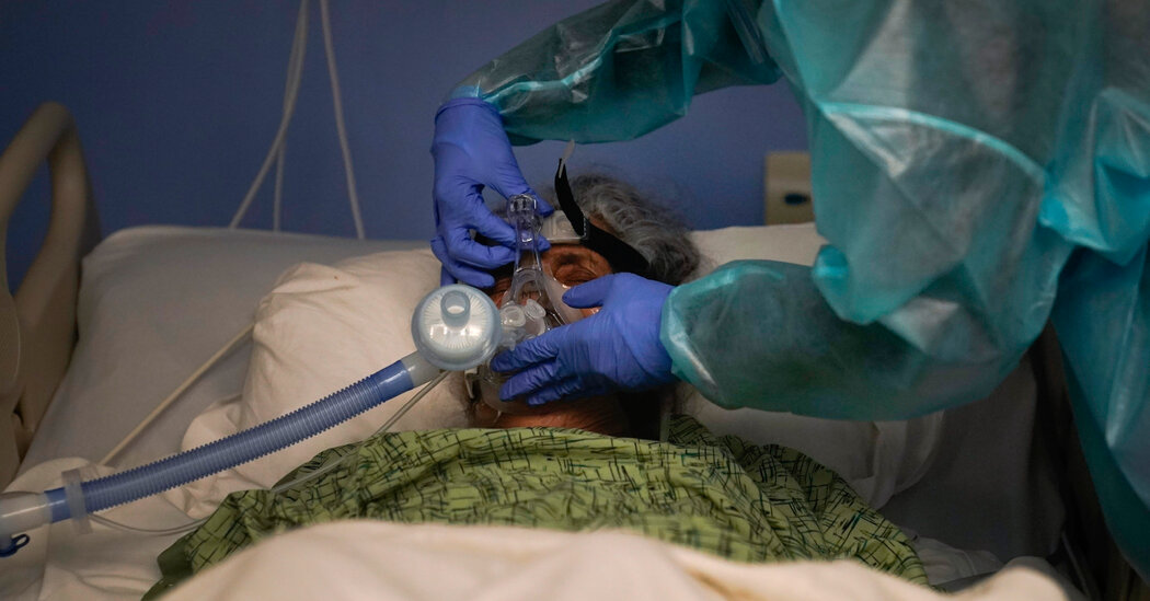
Covid Patients Coming Off Ventilators Can Take Weeks to Regain Consciousness

A new paper suggests that the combination of the virus and anesthesia plunges the brain into a prolonged state of quiet — like a freshwater turtle in winter.In March 2020, New York City’s hospitals filled up with patients desperately ill with Covid-19. In many cases, when their fluid-filled lungs could no longer give them oxygen, doctors sedated them and put them on ventilators.The patients who recovered were taken off the machines and anesthesia. Within a day or so, their doctors expected them to wake up.But that’s when the phone of Dr. Nicholas Schiff, a neurologist at Weill Cornell Medicine, started lighting up.“We’re starting to get all these weird consults,” Dr. Schiff recalled. “People have been liberated from anesthesia after surviving Covid, and they’re not waking up.”Dr. Schiff, who had spent 25 years treating disorders of consciousness, was perplexed by the influx of unconscious Covid patients. They took weeks, and sometimes months, to wake up. But then they usually regained full consciousness, with no sign of brain damage.Dr. Schiff and his colleagues have been trying to make sense of this strange phenomenon ever since. On Monday, he published a paper that proposes an answer. And the answer involves turtles.The brains of unconscious Covid patients bear a striking resemblance to those of turtles that spend the winter encased in ice, argued Dr. Schiff and his collaborator, Dr. Emery Brown, a computational neuroscientist at M.I.T. The turtles survive by putting their neurons into an unusual quiet state that lasts for months. Dr. Schiff and Dr. Brown believe that the combination of Covid and sedatives prompts a similar response in people.Read More on the Coronavirus PandemicWarnings of a ‘Tripledemic’: An expected winter rise in Covid cases appears poised to collide with a resurgent flu season and a third pathogen straining pediatric hospitals in some states.A Decline Among Seniors: Americans over 65 remain the demographic most likely to have received the original series of Covid vaccinations. But fewer are getting follow-up shots, surveys indicate.Updated Boosters: New findings show that Pfizer’s updated booster is better than its predecessor at increasing the antibody levels of people over age 55 against the most common version of the virus now circulating.Personality Changes: New research suggests that Covid’s disruption of social rituals and rites of passage have made people less extroverted, creative, agreeable and conscientious.If the theory holds up, it might point to new ways to save people from brain damage: by intentionally putting people into this state, rather than doing so by accident.“If it’s right, it can teach us how to protect and preserve the brain better,” Dr. Schiff said.Dr. Schiff discovered that his experience was not unusual. Many other neurologists were seeing Covid patients taking a very long time to wake up. In March of this year, Dr. Schiff, Dr. Brown and their colleagues published a study of 795 severe Covid patients with delayed recovery in three hospitals in New York City and Boston. One-quarter of the patients took 10 days or more after coming off a ventilator to follow simple commands like squeezing a doctor’s finger. After 23 days, 10 percent were still not conscious.But the analysis did not offer any easy answers about why they were experiencing such a long delay. The anesthetic drugs could not alone account for the long journey back to consciousness. “The time courses were absurd,” Dr. Schiff said.Brain damage can lead to months of minimal consciousness, but many of the Covid patients had healthy brains. “There was no expectation that there should be a problem,” Dr. Schiff said.For years, Dr. Schiff and Dr. Brown have been developing theories about what happens in the brain during comas, sleep and anesthesia. Now they turned their efforts to Covid. Their search for clues brought them unexpectedly to studies of turtles.Across the northern world, coldblooded turtles that live in freshwater have to survive chilly winters. They do so by spending months buried in frozen mud, barely breathing. Researchers who study turtles in laboratories have found that the animal prepares its brain for winter by flooding it with a chemical called GABA. The compound quiets the activity of neurons so that they don’t waste energy producing electrical pulses.“It’s like they auto-anesthetize themselves,” Dr. Brown said.Over the winter, the turtles produce distinctive brain-wave patterns, with isolated bursts of electricity separated by long stretches of silence. Just like the turtles, unconscious Covid patients produce brief bursts of electrical activity between long silences. And these patients typically received anesthetic drugs that mimic GABA.Dr. Schiff and Dr. Brown proposed that in response to GABA-like sedatives and the stress of Covid, human neurons shift to a quiet mode in which they don’t need much oxygen to survive. Even after the sedatives wear off, the brain can keep itself in this state for months.“I think it’s an intriguing analogy, definitely,” said Amanda Bundgaard, a postdoctoral researcher at the University of Cologne in Germany who studies turtle brains. But she cautioned against pushing the analogy too far at this point, because there’s so much left to understand about turtles.“One thing that maybe is a little bit problematic is that we don’t know how turtles wake up again from this,” she said.After months in a state of suspended animation, turtles jump back to normal by flooding their brains with oxygen. That’s astonishing, because the surge of oxygen ought to kill their neurons by triggering toxic chemical reactions.Some studies suggest that turtles soak up the extra oxygen with a chemical called neuroglobin. But it’s possible they use a number of other chemicals to create many lines of defense.“It’s exciting to have a new hypothesis to think about if it helps us create better outcomes for patients,” said Martin Monti, a neuroscientist at U.C.L.A. who was not involved in the study.In their paper, Dr. Schiff and Dr. Brown suggest that neurologists should check samples of cerebrospinal fluid taken from Covid patients as they returned to consciousness. Like turtles, they might release a surge of neuroglobin to protect their brains.“That would be a pretty good test of the hypothesis,” Dr. Monti said.He added that the hypothesis might also lead to new ways to prevent brain tissue from dying after strokes, heart attacks or even traumatic brain injuries. A combination of sedatives and other treatments might induce neurons to give themselves a turtlelike protection.“This could ultimately turn into a new tool in the toolbox to make patients not only survive, but recover as well as possible,” Dr. Monti said.

Read more →

Breast cancer: New treatment can significantly increase the efficacy of chemotherapy and prevent metastasis

A new treatment developed at Tel Aviv University may significantly enhance the efficacy of chemotherapy in breast cancer patients, reducing the risk for lung metastasis following chemo from 52% to only 6%. Conducted in an animal model, the study identified the mechanism that generates a cancer-promoting inflammatory environment in response to chemotherapy. Moreover, the researchers found that by adding an anti-inflammatory agent to the chemotherapy, metastasis can be prevented.
The study was led by Prof. Neta Erez of the Department of Pathology at TAU’s Sackler Faculty of Medicine, and researchers from her group: Lea Monteran, Dr. Nour Ershaid, Yael Zait, and Ye’ela Scharff, in collaboration with Prof. Iris Barshack of the Sheba Medical Center and Dr. Amir Sonnenblick of the Tel Aviv Sourasky (Ichilov) Medical Center. The paper was published in Nature Communications. The study was funded by ERC, the Israel Cancer Association, and the Emerson Cancer Research Fund.
Prof. Erez: “In many cases of breast cancer, surgical removal of the primary tumor is followed by a chemotherapy regimen intended to kill any remaining malignant cells — either left behind by the surgeon or already colonizing in other organs. However, while effectively killing cancer cells, chemotherapy also has some undesirable and even harmful side effects, including damage to healthy tissues. The most dangerous of these is probably internal inflammations that might paradoxically help remaining cancer cells to form metastases in distant organs. The goal of our study was to discover how this happens and try to find an effective solution.”
To this end, the researchers created an animal model for breast cancer metastasis. The animals received the same treatment as human patients: surgical removal of the primary tumor, then chemotherapy, followed by monitoring to detect metastatic relapse as early as possible. The disturbing results: metastatic tumors were detected in the lungs of a large percentage of the treated animals — similar to the percentage found in the control group.
To decipher these adverse effects, the researchers examined the animals’ lungs at an intermediate stage — when tiny micro-metastases may have already developed, but even advanced imaging technologies like CT cannot detect them. Prof. Erez: “In humans this interval between chemotherapy and detection of metastatic tumors is an inaccessible ‘black box’. Working with an animal model we could check what actually happens inside this ‘box’. We discovered a previously unknown mechanism: the chemotherapy generates an inflammatory response in connective tissue cells called fibroblasts, causing them to summon immune cells from the bone marrow. This in turn creates an inflammatory environment that supports the micro-metastases, helping them grow into full-fledged metastatic tumors. In this way, the chemotherapy, administered as a means for combating cancer, achieves the opposite result.”
The researchers also identified the mechanism through which fibroblasts recruit immune cells, and ‘train’ them to support the cancer. Prof. Erez: “We found that in response to chemotherapy, the fibroblasts secrete ‘complement proteins’ — proteins that mediate cell recruitment and intensify inflammation, often by summoning white blood cells to damaged or infected areas, a process called chemotaxis. When the immune cells reach the lungs, they create an inflammatory environment that supports cancer cells and helps them grow.”
To combat this newly discovered process, the researchers combined the chemotherapy administered to the animals with a drug that blocks the activity of complement proteins. The results were very encouraging: following the combined treatment the percentage of animals developing no metastases rose from 32% to 67%; and the percentage of those with extensive cancer colonization in their lungs decreased from 52% with regular chemotherapy to 6% when the inflammation inhibitor was added.
Prof. Erez concludes: “We discovered the mechanism behind a severe problem in the treatment of breast cancer: many patients develop metastatic tumors following removal of the primary tumor plus chemotherapy. We identified an inflammatory mechanism through which chemotherapy inadvertently supports the growth of metastatic tumors, and also discovered an effective solution: combining chemotherapy with an inflammation inhibitor. We hope that our findings will enable more effective treatment for breast cancer, and perhaps other types of cancer as well — to prevent metastatic relapse and save numerous lives worldwide.”
Story Source:
Materials provided by Tel-Aviv University. Note: Content may be edited for style and length.

Read more →

Automated texts decrease odds of rehospitalization

An occasional, simple “How are you feeling?” text from a primary care team can make a big difference in patients’ health after they are discharged from the hospital, according to a new JAMA Network Openstudy by researchers in the Perelman School of Medicine at the University of Pennsylvania.
After patients were discharged from a hospital following emergency care, researchers saw a significant decrease in hospital readmission among patients who received automated check-in text messages from their primary care team. Specifically, the researchers found a 55 percent decline in the likelihood that these patients would need to stay at the hospital again in the next month, and a 41 percent reduction in the odds that they would need emergency care of any kind over the next 30 days.
“In a fragmented health care landscape, relatively simple applications of technology can help patients feel more connected to their primary care practice,” said the study’s first author, Eric Bressman, MD, a fellow in the National Clinical Scholars Program at Penn. “This is especially important as patients recover from acute illness, as it reminds them that they have a medical home to which they can turn for support.”
As health systems across the United States seek to improve public health, address capacity concerns, and reduce costs, a special focus has been placed on preventing patients needing readmission to the hospital. Readmissions have been tied to poorer patient outcomes, including things like increased stress and higher mortality rates. One tool in the effort to decrease rehospitalization establishing a strong connection between patients and their primary care providers.
“Contact from a primary care practice can help patients feel more connected and enable them to access care in a timely manner,” said the study’s senior author Anna U. Morgan, MD, an assistant professor of Internal Medicine at Penn.
Amid the COVID-19 pandemic, Morgan became medical director of a program Penn Medicine established to enable patients who’d been initially hospitalized with the virus to recover at home called COVID Watch. The program used automated text messaging to check in with patients daily after discharge to ensure symptoms weren’t worsening. Those who did indicate new or worsening symptoms could be elevated to a hotline of practitioners for additional help.
Bressman and Morgan’s study focused on a similar program established for patients who were discharged after emergency care visits. When patients got a standard phone call check-in from their primary care practice two days after discharge, they were given an opportunity to enroll in the text messaging program. If a patient enrolled, the program automatically sent check-in text messages to the discharged patients at a regular but tapering-off cadence over a month. The program was designed to elevate any concerns conveyed in patients’ responses to the patient’s primary care practice.
Comparing data of patients who were enrolled (more than 400 people) with patients who did not participate in the program (more than 1,000), Bressman, Morgan, and their fellow researchers found that those in the text messaging program were 41 percent less likely to need any kind of acute care after discharge compared to those who didn’t get the texts. That included patients in the texting program being 55 percent less likely to need to go back for readmission and 33 percent less likely to go to the emergency department at all.
“This study adds to a growing body of evidence that connecting with patients through text messaging can help patients achieve better health outcomes and even save lives,” said Morgan.
In addition to the study’s mortality findings, an evaluation published last year that showed COVID Watch’s messages saved lives weekly during one of the pandemic’s worst periods.
“We hope this all will build toward the roll-out of more applications of digital medicine that bridge gaps in care and offer patients easier pathways to connect with their primary care team,” said Bressman.
This study was funded in part by UnitedHealth Group.

Read more →

Motivation is affected by oxidative stress, nutrition can help

In life, motivation can be the difference between success and failure, goal-setting and aimlessness, well-being and unhappiness. And yet, becoming and staying motivated is often the hardest step, a problem which has prompted much research.
A very small part of that research has looked into the question of metabolism. “Do differences in metabolites in the brain affect our capacity for motivation?” asks Professor Carmen Sandi at EPFL’s School of Life Sciences. “If that is the case, could nutritional interventions that can affect metabolite levels be an effective vehicle to improve motivated performance?”
Sandi’s group, with their colleagues at the Nestlé Institute of Health Sciences, have now published a study that shines the first light into answering that question. The researchers focused on an area deep into the brain called the “nucleus accumbens,” which is known to play a major role regulating functions like reward, reinforcement, aversion, and not least, motivation.
Metabolism and oxidative stress in the brain
The idea behind the study was that the brain itself — like all tissues in our body — is subjected to constant oxidative stress, as a result of its metabolism.
What is oxidative stress? As cells “eat” various molecules for fuel, they produce a number of toxic waste products in the form of highly reactive molecules collectively known as “oxidative species.” Of course, cells have a number of mechanisms in place to clear oxidative species out, restoring the cell’s chemical balance. But that battle is ongoing, sometimes that balance is disturbed and that disturbance that’s what we call “oxidative stress.”
The glutathione connection

Read more →

Converging and diverging immune factors that may predispose people to HIV and HSV

Investigators at Brigham and Women’s Hospital, a founding member of the Mass General Brigham healthcare system, analyzed longitudinal samples of cervical and serum biomarker levels for immune activation, before and after subjects acquired HSV-2. They found that altered levels of specific biomarkers in the mucosa and serum were associated with HSV-2 acquisition only, while others overlapped with biomarkers and combinations predictive of HIV-1 acquisition.
Genital herpes, caused by the herpes simplex virus-2 (HSV-2), is a known risk factor for HIV acquisition: people infected with HSV are three times more likely to acquire HIV. But do HIV predictors and other risk factors in the immune system and cervical mucosa predispose people to acquiring HSV-2? To answer this question, investigators at the Brigham analyzed longitudinal samples of cervical and serum biomarker levels for immune activation, before and after subjects acquired HSV-2. They found that altered levels of specific biomarkers in the mucosa and serum were associated with HSV-2 acquisition only, while others overlapped with biomarkers and combinations predictive of HIV-1 acquisition. This study helps highlight the converging and diverging factors predisposing one to both viral diseases.
“HSV-2 infections, which cause genital herpes, have a high global prevalence especially in regions of Sub-Saharan Africa that are at the center of the HIV pandemic, where as much as 80 percent of women are infected with HSV-2,” said corresponding author Raina Fichorova, MD, PhD, of the Department of Obstetrics and Gynecology. “By identifying molecular predictors of HSV-2 risk, we help provide targets for future development of preventive treatments.”
Story Source:
Materials provided by Brigham and Women’s Hospital. Note: Content may be edited for style and length.

Read more →