Unexpected cognitive deteriorations in epilepsy

In severe epilepsies, surgical intervention is often the only remedy — usually with great success. While neuropsychological performance can recover in the long term after successful surgery, on rare occasions, unexpected declines in cognitive performance occur. Researchers at the University of Bonn have now been able to show which patients are at particularly high risk for this. Their findings have been published in the journal “Annals of Neurology.” They may help identify affected individuals for whom surgery should be avoided. Instead, new treatment prospects are emerging for these patients.
An epilepsy disorder cannot always be controlled with medication. In such cases, sufferers should seek advice as to whether surgery is an option. During the procedure, surgeons selectively remove damaged brain tissue from which seizures originate. This often provides a complete cure for the patients.
“Surgery has revolutionized the treatment of severe epilepsies,” explains Dr. Juri-Alexander Witt, neuropsychologist at the Department of Epileptology at the University Hospital Bonn. “However, with epilepsy surgery, there is always a risk of cognitive decline. That said, long-term studies have shown that brain performance can recover in the long term after successful surgery.”
Unexpected drop in performance months after surgery
However, in a small proportion of sufferers, unexpected drops in performance do not occur until months or years after the surgery. “We wanted to know the reason behind this,” says Annika Reimers, a doctoral student at the Institute of Neuropathology in Prof. Albert Becker’s research group.
The researchers were helped by the University Hospital Bonn’s extensive experience in epilepsy surgery: They were able to draw on brain tissue samples that had in some cases been taken decades ago. “We analyzed a total of 24 samples from men and women who had been diagnosed with serious cognitive decline months or even years after surgery,” Reimers explains.
In the course of this, the researchers came across a striking finding: In those affected, the removed tissue was damaged by secondary disease at the time of surgery — either through inflammation or incipient Alzheimer’s dementia-like. “With these pre-existing conditions, the body’s defenses are particularly active,” says Becker, who is also a member of the Transdisciplinary Research Area (TRA) “Life and Health.” “It’s possible that the trauma of the surgical procedure further stimulates the immune system in the brain to attack healthy brain tissue.”
Tests provide guidance on when surgery should be avoided
The researchers now plan to study samples from other epilepsy centers to corroborate their finding. The results may help identify affected individuals for whom surgery should be avoided. “We currently have various diagnostic methods at hand that we can use for this purpose,” emphasizes Dr. Juri-Alexander Witt.
For instance, all patients complete an extensive battery of psychometric tests prior to surgery to determine their cognitive performance. When considered together with brain scanner images and the examination of cerebrospinal fluid by means of a lumbar puncture, some of the results already allow conclusions to be drawn about concomitant diseases. “If such tests indicate inflammation or the onset of neurodegenerative disease, entirely new treatment options emerge, such as anti-inflammatory pharmaceuticals, which may make surgery unnecessary,” Becker says.
Funding:
The study was funded by the German Research Foundation (DFG) and the Else Kröner-Fresenius-Foundation within the framework of the NeuroImmunology doctorate program.
Story Source:
Materials provided by University of Bonn. Note: Content may be edited for style and length.

Read more →

Chemical coatings can affect microparticles 'swimming' in mucus solutions, study shows

Collaborative research between SMU nanorobotics authority MinJun Kim’s Biological Actuation, Sensing, and Transport (BAST) Lab and international research and engineering company ARA has demonstrated for the first time that certain chemical coatings, applied to micro/nanoparticles, can alter their swimming propulsion within biological fluids.
The joint research, “Spontaneous symmetry breaking propulsion of chemically coated magnetic microparticles,” has been published in Scientific Reports.
Designing specialized surface coatings to generate specific propulsion properties will provide new approaches to drug delivery strategies, the study concludes. Being able to navigate microparticles quickly will support drug deployment when delivery speed is critical for patient recovery. In addition, being able to precisely navigate these “swimming” microparticles will allow them to travel through complex fluids and tissue environments to targeted locations in the human body.
“Thanks to SMU’s partnership, we will continue to push the boundaries of microrobotics research and look forward to sharing our ongoing work with the scientific community,” said Louis William Rogowski, lead microrobotics investigator at ARA. “We are honored to have our joint research published in Scientific Reports.”
Rogowski, Kim and their team members were able to demonstrate that changing the surface chemistry of microparticles can dynamically change propulsion behavior.
“We are excited to see the feasibility of chemically coated magnetic microparticles for precise navigation in bodily fluidic environments,” said Kim, the Robert C. Womack Chair in SMU’s Lyle School of Engineering and principal investigator of the BAST Lab. “We will continue to work together to develop a new type of microrobotics for targeted drug delivery systems.”
For this study, biotin, Biotin-PEG3-amine and biotin chitosan were chemically applied to the surface of microparticles. Coated microparticles were then suspended in mucus synthesized from porcine stomach mucins (glycoproteins found in mucus) and navigated with rotating magnetic fields using a spontaneous symmetry breaking propulsion mechanism. The surface coatings altered the propulsion behavior of microparticles, depending on both magnetic field properties and localized mucus properties.
Next steps, say the researchers, include coating microparticles with an actual pharmaceutical compound and measuring uptake within live environments using “swarms” of microparticles, or examining cellular membrane interactions. Designing specialized surface coatings to generate specific propulsion properties will also provide new approaches to drug delivery strategies. The authors hope the study will increase interest into microparticle-based propulsion mechanisms and help provide novel innovations to targeted drug delivery applications.
Story Source:
Materials provided by Southern Methodist University. Note: Content may be edited for style and length.

Read more →

Researchers reveal a new ALS hallmark, shedding light on a likely trigger of neurodegeneration

New research from the Institute of Psychiatry, Psychology & Neuroscience (IoPPN) at King’s College London has identified a new hallmark of the neurodegenerative disorder, amyotrophic lateral sclerosis (ALS), revealing a likely molecular trigger of the disease.
The study, published in Nature Communications, found that loss of the RNA processing protein, SFPQ, in motor neurons results in their ‘dying back’. This involves destruction of the axon (the part of the neuron responsible for connecting to and communicating with the rest of the body) and later, cell body death.
Using zebrafish, the investigators explored what happens inside SFPQ-depleted motor neurons before they degenerate. They found that draft messenger-RNAs (mRNAs) that are normally regulated by SFPQ are only partially edited and become truncated. To their surprise, these defective mRNAs are not degraded but instead stably localise to axons, where they accumulate and interfere with normal function. This is the first study to show the cascade of molecular events in axons triggered by loss of the protein.
As loss of SFPQ is strongly associated with ALS, the authors took their findings from the zebrafish and analysed data from ALS patients. They found that the unusual defective mRNAs are enriched in patient neurons, pointing to these axonal mRNAs to be a cause for degeneration in the disease.
Corinne Houart, Professor of Developmental Neurobiology at King’s IoPPN and the study’s lead author said, “Finding the same mRNA abnormalities in zebrafish SFPQ mutant and in human ALS neurons is opening a new path in understanding the neurodegenerative process. Our findings as well as those of international colleagues show that controlling mRNA diversity in axons is essential of neuronal health. Uncovering the local changes in mRNA regulation in neurons affected by aging or neurological disorders will provide great progress in tackling these issues.”
It’s estimated that around 5000 people in the UK live with ALS at any one time. The average survival time following diagnosis is between two to five years, and as yet there is no available cure. The findings open a possible new avenue of therapy in ALS patients, aimed at restoring SFPQ function in neurons. In doing so, one may be able to prevent their degeneration.
Dr. Richard Taylor, the study’s first author said, “We were surprised to find that these abnormal mRNAs, forming upon loss of SFPQ function, are not degraded in the nucleus. Instead, contrary to dogma they seem to escape and specifically localise to axons. Their effects either at the RNA level, or protein level if translated, are likely detrimental to axon integrity and may initiate the “dying back” sequence of degeneration observed in ALS neurons.”
The researchers will now turn their focus to determining the mechanisms by which these defective mRNAs are toxic in ALS patient axons, and will investigate the effect of introducing normal SFPQ into patient iPSC-derived degenerating neurons.
Story Source:
Materials provided by King’s College London. Note: Content may be edited for style and length.

Read more →

Powerful new tool to advance genomics, disease research

UVA Health researchers have developed an important new tool to help scientists sort signal from noise as they probe the genetic causes of cancer and other diseases. In addition to advancing research and potentially accelerating new treatments, the new tool could help improve cancer diagnosis by making it easier for doctors to detect cancerous cells.
Developed by UVA’s Chongzhi Zang, PhD, and his team and collaborators, the new tool is a mathematical model that will help ensure the integrity of “big data” about the building blocks of our chromosomes, genetic material called chromatin. Chromatin — a combination of DNA and protein — plays an important role in directing the activity of our genes. When chromatin goes wrong, it can turn a healthy cell into cancer or contribute to other diseases.
Scientists now can study chromatin within individual cells using a cutting-edge technology called “single-cell ATAC-seq,” but this generates a tremendous amount of data, including much noise and bias. Zang’s new tool cuts through that, saving scientists from false leads and wasted efforts.
As the best of times, large-scale, single-cell genomics research is like “hunting a needle in a haystack,” Zang says. But his new tool will make it much easier by clearing away a lot of bad hay.
“Using the traditional way of analyzing the data, you might see some patterns that look like real signals of a particular chromatin state, but they are actually fake due to the bias of the experimental technology itself. Such fake signals can confuse scientists,” said Zang, a computational biologist with UVA’s Center for Public Health Genomics and UVA Health Cancer Center. “We developed a model to better capture and filter out such fake signals, so that the real needle we are looking for can more easily stand out of the hay.”
About the Genomics Tool
Zang’s new tool adapts a model from number theory and cryptology called “simplex encoding.” He and his colleagues used that to code DNA sequences into mathematical forms and, ultimately, convert the complex genome sequence into a much simpler mathematical form. They can then compare different forms to detect bias and noise in the sequence data that cannot be found easily using conventional approaches.

Read more →

Discovery suggests new way to target mantle cell lymphoma

A form of blood cancer known as mantle cell lymphoma is critically dependent on a protein that coordinates gene expression, such that blocking its activity with an experimental drug dramatically slows the growth of this lymphoma in preclinical tests, according to a study from Weill Cornell Medicine researchers.
The discovery, reported Oct. 25 in the Journal of Clinical Investigation, could lead to new mantle cell lymphoma drugs as well as a better understanding of how this type of lymphoma develops.
“There’s a strong need for better therapies against mantle cell lymphoma, and our findings suggest that inhibition of this protein, called FOXO1, could be an effective new strategy to try alone or in combination with existing drugs,” said co-senior author Dr. Jihye Paik, associate professor of pathology and laboratory medicine and a member of the Sandra and Edward Meyer Cancer Center at Weill Cornell Medicine.
Lymphomas are cancers that arise in lymph nodes, small organs where immune cells gather to intercept infectious pathogens. Mantle cell lymphomas (MCLs) arise from immune cells called B cells, which make antibodies, in areas of lymph nodes known as “mantle zones.” Most cases are diagnosed in men who are in their 60s or 70s. MCL is relatively rare, with only about 2,000 new cases per year in the United States, and it often progresses slowly, but it generally recurs after therapy, and is considered virtually incurable.
In the study, the researchers used CRISPR/Cas9 gene-editing technology on arrays of MCL cells grown in the laboratory to block 1,427 different transcription factor proteins. Transcription factors are proteins that bind to DNA and work as master programmers of gene activity. Many cancers depend on the activities of particular transcription factors, although traditionally they have been hard to target with drugs.
The screening process revealed several transcription factors whose disruption caused a severe slowdown in MCL cell division, without slowing the growth of other cell types. The researchers discovered in further experiments that one of these, FOX01, is responsible for driving the activities of the others — and essentially works as a critical factor sustaining the pattern of gene activity that defines MCL cells.
Drs. Paik and Zheng soon got in touch with scientists at a biotechnology company, Forkhead Biotherapeutics, which has been trying to develop FOX01-inhibiting compounds for possible use in treating type 1 diabetes. Using an experimental FOX01 inhibitor from the company, the researchers found that it had similar effects on MCL cells as blocking FOX01 by genetic means. The compound also significantly extended survival in a mouse model of MCL.
FOX01 is critical for the development of some normal cell types. Prior studies also have found evidence that FOX01 helps to suppress, rather than promote, some other cancers. Yet in this study, adult mice tolerated a month of FOX01-inhibitor treatment with no major side effects.
“This has the potential to be a relatively safe strategy for treating MCL,” said Dr. Hongwu Zheng, the other co-senior author of the study and an assistant professor of research in pathology and laboratory medicine at Weill Cornell Medicine.
The researchers plan to continue their preclinical investigations by further optimizing FOXO1 inhibitors and looking for a suitable combination with other drugs for more potent and durable responses.
Story Source:
Materials provided by Weill Cornell Medicine. Note: Content may be edited for style and length.

Read more →

HIV infection leaves a 'memory' in cells

Though antiretroviral therapy has made HIV a manageable disease, people living with HIV often suffer from chronic inflammation. This can put them at an increased risk of developing comorbidities such as cardiovascular disease and neurocognitive dysfunction, impacting the longevity and quality of their lives. Now, a new study in Cell Reports explains why chronic inflammation may be happening and how suppression or even eradication of HIV in the body may not resolve it.
In the study, researchers from the George Washington University show how an HIV protein permanently alters immune cells in a way that causes them to overreact to other pathogens. When the protein is introduced to immune cells, genes in those cells associated with inflammation turn on, or become expressed, the study showed. These pro-inflammatory genes remain expressed, even when the HIV protein is no longer in the cells. According to the researchers, this “immunologic memory” of the original HIV infection is why people living with HIV are susceptible to prolonged inflammation, putting them at greater risk for developing cardiovascular disease and other comorbidities.
“This research highlights the importance of physicians and patients recognizing that suppressing or even eliminating HIV does not eliminate the risk of these dangerous comorbidities,” Michael Bukrinsky,professor of microbiology, immunology, and tropical medicine at GW’s School of Medicine and Health Science and lead author on the study, said. “Patients and their doctors should still discuss ways to reduce inflammation and researchers should continue pursuing potential therapeutic targets that can reduce inflammation and co-morbidities in HIV-infected patients.”
For the study, the research team isolated human immune cells in vitro and exposed them to the HIV protein Nef. The amount of Nef introduced to the cells is similar to the amount found in about half of HIV-infected people taking antiretrovirals whose HIV load is undetectable. After a period of time, the researchers introduced a bacterial toxin to generate an immune response from the Nef-exposed cells. Compared to cells that were not exposed to the HIV protein, the Nef-exposed cells produced an elevated level of inflammatory proteins, called cytokines. When the team compared the genes of the Nef-exposed cells with the genes of the cells not exposed to Nef, they identified pro-inflammatory genes that were in a ready-to-be-expressed status as a result of the Nef exposure.
According to Bukrinsky, the findings in this study could help explain why certain comorbidities persist following other viral infections, including COVID-19.
“We’ve seen this pro-inflammatory immunologic memory reported with other pathogenic agents and often referred to as ‘trained immunity,'” Bukrinsky explains. “While this ‘trained immunity’ evolved as a beneficial immune process to protect against new infections, in certain cases it may lead to pathological outcomes. The ultimate effect depends on the length of this memory, and extended memory may underlie long-lived inflammatory conditions like we see in HIV infection or long COVID.”
The paper, “Extracellular vesicles carrying HIV-1 Nef induce long-term hyperreactivity of myeloid cells,” will be published in Cell Reports on November 14. The National Institute of Health’s National Heart, Lung, and Blood Institute supported this research.
Story Source:
Materials provided by George Washington University. Note: Content may be edited for style and length.

Read more →

Remdesivir reduces COVID-19 mortality in a real-world setting

The COVID-19 pandemic has led to an explosion of clinical research resulting in the development of a variety of vaccines and treatments, although the efficacy of some remains controversial. Now, researchers from Japan report that remdesivir, a drug whose effectiveness has been debated, appears to make a big difference in Japanese patients with COVID-19 who received corticosteroids in the ICU.
In a study published in September in the Journal of Medical Virology, researchers from Tokyo Medical and Dental University (TMDU) have revealed that remdesivir can reduce mortality in Asian patients if administered shortly after they begin showing COVID-19 symptoms.
Several studies have already shown that remdesivir can shorten recovery time in patients with COVID-19, although there are conflicting reports on whether the drug prevents patients from dying. In addition, previous trials did not focus on patients who required breathing support while in the ICU.
“Given the inconsistent evidence regarding the survival benefit it confers, we sought to investigate the effectiveness of remdesivir in patients with COVID-19, who were admitted to an ICU in Japan,” says Mariko Hanafusa, first author of the study. “All of these patients were being treated with corticosteroids for pneumonia, and some were receiving mechanical assistance for breathing.”
The researchers analyzed the medical records of 168 patients with COVID-19 admitted to the ICU at TMDU Hospital between April 2020 and November 2021. The patients were divided into groups based on whether or not they were also treated with remdesivir.
“The results showed a clear difference in patient survival based on when they received treatment with remdesivir,” states Takeo Fujiwara, senior author of the study. “In-hospital mortality rates were significantly lower in ICU patients who received remdesivir and corticosteroids within 9 days of symptom onset than in patients whose treatment with remdesivir started 10 or more days after they first developed symptoms.”
A small number of patients experienced adverse events such as a rash, requiring them to stop taking remdesivir, while a greater proportion experienced acute kidney injury or liver injury but were able to continue treatment.
“Our findings suggest that, at least in a largely Japanese patient population with severe to critical COVID-19, early treatment with remdesivir and corticosteroids is associated with decreased mortality,” says Hanafusa.
Given the survival benefit demonstrated in this study, the time that has elapsed since symptom onset should be considered when using remdesivir to treat patients who are critically ill with COVID-19. The varying effectiveness of remdesivir at different time points may reflect increasing viral load and lung damage over time, and could help explain why the effectiveness of this drug remains controversial.
Story Source:
Materials provided by Tokyo Medical and Dental University. Note: Content may be edited for style and length.

Read more →

Gratitude for Biomedical Progress and All Those Who Make It Possible

Credit: Shutterstock/Rawpixel.com

It’s good for our health to eat right, exercise, and get plenty of rest. Still, many other things contribute to our sense of wellbeing, including making it a point to practice gratitude whenever we can. With this in mind, I can’t think of a better time than Thanksgiving to recognize just a few of the many reasons that I—and everyone who believes in the mission of the National Institutes of Health (NIH)—have to be grateful.

First, I’m thankful for the many enormously talented people with whom I’ve worked over the past year while performing the duties of the NIH Director. Particular thanks go to those on my immediate team within the Office of the Director. I could not have taken on this challenge without their dedicated support.

I’m also gratified by the continued enthusiasm and support for biomedical research from so many different corners of our society. This includes the many thousands of unsung, patient partners who put their time, effort, and, in some cases, even their lives on the line for the sake of medical progress and promising treatment advances. Without them, clinical research—including the most pivotal clinical trials—simply wouldn’t be possible.

I am most appreciative of the continuing efforts at NIH and across the broader biomedical community to further enable diversity, equity, inclusion, and accessibility within the biomedical research workforce and in all the work that NIH supports.

High on my Thanksgiving list is the widespread availability of COVID-19 bivalent booster shots. These boosters not only guard against older strains of the coronavirus, but also broaden immunity to the newer Omicron variant and its many subvariants. I’m also tremendously grateful for everyone who has—or soon will—get boosted to protect yourself, your loved ones, and your communities as the winter months fast approach.

Another big “thank you” goes out to all the researchers studying Long COVID, the complex and potentially debilitating constellation of symptoms that strikes some people after recovery from COVID-19. I look forward to more answers as this work continues and we certainly couldn’t do it without our patient partners.

I’d also like to express my appreciation for the NIH’s institute and center directors who’ve contributed to the NIH Director’s Blog to showcase NIH’s broad and diverse portfolio of promising research.

Finally, a special thanks to all of you who read this blog. As you gather with family and friends to celebrate this Thanksgiving holiday, I hope the time you spend here gives you a few more reasons to feel grateful and appreciate the importance of NIH in turning scientific discovery into better health for all.

Read more →

Why Did the Chicken Cross the Barn? To Sign Up for the Scientific Study.

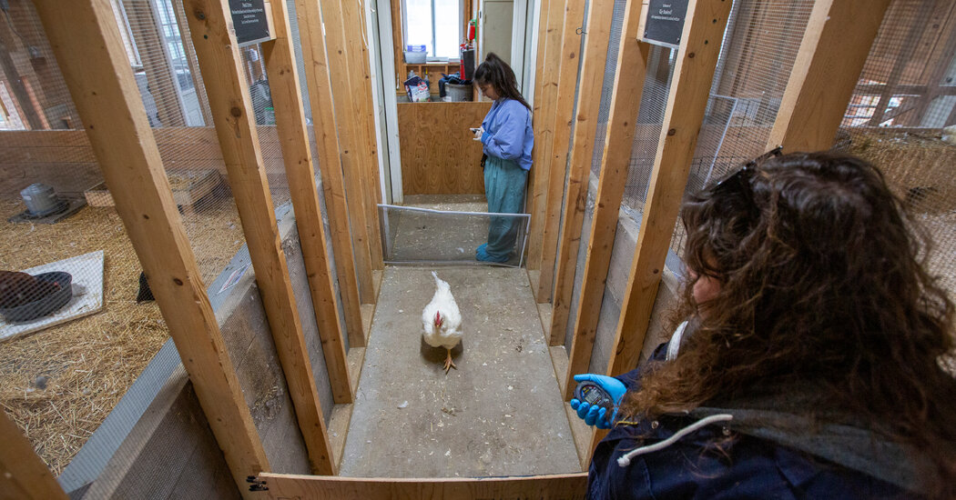
A farm sanctuary in New York is investigating the inner lives of cows, pigs and chickens — but only if they volunteer.WATKINS GLEN, N.Y. — It was a crisp October day at Farm Sanctuary, and inside the small, red barn, the chicken people were restless.A rooster, or maybe two, yodeled somewhere out of sight. A bruiser of a turkey strutted through an open door, tail feathers spread like an ornamental fan. And a penned flock of white-feathered hens emitted tiny, intermittent squeaks, an asynchronous symphony of chicken sneezes.The hens were experiencing a flare-up of a chronic respiratory condition, said Sasha Prasad-Shreckengast, the sanctuary’s manager of research and animal welfare, who was preparing to enter the chicken pen. She donned gloves and shoe covers, threw on a pair of blue scrubs and then slipped inside, squatting to bring herself face-to-face with the first hen who approached.“Who are you?” she cooed.Ms. Prasad-Shreckengast meant the question literally. She was trying to find the birds that were enrolled in her study: an investigation into whether chickens — animals not often heralded for their brainpower — enjoy learning.But her question was also the big philosophical one driving the new, in-house research team at Farm Sanctuary, a nonprofit that has spent more than 35 years trying to end animal agriculture.They have their work cut out for them: The United States alone keeps more than 90 million cattle and slaughters more than 9 billion chickens (and 200 million turkeys) a year. But there are some signs of a societal shift. In a 2019 Gallup poll, nearly one in four Americans said that they had curbed their consumption of meat. A jury recently acquitted activists who ferried two piglets away from a factory farm. Fast-food giants are adding faux meat to the menu, and just last week the U.S. Food and Drug Administration gave the green light to lab-grown chicken.And a growing body of research suggests that farmed species are brainy beings: Chickens can anticipate the future, goats appear to solicit help from humans, and pigs may pick up on one another’s emotions.But scientists still know far less about the minds of chickens or cows than they do about those of apes or dogs, said Christian Nawroth, a scientist studying behavior and cognition at the Research Institute for Farm Animal Biology in Germany. “I’m still baffled how little we know about farm animals, given the amount or the numbers that we keep,” he said.Farm Sanctuary, which was founded in 1986, has always held that farm animals are sentient beings, even referring to its feathered and four-legged residents as “people.”“They have their own desires, and their own wants and preferences and needs, and their own inner lives — the same way that human people do,” said Lauri Torgerson-White, the sanctuary’s director of research.Now, the sanctuary is trying to collect enough data to convince the general public of the humanity of animals.“Our hope,” Ms. Torgerson-White said, “is that through utilizing really rigorous methodologies, we are able to uncover pieces of information about the inner lives of farmed animals that can be used to really change hearts and minds about how these animals are used by society.”The sanctuary is conducting the research in accordance with its own strict ethical standards, which include giving the animals the right to choose whether or not to participate in studies. Consequently, the researchers have sometimes found themselves grappling with the very thing that they are keen to demonstrate: that animals have minds of their own.And today, the birds in “West Chicken” seemed a bit under the weather. Ms. Prasad-Shreckengast crossed her fingers that a few of them might still be up for a brief demonstration.Understand Post-Traumatic Stress DisorderThe invasive symptoms of PTSD can affect combat veterans and civilians alike. Early intervention is critical for managing the condition.Understanding E.M.D.R.: The once-experimental trauma treatment might look bizarre, but some clinicians say it’s highly effective against PTSD. Here’s how the therapy works.Removing the Stigma: Misconceptions about how PTSD develops and its symptoms, can prevent people from seeking treatment.Psychedelic Drugs: As studies continue to point to the therapeutic value of substances like MDMA, veterans are becoming unlikely advocates for their decriminalization.Healing Power: Despite weight lifting being associated with violent bursts of brawn, many people find pumping iron to be a valuable tool in addressing PTSD.“Hopefully,” she said, “people will be feeling like — chicken people will be feeling like — they’re eager and interested in participating.”‘Somebody, not something’Mia, a Farm Sanctuary resident, was rescued from a farm with her five piglets two years ago.Farm Sanctuary began not as a home for rescued animals but with a group of young activists working to expose animal cruelty at farms, stockyards and slaughterhouses.“We lived in a school bus on a tofu farm for a couple of years,” said Gene Baur, the president and co-founder of the organization. But in the course of its investigations, the group kept stumbling upon “living animals left for dead,” he recalled. “And so we started rescuing them.”They ultimately opened sanctuaries in New York and California, establishing educational programs and political advocacy campaigns. (They raised money, in part, by selling veggie hot dogs at Grateful Dead concerts.)And in 2020, the organization, which now houses about 700 animals, began assembling an internal research team. The goal was to assemble more evidence that, as Mr. Baur put it, “these animals are more than just pieces of meat. There’s emotion there. There is individual personality there. There’s somebody, not something.”The research team worked with Lori Gruen, an animal ethicist at Wesleyan University, to develop a set of ethics guidelines. The goal, Dr. Gruen explained, was to create a framework for conducting animal research “without dominance, without control, without instrumentalization.”Among other stipulations, the guidelines prohibit invasive procedures — forbidding even blood draws unless they are medically necessary — and state that the studies must benefit the animals. And participation? It’s voluntary.“Residents must be recognized as persons,” the guidelines state, “and always be provided with choice and control over their participation in an experimental study.”A medicine checklist for the animal residents.The idea is not entirely novel. Zoo animals, for instance, are often trained to cooperate in their own health care, as well as in studies that might stem from it. But such practices remain far from the norm.For the researchers at Farm Sanctuary, voluntary participation was not only an ethical imperative but also, they thought, a path to better science. Many prior studies have been conducted on farms or in laboratories, settings in which stress or fear might affect animals’ behavior or even impair their cognitive performance, the researchers note.“Our hope is that they’re able to tell us more about what the upper limits are for their cognition and emotional capacities and social structures because of the environment that they’re in and because of the way we are performing the research,” Ms. Torgerson-White said.Although the approach is unconventional, outside scientists described the sanctuary’s ethical guidelines as admirable and its research questions as interesting.“The idea that you could study these species, who are usually only studied in sort of pseudofarm conditions, in more naturalistic environments that actually meet not just their needs but even their most arcane preferences — I think they’re right,” said Georgia Mason, who directs the Campbell Center for the Study of Animal Welfare at the University of Guelph. “I think that really allows you to do something special.”Putting a wing upMs. Prasad-Shreckengast opening a gate for hens to voluntarily enter the experimental area.The researchers decided to start with a study on the much-maligned chicken and the birds’ emotional response to learning. “We call it ‘The Joys of Learning,’ but we don’t know that for sure, that they’re going to experience joy,” Ms. Torgerson-White said. “That’s our hypothesis.”To recruit their avian volunteers, Ms. Prasad-Shreckengast and her colleague, Jenna Holakovsky, worked slowly and methodically. They started last fall by spending a few days just sitting in the chicken pen, before opening the door to the hallway where the experiment would eventually take place.Then, they began adding elements of the experimental infrastructure — a window screen, a piece of plywood — and doling out food pellets to any birds brave enough to approach. After about three weeks, they had the entire experimental arena set up and 13 birds who regularly chose to enter it, becoming their volunteer chicken corps.The researchers offered some of these chickens an opportunity to learn something new — how to knock a lid off a bowl — and assessed their overall emotional states, using what is known as a judgment bias test. The test, variations of which have been used with a wide variety of species, involved measuring how quickly the chickens approached a mysterious bowl and its unknown contents.The theory was that a chicken in a generally positive mood would be more likely to assume that the bowl contained something good, like food, and would stride toward it more quickly than a down-in-the-dumps chicken would.A hen being weighed at Farm Sanctuary.So far, the researchers have tested eight chickens, half of whom were in the control group, and it is too early to draw firm conclusions about chickenkind. (The original group of recruits dwindled after one bird died, another failed to meet the study criteria, and three others dropped out — in one case, to spend time in the nest box instead. “I think she really just was highly motivated to sit on some eggs,” Ms. Prasad-Shreckengast said.)But the preliminary data suggest that learning did seem to boost the mood of some of the birds. (Here’s looking at you, Shirley and Murielle.)Then there was Yoshi, who had attempted to bypass the learning challenge altogether. Instead of completing the task for her reward, she went straight for the food, trying to hop over the intervening window screen. Although Yoshi did eventually deign to complete the task, she did not seem to enjoy the experience. She probably found it frustrating, Ms. Torgerson-White said: “She knows how to jump over screens, so why did she need to perform this task?”The researchers were initially disappointed by the result, but they were also charmed by Yoshi’s intransigence, viewing it as evidence of her individual personality.Personality remains a tricky issue. By limiting their study to chickens who, in essence, raised their wings to volunteer, they may have enrolled an unusually bold group of birds, potentially skewing their results. So the researchers are now administering personality assessments and may try to repeat the study with more birds.“Can they work out protocols to get all the chickens so calm and used to them that all the chickens volunteer?” Dr. Mason wondered. “Then their problem is solved.”Barnyard bluesLizzie, a porcine resident who has been generous with saliva samples.The researchers are also investigating whether farmed animals can develop symptoms akin to post-traumatic stress disorder — and, if so, whether spending time in a sanctuary helps them heal.“As a part of a normal life of a farmed animal, honestly, almost no matter the species, they are undergoing or experiencing the types of trauma that human psychologists use to diagnose PTSD,” Ms. Torgerson-White said.Some of the sanctuary’s residents have escaped from slaughterhouses or suffered serious injuries on farms, and scientists have reported PTSD-like symptoms in elephants and chimpanzees exposed to violence or abuse.“If PTSD exists in humans, then clearly it will exist in other species as well,” said Donald Broom, an emeritus professor of animal welfare at the University of Cambridge. “So to look into that would be an interesting thing to do.”The study is primarily observational, involving a careful analysis of the behavior of new residents, such as Bella, a Holstein who arrived at the sanctuary this fall after watching her companion, a steer named Buck, be euthanized. But the team is also measuring the animals’ cortisol levels, inviting residents to cough up some saliva samples.Lizzie and Robbie, a bonded pig pair with bristly coats and a fondness for mouthing visitors’ shoelaces, were absolute champs, happily slobbering all over the big cotton swabs proffered by the scientists. But Hayes, a steer with impossibly fuzzy ears, showed absolutely no interest in mouthing the swabs, not even when the researchers tried to sweeten the deal with molasses.Hayes, the steer with the impossibly fuzzy ears, nuzzling with Ms. Prasad-Shreckengast.“He had just gotten access to pasture for the first time in his life, and nothing, not even molasses, was more interesting or exciting than grazing,” said Ms. Prasad-Shreckengast, whom Hayes nuzzled affectionately when she stopped by the pasture.Some of their studies may not pan out, the researchers acknowledged, and their methods are still evolving. There are some clear areas for improvement: They did not conduct the chicken study “blind,” which means that they knew which chickens were in the control group and which were not. As a result, the researchers could have unconsciously influenced the birds’ behavior, especially if they were hoping for a specific result.“We did our best to avoid unintentional cuing by remaining still, keeping our heads down and stepping away from the testing arena when possible,” Ms. Prasad-Shreckengast said. But, she acknowledged, “We recognize this is a limitation of our study design and plan to address it in our eventual manuscript.”The researchers may be unusually upfront about their mission and values, but they are not alone in bringing a point of view to their work, Dr. Gruen, the animal ethicist, noted. After all, many biomedical scientists have made their own calculations that the possibility of alleviating human suffering outweighs the suffering that lab animals experience. “Values enter into scientific practice at every level,” Dr. Gruen said. “I don’t think it’s unusual that the values are there — I think it’s unusual that those values are there.”The sanctuary said it was committed to publishing its results, no matter what they are. The scientists also run their research proposals through an advisory committee, a group of six outside experts tasked with ensuring that the studies are both ethically and scientifically sound.“To be ethical,” said Becca Franks, an animal welfare scientist at New York University and a member of the committee, “to spend people’s time and energy and money on this and engage with the animals, the science also has to be good science.”Small stepsHolly, a sheep, has a prosthetic to help increase mobility.The researchers are working to expand their PTSD study to animals living on other farm sanctuaries, with financial support from the American Society for the Prevention of Cruelty to Animals, which also provided funding for the chicken study. Next year, the researchers hope to explore aspects of animal culture, as well as the emotional lives of turkeys. And they are eager to spread the word about their ethics guidelines, which they hope other animal researchers will adopt.“If they can show this model works, I think that could really motivate more people to try it,” Dr. Mason said.Although the sanctuary wants to end animal agriculture, other scientists view this kind of research as a path to improving the system. If chickens enjoy learning, for instance, then poultry farmers should give their birds opportunities to do just that, Dr. Broom said.“I’m not against the use of animals for a variety of purposes,” he said. “But I’m very strongly in favor of providing for needs in such a way that the welfare of each individual animal is good.”How will the sanctuary’s staff members feel if their work is used to tweak, rather than eliminate, the existing system? “If we can lessen the suffering of animals in the near term, I think that is positive,” Mr. Baur said. “However, we don’t want to further entrench the idea that these animals are here for us to be exploiting.”Changing public attitudes and societal practices is a long-term project, Ms. Torgerson-White acknowledged. But she and her colleagues are trying to nudge it along from the pastures in Watkins Glen, where the animals are people and the residents are not scientific subjects but research partners.“We’re not extracting information or knowledge from them,” Ms. Prasad-Shreckengast said. “Together, we’re learning, and they’re teaching us what they want and what they’re capable of.”

Read more →

Researchers move closer to better care for life-threatening pregnancy condition

Oregon State University scientists have produced a proof of concept for a new and better way of caring for women facing the life-threatening situation of ectopic pregnancy, which occurs when a fertilized egg implants somewhere other than the lining of the uterus.
Olena Taratula of the OSU College of Pharmacy and Leslie Myatt of Oregon Health & Science University led a team of researchers that used pregnant mice to develop a novel nanomedicine technique for diagnosing and ending ectopic pregnancies, which are non-viable and the leading cause of maternal death in the first trimester.
Findings were published in the journal Small.
The study is important because 2% of all pregnancies in the United States, and between 1% and 2% worldwide, are ectopic, the authors note. In the U.S. alone that translates to approximately 100,000 ectopic pregnancies annually.
About 98% of ectopic implantations happen in the fallopian tubes, putting women at risk of hemorrhage and death. Complicating matters are a high misdiagnosis frequency — ultrasound yields an incorrect diagnosis 40% of the time — combined with a 10% failure rate of the primary drug, methotrexate, used to end an ectopic pregnancy.
Roughly 70 women in the U.S. die each year from ectopic pregnancies, which are responsible for 10% of all pregnancy-related deaths. Women who survive often struggle with a range of issues resulting from diagnosis and treatment, Taratula said.

Read more →