Novel spatial-omics technology enables investigation of diseases at their early stages

How can you trace a single diseased cell in an intact brain or a human heart? The search resembles looking for a needle in a haystack. The teams of Ali Ertürk at Helmholtz Munich and LMU Munich and Matthias Mann at the Max Planck Institute of Biochemistry in Martinsried near Munich have now developed a new technology named DISCO-MS that solves the problem. DISCO-MS uses robotics technology to obtain proteomics data from ‘sick’ cells precisely identified early in the disease.
Most diseases are asympotomatic initially and the affected persons usually still feel fine — symptoms are not yet present, or still too mild to realize. However, a change has already happened within the body: a virus may have started replicating, or a rogue cell might have divided more often than it should have. But how can these changes be perceived?
Researchers face a similar dilemma when they are studying the early development of diseases. Even when working with animal models, scientists can rarely pinpoint the small sites of disease initiation or characterize the exact molecular changes that drive disease progression.
With the development of DISCO-MS by the research teams of Ertürk and Mann in Germany, this task has become much easier. DISCO-MS combines methods to turn mouse and human tissues transparent with the latest robotics and proteomics technologies to determine their molecular makeup.
DISCO-MS: Transparency to detect early molecular changes
DISCO-MS starts with the so-called DISCO tissue clearing, which renders the mouse body or human organs transparent — making them accessible to imaging. Thereby, fluorescently labeled cells can be readily identified in intact tissues of specific sites using high-resolution three-dimensional microscopy.
Once the regions of interest have been identified, they are isolated using a new robotics technology called DISCO-bot, developed by mechanical engineer Furkan Öztürk, a Ph.D. student in Ertürk’s lab. The robot-assisted extracted tissues are processed for their proteome analysis using advanced mass spectrometry (MS) methods developed by Andreas-David Brunner, a former Ph.D. student in Mann’s lab. This high-tech approach allows complete molecular characterization of any desired tissue region identified in 3D in whole mouse bodies or human organs.
Early detection catches the diseases
To showcase the method’s power, first author Harsharan Singh Bhatia and colleagues applied DISCO-MS to Alzheimer’s disease (AD) mouse model and to atherosclerotic plaques (pathological hardening and narrowing of blood vessels) in the human heart. In the tissue samples of the AD model, the team applied artificial intelligence (AI) to identify the typical AD plaques at the early stages of the disease, which had been difficult to detect by any other method. Subsequent proteomics analyses of the plaques provided an unbiased and large scale study of proteins affected in AD, revealing new molecular players that could be biomarkers for Alzheimer’s disease.
In the human heart, the researchers were interested in the composition of the tissues around atherosclerotic plaques, which were quickly visible after tissue clearing. AI detection and robotics extraction of the tissues again allowed the identification of dysregulated molecular pathways in human heart cells related to aortic plaques. These results are key findings, as they form the basis for potential therapeutic targets.
DISCO-MS is the first spatial-omics technology in intact 3D volumes and accelerates studying complicated diseases ranging from cancer to metabolic disorders. As DISCO-MS works with pre-clinical and clinical tissues, it enables the study of diseases at their earliest stages and, subsequently, the development of potential new therapeutics.
Story Source:
Materials provided by Helmholtz Munich. Note: Content may be edited for style and length.

Read more →

COVID-19 booster increases durability of antibody response, research shows

New research from the University of Virginia School of Medicine speaks to the benefits of a COVID-19 booster.
The new findings shed light on how mRNA boosters — both Pfizer and Moderna — affect the durability of our antibodies to COVID-19. A booster, the researchers report, made for longer-lasting antibodies for all recipients, even those who have recovered from a COVID-19 infection.
“These results fit with other recent reports and indicate that booster shots enhance the durability of vaccine-elicited antibodies,” said senior researcher Jeffrey Wilson, MD, PhD, of UVA Health’s Division of Asthma, Allergy and Immunology.
Tracking COVID-19 antibodies
Wilson and his collaborators looked at antibody levels following a booster in 117 UVA employee volunteers and compared those results with the levels seen in 228 volunteers after their primary vaccination series. Antibody levels one week to 31 days after the primary series and booster were similar, but the boosted antibodies stuck around longer regardless of whether the person had had COVID-19.
“Our initial thought was that that boosters would lead to higher antibody levels than the primary vaccine series, but that was not what we found,” said researcher Samuel Ailsworth, the first author of a new scientific paper outlining the findings. “Instead, we found that the booster led to longer lasting antibodies.”
Antibody levels naturally decline over time after an infection or after vaccination, but higher levels are thought to be more protective. Thus, longer-lasting antibodies would be expected to provide more sustained immunity against severe COVID-19.

Read more →

Epigenetics breaks into the clinical practice of cancer

Dr. Manel Esteller and Dr. Verónica Dávalos, researchers at the Josep Carreras Leukaemia Research Institute, describe in a new article the impact of epigenetics on cancer treatment and how it has become a crucial tool to improve early detection, predict disease progression and become a target for new treatments.
In the early 1980s, the first changes in DNA related to a chemical modification called methylation were discovered, followed by the discovery in the mid-1990s of the first tumor suppressor genes inactivated by these modifications of the genetic material. The early 2000s saw the first use of these altered marks as a biomarker of cancer disease, as well as the first uses of drugs against them.
In parallel, the first chemical modifications were detected in proteins called histones, where DNA wraps around them like a pearl necklace. All this “decoration” of DNA and its regulatory proteins define the field of Epigenetics.
Now, on an article published in the journal “CA: A Cancer Journal for Clinicians,” Dr. Manel Esteller, Director of the Josep Carreras Leukaemia Research Institute (IJC), ICREA Research Professor and Professor of Genetics at the University of Barcelona, and researcher Dr. Verónica Dávalos explain its impact on the clinical management of cancer patients. 
“Epigenetics has gone from being a purely basic research discipline focused on studying how gene expression is controlled to a tool to improve early detection, predict the evolution of the disease and become a target for new treatments,” -says Dr. Esteller and adds — “One of the most outstanding aspects of its clinical translation is its use in liquid biopsy, as well as helping in the classification of tumor types, for example to correctly diagnose the types of tumors derived from the brain, skeletal muscles, joints, bones or of unknown origin. But, in addition to this aspect, DNA methylation profiles are approved to determine the efficacy of treatment in brain tumors and other tumor pathologies.”
Perhaps one the most attractive aspect for the medical oncologist is the use of epigenetic drugs to treat cancer. There are currently nine drugs against various epigenetic marks (DNA methylation and histone methylation and acetylation) approved for clinical use in various types of leukemia, lymphoma and blood diseases, as well as tumors of the soft tissues. According to Esteller, “these treatments are usually very well tolerated by patients and more than killing the tumor cell, they stop its growth, as if it were a tamed beast.”
Epigenetic drugs are a clinical reality and they are already providing benefits for patients, but research continues and, currently, there is a whole new generation of epigenetic drugs in different phases of clinical trials that, alone or combined with immunotherapy, can make a positive difference in many patients.
Story Source:
Materials provided by Josep Carreras Leukaemia Research Institute. Note: Content may be edited for style and length.

Read more →

Cystic fibrosis drug could help treat pneumonia

Pathogens such as SARS-CoV-2 and pneumococcus can cause severe pneumonia. If the airways then fill with fluid, the patient risks developing acute respiratory distress syndrome. Researchers at Charité — Universitätsmedizin Berlin have now discovered the molecular mechanisms that trigger fluid accumulation in the lungs. This also led them to discover a potential new therapy: A cystic fibrosis drug proved effective in their laboratory experiments, raising hope that this could be used to treat pneumonia regardless of the pathogen that caused it. The study has been published in the journal Science Translational Medicine.
Pneumonia is the most common cause of fluid buildup in the lungs. This condition, known as pulmonary edema, results in parts of the airspaces filling with fluid instead of air, which prevents them from doing their job of exchanging gases. Patients struggle to breath and their body can’t get enough oxygen. The diagnosis is acute respiratory distress syndrome, or ARDS. “Despite cutting-edge medical procedures, roughly 40 percent of patients with ARDS die in intensive care. The problem is that antibiotics, antivirals, and immune modulating therapies rarely work well enough,” says study leader Prof. Dr. Wolfgang Kuebler, Director of the Institute of Physiology at Charité. “That’s why we took a very different approach in our study. Instead of focusing on the pathogen, we focused on strengthening the barrier function of the blood vessels in the lungs.” This makes sense, as they are the source of the fluid in pulmonary edema. The lung vessels become permeable, allowing fluid from the blood to flow into the surrounding tissue — and thereby flood the airspaces.
But what actually causes this? What are the underlying molecular mechanisms? A Charité research team led by Prof. Kuebler set out to answer these questions. They performed experiments using cells, lung tissue, and isolated lungs. The study centered on the CFTR chloride channel, which scientists know is mainly found in the mucosal cells of our airways. There, it plays a major role in keeping our mucus thin so it can drain away easily. The researchers have now shown for the first time that cells in the blood vessels of the lungs also have CFTR and that its presence is drastically reduced in pneumonia.
To find out what role CFTR plays in the pulmonary vessels and what is happening at the molecular level when the chloride channel is lost, the researchers blocked the channel with an inhibitor and dictated the number of chloride ions in the cells. They then used a special imaging technique known as immunofluorescence imaging: “We saw that inhibiting CFTR triggered a molecular cascade that ultimately causes the lung’s blood vessels to begin leaking,” says Dr. Lasti Erfinanda, who also works at the Institute of Physiology and is the study’s lead author. “So CFTR actually does play a very key role in the development of pulmonary edema.”
The study findings indicate that the loss of CFTR causes chloride to accumulate in the cells because it stops being transported out of them. The excess chloride triggers signaling that ends with an uncontrolled flow of calcium into the cells via a calcium channel. “The increased calcium concentration then causes the vascular cells to contract — much like the effect that calcium has on muscle cells,” explains Prof. Kuebler. “This results in gaps between the cells — which allows fluid to spill out of the blood vessels. Chloride channels are therefore crucial in maintaining the barrier function of the pulmonary vessels.”
The research team then addressed another question: How could they attenuate or prevent the pneumonia-induced loss of chloride channels in the pulmonary vessels? To answer this, the researchers used a therapeutic agent that is classed as a CFTR modulator and currently used to treat cystic fibrosis. In cystic fibrosis patients, a genetic mutation prevents the CFTR chloride channel from working properly in the mucosal cells of the airways, resulting in very viscous mucus. “Ivacaftor is a drug that increases the chances of the chloride channel opening, which helps the mucus to flow through the airways,” says Dr. Erfinanda. “We wanted to see if it would also have a positive effect on the cells in the blood vessels of the lungs.”
Ivacaftor did make the chloride channels more stable: it led to less degradation in the channels than that typically caused by the lung’s inflammatory processes. Experiments on animal models showed the same effect: treatment with ivacaftor increased the probability of surviving severe pneumonia, reduced lung injury, and resulted in much milder symptoms and a much better general condition than without the drug. “We really weren’t expecting it to work so well,” says Prof. Kuebler. “We hope our findings will pave the way for clinical trials to test the efficacy of CFTR modulators in pneumonia patients. If this promising, pathogen-independent therapy finds its way into clinical practice, it could benefit a huge number of patients and prevent pneumonia from becoming life-threatening — even in the case of unknown pathogens.”
Prof. Kuebler and his team are now planning research projects aimed at developing other potential therapies based on the CFTR signaling pathway. They are also going to research which patients have an elevated risk of developing ARDS, so they can provide these patients with preventive, personalized treatment.
Story Source:
Materials provided by Charité – Universitätsmedizin Berlin. Note: Content may be edited for style and length.

Read more →

Fatty liver disease endangers brain health

In a study examining the link between non-alcoholic fatty liver disease (NAFLD) and brain dysfunction, scientists at the Roger Williams Institute of Hepatology, affiliated to King’s College London and the University of Lausanne, found an accumulation of fat in the liver causes a decrease in oxygen to the brain and inflammation to brain tissue — both of which have been proven to lead to the onset of severe brain diseases.
NAFLD affects approximately 25% of the population and more than 80% of morbidly obese people. Several studies have reported the negative effects of an unhealthy diet and obesity can have on brain function however this is believed to be the first study that clearly links NAFLD with brain deterioration and identifies a potential therapeutic target.
The research, conducted in collaboration with Inserm (the French National Institute of Health and Medical Research) and the University of Poitiers in France, involved feeding two different diets to mice. Half of the mice consumed a diet with no more than 10% fat in their calorie intake, while the other half’s calorie intake contained 55% fat; intended to resemble a diet of processed foods and sugary drinks.
After 16 weeks researchers conducted a series of tests to compare the effects of these diets on the body and more specifically, on the liver and the brain. They found that all mice consuming the higher levels of fat were considered obese, and developed NAFLD, insulin resistance and brain dysfunction.
The study which was funded by the University of Lausanne and Foundation for Liver Research also showed that the brain of mice with NAFLD suffered from lower oxygen levels. This is because the disease affects the number and thickness of the brain blood vessels, which deliver less oxygen to the tissue, but also due to specific cells consuming more oxygen while the brain is becoming inflamed. These mice were also more anxious and showed signs of depression.
By comparison, the mice consuming the healthy diet did not develop NAFLD or insulin resistance, they behaved normally, and their brain was completely healthy.
“It is very concerning to see the effect that fat accumulation in the liver can have on the brain, especially because it often starts off mild and can exist silently for many years without people knowing they have it,” said lead author Dr Anna Hadjihambi, sub-team lead in the Liver-Brain Axis group at the Roger Williams Institute of Hepatology and honorary lecturer at King’s College London.
To try and combat the dangerous effect that NAFLD has on the brain, the scientists bred mice with lower levels of a whole-body protein known as Monocarboxylate Transporter 1 (MCT1) — a protein specialised in the transport of energy substrates used by various cells for their normal function.
When these mice were fed the same unhealthy fat- and sugar-rich diet as those in the initial experiment, they had no fat accumulation in the liver and exhibited no sign of brain dysfunction — they were protected from both ailments.
“Identifying MCT1 as a key element in the development of both NAFLD and its associated brain dysfunction opens interesting perspectives,” said Professor Luc Pellerin, director of the Inserm U1313 research unit at the University of Poitiers in France and senior researcher in the study. “It highlights potential mechanisms at play within the liver-brain axis and points to a possible therapeutic target.”
Dr Hadjihambi added: “This research emphasises that cutting down the amount of sugar and fat in our diets is not only important for tackling obesity, but also for protecting the liver to maintain brain health and minimise the risk of developing conditions like depression and dementia during ageing, when our brain becomes even more fragile.
Story Source:
Materials provided by King’s College London. Note: Content may be edited for style and length.

Read more →

Physical fitness a demographic watershed

Sedentary behavior, a large waist circumference, and advanced age: These factors are clearly associated with inferior physical fitness among people aged 50 to 64. In a study with over 5,000 participants, investigating the correlations in detail, major fitness disparities are shown.
Fitness is a vital factor for performance in sports, but also for the stamina required for exercising and leading an active everyday life. Previous studies have shown a strong connection between good fitness and various sickness and health outcomes, including reduced risk of cardiovascular disease.
The present study, published in the scientific journal BMJ Open, involved 5,308 participants aged 50-64, 51% of whom were women. The article describes how maximal oxygen uptake (commonly known as VO2 max), a common measure of fitness, varies from one demographic group to another.
The variables evaluated were sociodemographic (age, gender, education, etc.), lifestyle factors, perceived health, body measurements, disease prevalence, and self-appraised physical activity and sedentariness measured with an accelerometer.
Highly uneven fitness distribution
Every participant completed a cycling fitness test, while wearing an accelerometer on an elastic band around the waist. The purpose was to collect a week’s measurements of the frequency, duration, and intensity level of individuals’ exertion, both on an everyday basis and during training sessions, if any.

Read more →

Fewer infectious particles from children's lungs

Children exhale significantly fewer potentially infectious particles than adults — at least this is true for the small respiratory droplets that are predominantly produced in the lungs. This is a key finding of a study conducted by the Max Planck Institutes for Dynamics and Self-Organization (MPI-DS) and for Chemistry in collaboration with the University Göttingen Medical Center (UMG). The researchers investigated the concentrations of aerosol particles and droplets emitted with the breath when breathing, speaking, singing and shouting, using measurements on 132 people of all ages. The findings help to understand how the spread of diseases like influenza or Covid-19 can be contained.
Infectious diseases are often transmitted via particles exhaled from infected persons. Yet, the size of such aerosol particles varies greatly, depending on its origin in the respiratory tract. In the lung, mostly small particles with less than five microns — that is five thousandths of a millimeter — are produced, also known as PM5. In contrast, bigger particles are produced in the upper respiratory tract. As the measurements showed, children exhale far less smaller particles than adults. “We found that the concentration of small particles below five microns increases with age and is particularly low in children. As a result, adults are much more likely to trigger spreading if the infection is only in the lower respiratory tract,” says Mohsen Bagheri, research group leader and lead author of the study at the MPI-DS. Notably, larger particles that originate in the throat are spread by children and adults to the same extent, according to the study. The researchers did not find a correlation between the concentration of exhaled particles and the person’s gender, weight, fitness or smoking habits.
Vocalization activities increase small particle concentration
In this comprehensive study, the researchers recorded data from 132 healthy volunteers. The study also included children and adolescents between 5 and 18 years about which very little data were available. They used various instruments installed in a clean room to measure the full range of particle sizes exhaled: from a tenth of a micrometer to a quarter of a millimeter. The participants performed different vocalization activities such as singing, speaking and shouting for 20 minutes in total. “Vocalization and age are shown to be independent risk factors for particle production,” reports Prof. Simone Scheithauer from the Department of Infection Control and Infectious diseases at the UMG.
The volume of the exhaled particles determines potential infection risk
Although human drops and aerosols contain mostly small particles, larger particles constitute the major part of the total volume that can contain pathogens. “If the pathogen mainly resides in the upper respiratory tract, the large particles are by far the main transmitter of the disease,” explains Eberhard Bodenschatz, director at the MPI-DS. “It is thus important to consider the localization of the infectious particle in the respiratory tract to decide on appropriate protection,” he continues. “For example, the current Omicron variant of the Coronavirus seems to be localized more to the upper respiratory tract and this is why even simple filtering face masks are a great protection.”
Protective measures depend on the localization of the pathogen
In contrast, Infectious diseases that reside mainly in the lungs will primarily be transmitted via small particles. Since their production increases with age, children are less likely to transmit such diseases than adults, according to the study. To prevent airborne transmission of lung diseases, wearing of well-fitted and high-efficiency facial masks can thus be an effective measure to avoid disease transmission, especially for adults.
Story Source:
Materials provided by Max Planck Institute for Dynamics and Self-Organization. Note: Content may be edited for style and length.

Read more →

High-energy X-rays leave a trace of destruction in bone collagen

It has long been known that beyond a certain dose, X-rays damage living tissue, so there are clear medical indications for X-rays to keep radiation exposure to a minimum. In basic research on the properties and characteristics of mineralised tissue samples such as bone, researchers rely on increasingly powerful X-ray sources.
Bones from fish and mammals
“Until now, the motto has actually been: more flux and higher energy is better, because you can achieve greater depth of field and higher resolution with more intense X-rays,” says Dr. Paul Zaslansky from Charité-Universitätsmedizin. Zaslansky and his team have now analysed bone samples from fish and mammals at the MySpot beamline at BESSY II.
BESSY II generates a well characterized broad-range of X-rays, precisely focused in an intermediate energy range which allows insights into the finest structures and even chemical and physical processes in materials. “Thanks to sensitive detectors and rather mild irradiation conditions in BESSY II as compared with harder X-ray synchrotron sources, we were able to demonstrate on our various bone samples that collagen fibres become damaged by the irradiation absorption in the mineral nanocrystals,” Zaslansky summarises the results of the study.
Imaging the protein fibers
“We examined the samples under Second-Harmonic Generation laser-scanning microscopy for the imaging the protein fibers” explains first author Katrein Sauer, who is doing her doctorate in Zaslansky’s team. Together with HZB expert Dr. Ivo Zizak, she irradiated bone samples from pike fish, pigs, cattle and mice with precisely calibrated X-ray light.
Trail of destruction
The beams left a trail of destruction that is clearly visible in the confocal and electron microscopy images. “The high-energy photons from the X-ray light trigger a cascade of electron excitations. Ionisaton of calcium and phosphorus in the mineral then damages proteins like collagen in bone,” Sauer says. Break-down of collagen increases with the duration of the irradiation, but also shows up even with short irradiation at high flux.
Minimal doses for research on living materials
“X-ray methods are considered non-destructive in materials research, but at least for research on bone tissues this is not true,” says Zaslansky. “We have to be more careful in basic medical research that we don’t damage the very structures we actually want to analyse.” So, as everywhere in medicine, and even when there are no living tissues and DNA to damage, it comes down to using a minimal dose to get the insights that reflect the material condition without causing damage.
Note:
The X-rays produced at BESSY II are about ten thousand times more intense than X-rays used for medical examinations (for X-rays of a broken leg, the German Federal Office for Radiation Protection gives a dose of 0.01 millisievert). X-ray methods are extremely useful for medical examinations.
Story Source:
Materials provided by Helmholtz-Zentrum Berlin für Materialien und Energie. Note: Content may be edited for style and length.

Read more →

Head trauma, PTSD may increase genetic variant's impact on Alzheimer's risk

The medical community has never researched the simultaneous impact of post-traumatic stress disorder (PTSD), traumatic brain injury (TBI), and genetic risk factors in a large cohort … until now.
In a study of Veterans led by Dr. Mark Logue, a statistician in the National Center for PTSD at the VA Boston Healthcare System, researchers concluded that PTSD, TBI, and the ε4 variant of the APOE gene showed strong associations with Alzheimer’s Disease and related dementias (ADRD).
The researchers first found a greater percentage of ADRD in Veterans with PTSD and in those with TBI, relative to those without, as well as higher rates of ADRD in Veterans who had inherited the ε4 variant. Logue and his team then looked for interactions between the ε4 variant, PTSD, and TBI using a mathematical model.
The study found an increase in risk due to PTSD and TBI in Veterans of European ancestry who inherited the ε4 variant. In Veterans of African ancestry, the impact of PTSD didn’t vary as a function of ε4, but the TBI effect and interaction with ε4 was even stronger. Other studies have suggested that ε4 may magnify the effects of a head injury and/or combat-related stress.
“These additive interactions indicate that ADRD prevalence associated with PTSD and TBI increased with the number of inherited APOE ε4 alleles,” Logue and his colleagues wrote. “PTSD and TBI history will be an important part of interpreting the results of ADRD genetic testing and doing accurate ADRD risk assessment.”
Capitalizing on VA’s Million Veteran Program
The researchers carried out the study by accessing data from VA’s Million Veteran Program (MVP), one of the world’s largest databases of health and genetic information. MVP is aimed at learning how genes, lifestyle, and military exposures affect health and illness, with more than 900,000 Veterans enrolled in its climb to 1 million and beyond.

Read more →

‘Major Trustee, Please Prioritize’: How NYU’s E.R. Favors the Rich

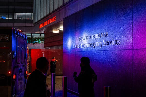
In New York University’s busy Manhattan emergency department, Room 20 is special.Steps away from the hospital’s ambulance bay, the room is outfitted with equipment to perform critical procedures or isolate those with highly infectious diseases.Doctors say Room 20 is usually reserved for two types of patients: Those whose lives are on the line. And those who are V.I.P.s.In September 2021, doctors were alerted that Kenneth G. Langone, whose donations to the university’s hospital system had led it to be renamed in his honor, was en route. The octogenarian had stomach pain, and Room 20 was kept empty for him, medical workers said. Upon his arrival, Mr. Langone was whisked into the room, treated for a bacterial infection and sent home.The next spring, Senator Chuck Schumer accompanied his wife, who had a fever and was short of breath, to the emergency room. As sicker patients were treated in the hallway, the couple were ushered into Room 20, where they received expedited Covid-19 tests, according to workers who witnessed the scene. The tests came back negative.NYU Langone denies putting V.I.P.s first, but 33 medical workers told The New York Times that they had seen such patients receive preferential treatment in Room 20, one of the largest private spaces in the department. One doctor was surprised to find an orthopedic specialist in the room awaiting a senior hospital executive’s mother with hip pain. Another described an older hospital trustee who was taken to Room 20 when he was short of breath after exercising.The privileged treatment is part of a broader pattern, a Times investigation found. For years, NYU’s emergency room in Manhattan has secretly given priority to donors, trustees, politicians, celebrities, and their friends and family, according to 45 medical workers, internal hospital records and other confidential documents reviewed by The Times.On hospital computers, electronic medical charts sometimes specify whether patients have donated to the hospital or how they are connected to executives, according to screenshots taken by frustrated doctors in recent years and shared with The Times.“Major trustee, please prioritize,” said one from July 2020.Dozens of doctors said they felt pressure to put V.I.P.s first. Many witnessed such patients jumping ahead of sicker people for CT scans and M.R.I.s. Some said medical specialists, often in short supply, were diverted from other cases to attend to mild complaints from high-priority patients.Many hospitals offer exclusive concierge services to the rich. But emergency rooms are built around the premise of medical triage: that the sickest patients, regardless of their ability to pay, are treated first. Everyone else has to wait.At NYU Langone, one of the country’s pre-eminent medical institutions, some doctors said that process had been upended.“As emergency department doctors, we have two important skills: triage and resuscitation,” said Dr. Kimbia Arno, who worked in the emergency room in 2020 and 2021. “This system is in direct defiance of what we do and what we were trained to do.”“The stress on providers is harmful,” said Dr. Anand Swaminathan, a physician in the emergency room from 2009 to 2018. “It’s the fact that I am getting multiple calls, from multiple people, asking me to drop everything to treat a V.I.P.”Eleven doctors told The Times that they had resigned from the emergency department in part because they objected to favoring V.I.P.s.Some residents — doctors in their first years of practice — complained to the national organization that accredits medical training programs. The frustrations included NYU’s “special treatment” of trustees, donors and their families, according to documents reviewed by The Times. The group’s subsequent investigation confirmed that some doctors “felt pressured to see V.I.P. patients first” and that they “experience a sense of fear and intimidation and retaliation for not expediting V.I.P. patient care.”The Internal Revenue Service requires nonprofit hospitals like NYU, which avoids $250 million a year in taxes, to benefit their communities. A primary way to meet the requirement is to run an emergency room that is open to everyone.But at NYU, poor people sometimes struggle to be seen. For example, ambulance workers said nurses in the emergency room routinely discouraged them from dropping off homeless or intoxicated patients. Instead, they were often shuttled to nearby Bellevue, a strained public hospital that primarily treats the poor.A Times series this year has found that many nonprofit hospitals have strayed from their charitable roots to maximize profits. Giant hospital systems illegally sent exorbitant bills to Medicaid patients. They used hospitals in poor neighborhoods to qualify for steep drug discounts, funneling the proceeds into wealthier neighborhoods. Others cut staff to dangerously low levels.NYU Langone denies that its emergency room favors V.I.P.s, but dozens of doctors said they felt pressured to put such patients first.Ashley Gilbertson for The New York TimesNYU’s chief of hospital operations, Dr. Fritz François, denied that the hospital favored donors, trustees and other prominent patients. He said that patients received treatment based on how sick they were, regardless of their wealth or status, and that the emergency room treated many low-income and homeless patients.“We do not have a V.I.P. program,” Dr. François wrote in a letter to The Times. “We do not have V.I.P. patients. We do not have V.I.P. floors. We do not have V.I.P. rooms. We do not have V.I.P. clinical teams. We do not offer V.I.P. care.”Lisa Greiner, a spokeswoman for NYU Langone, confirmed that Mr. Langone had been treated in Room 20, which she said was “absolutely appropriate” based on his symptoms. She said the room served a variety of purposes, including privacy. She said no patient, including Mr. Langone, “has ever been treated in an isolated room at the expense of any other patient’s care.”Mr. Langone said, “As a matter of personal integrity I have never asked for any special treatment at the hospital, and they have never offered.”Angelo Roefaro, a spokesman for Mr. Schumer, said the protocol for the senator’s security detail was “to have the senator stay, whenever possible, in a secure location.”Andrew C. Phillips, a lawyer for NYU, said some of the doctors who had spoken to The Times were motivated to disparage the hospital. Dr. Arno, for example, had been in a fellowship program and was passed over for a permanent job, he said. Mr. Phillips also said Dr. Swaminathan had never voiced concerns to hospital leaders about V.I.P.s.Dr. François acknowledged that NYU’s electronic medical records sometimes included notations describing patients as “friends and family.” But he said these labels were available for all hospital employees — even the cousins of security guards and housekeepers — and enabled employees to pay courtesy visits to such patients.“Our friends and family do not receive different or better medical care,” Dr. François wrote. He added, “Our friends and family don’t skip the triage process, don’t jump any lines, don’t get placed in any special rooms or floors and don’t get fed any differently.”Dozens of doctors and other emergency room staff said that, when it came to many V.I.P.s, that was simply not true.An E.R.’s TransformationKenneth G. Langone, a Home Depot founder, helped NYU’s hospital system raise billions of dollars.Dave Kotinsky/Getty ImagesIn 2007, the New York University Medical Center was in grave financial trouble.Were it not for royalties from an arthritis drug developed by one of its researchers, the hospital would have lost $150 million that year. The patent’s expiration was looming.A lifeline came from Mr. Langone, the founder of Home Depot and chairman of the hospital’s board of trustees. He and his wife donated $100 million in 2008, matching a contribution they had made eight years earlier. The medical center was renamed NYU Langone.Mr. Langone became known not just for his own philanthropy — he donated another $100 million in 2019 — but also his ability to persuade other wealthy New Yorkers to donate. Over the ensuing years, he helped the hospital raise $3 billion.In 2012, the run-down emergency room, on the East River in Midtown Manhattan, was destroyed by Hurricane Sandy. It reopened two years later with more space and a new name, the Ronald O. Perelman Center for Emergency Services, named for the billionaire who financed its construction.The emergency department’s longtime chair, an outspoken champion of serving the needy, stepped down in 2015. Around then, several doctors said, they began receiving requests from administrators to give priority to V.I.P.s.“Suddenly, we started getting these phone calls that X person is coming in, they are X relation to board member, and we were given the strong sense that you had to push them to the front of the line,” said Dr. Swaminathan, who worked in the emergency room at the time.NYU was not the only prestigious nonprofit hospital system finding ways to cater to donors and other wealthy patients.In San Francisco, the UCSF Medical Center rewarded donors with faster access to top cardiologists. Stanford Medical Center gave wealthy patients red blankets to distinguish them from everyone else. (Spokeswomen for those medical centers said they no longer provided such perks.)Today, top New York hospitals like Mount Sinai and NewYork-Presbyterian/Weill Cornell offer luxury accommodations and personal concierge services to patients who can afford them.And emergency room workers at several elite academic medical centers said in interviews that, as at NYU, administrators sometimes requested expedited treatment for well-connected patients.“The hospitals are acting as businesses,” said Dr. Renee Hsia, a professor at the University of California, San Francisco, who researches emergency room care. “They can often garner much more revenue from these patients that are huge donors.”‘Drop Everything’Hospital workers were alerted before V.I.P.s arrived at NYU’s emergency room.Ashley Gilbertson for The New York TimesThe V.I.P. experience in NYU’s Manhattan emergency room starts before the patient arrives.Trustees can use a dedicated phone number — the Trustee Access Line — to alert the hospital they are coming. Administrators then call, text and send messages notifying doctors that a high-priority patient is en route, according to 30 doctors. Doctors said that even when those messages did not explicitly seek priority treatment, that was how they were interpreted.“Just a heads up that a VIP/trustee is coming to the ED per notification from the Dean’s office and to keep an eye out for her,” one doctor wrote in an electronic chat in August 2021, referring to the emergency department. The Times reviewed a screenshot of the exchange.Ms. Greiner, the NYU spokeswoman, said the trustee line “does not entitle any member to better or prioritized care.” She said that the V.I.P. reference in the 2021 message was “colloquial and does not correspond with any special protocol at our hospital,” and that the sender did not “ask for or expect special care, line cutting or anything of the sort.”Doctors said they were sometimes required to carry a hospital-issued iPhone that, among other things, was logged into an email chain that alerted them to incoming V.I.P.s.“It didn’t matter how busy it was,” said Dr. Uché Blackstock, who worked in the emergency room from 2010 to 2019. “A V.I.P. was coming, and we had to drop everything.” She left NYU partly because of frustration with the preferential treatment, she said.Ms. Greiner said that Dr. Blackstock had never complained to the hospital about improper prioritization of patients and that Dr. Blackstock had herself alerted colleagues on a few occasions when her family or friends were in the emergency room. In response, Dr. Blackstock said there was a distinction between what she had done and what she and others perceived as institutional pressure to swiftly treat V.I.P.s.Some patients’ electronic medical charts included reminders about their V.I.P. status, according to screenshots captured by emergency room doctors and shared with The Times.The New York Times “NYUMC BOARD OF TRUSTEE AND IMMEDIATE FAMILY,” read one note.Another: “She is a donor and a prospect for a planned gift.”A third: “Escort Needed” and “Daughter of Trustee.” (Some V.I.P.s were assigned employees to stand by to transport them around the hospital, according to 13 medical workers. Ms. Greiner denied that.)The New York TimesTwo members of NYU Langone’s board of trustees said in interviews that they had received swift, excellent care at the emergency room. They believed everyone got such treatment.“I didn’t have to wait around for long hours for someone to come talk to me as happens in other emergency rooms,” said Bernard Schwartz, who said he had donated more than $30 million to NYU Langone. “I think that’s for all patients.”Mr. Schwartz said he did not think his medical record identified him as a trustee. But he presumed that doctors knew who he was.“I would be upset if that were not true,” he said.Delayed ResuscitationsNYU’s emergency room often has more than 100 patients at once but only 40 curtained beds, leaving many patients to be treated in the hallways.None of the doctors The Times interviewed had ever seen that happen with a V.I.P.One Thursday night in April 2018, workers in the emergency room got an alert that Mr. Langone would be arriving in about 20 minutes. They had to figure out where to put other patients to ensure that he could have a private room, according to two medical workers with direct knowledge of what happened. When he arrived with a two-centimeter cut on his thumb, doctors quickly stitched him up.Ms. Greiner said no other patients were awaiting care during Mr. Langone’s visit. The two workers told The Times that the emergency room had been as busy as usual.Dr. Michelle Romeo, who worked in the emergency room until 2021, recalled a famous actor’s jumping to the front of the line for a CT scan.Ashley Gilbertson for The New York TimesEmergency room workers said these arrangements for V.I.P.s sometimes delayed critical care for sicker patients.In late 2019, doctors were racing to rescue a patient in cardiac arrest. One pushed the gurney toward one of the private rooms meant for life-or-death emergencies. Another sat atop the unconscious patient, performing chest compressions. When they arrived at the room, they could not enter — a V.I.P. occupied it. The patient survived, but two workers who witnessed the episode said the delay could have been deadly.Ms. Greiner said, “Without the patient’s information, we cannot investigate this claim other than to say that at NYU Langone, there is one standard of care for all patients.”The Times identified many similar examples.For example, a relative of someone on the hospital’s leadership team went into the emergency room with chest pain and was promptly taken to a private room, even as a man experiencing a life-threatening emergency — a blockage of blood to one of his limbs — was put in the hallway, according to the accreditation group’s investigation.Another time, at the instruction of a hospital administrator, a V.I.P. patient with asymptomatic Covid was seen by pulmonology and infectious-disease specialists who had to be pulled away from sicker patients, according to two medical workers with direct knowledge of the case.Ms. Greiner said that The Times had not provided enough information for her to be able to respond definitively, but that the asymptomatic patient might have had an underlying illness.Dr. Michelle Romeo, who was a resident in the emergency room from 2017 until 2021, recalled when a famous actor with a headache and low-grade fever jumped to the front of the line for a CT scan, cutting off a nursing home resident who had possible sepsis and had been waiting for three hours.The actor requested a spinal tap, which Dr. Romeo believed was unnecessary. A supervisor instructed her to do it anyway, she said.Both tests showed nothing wrong with the patient.Mr. Phillips, the lawyer for NYU, said Dr. Romeo had an incentive to criticize the hospital because she had not been offered a full-time position after her residency. Dr. Romeo said she believed she had not been offered the job because she had been outspoken about issues including the treatment of V.I.P.s.A Public ShamingOver the years, doctors in NYU’s emergency room came to believe there could be career-threatening consequences if well-connected patients were dissatisfied with their treatment.In October 2019, Dr. Joe Bennett was at the end of what’s known as a shift-change huddle, updating his colleagues on the patients he was handing off, when a frustrated V.I.P. approached him. The V.I.P. demanded that a family member immediately receive a CT scan, according to a doctor who witnessed the encounter and two others who were briefed on the matter.Dr. Bennett explained that a sicker patient was the priority but that the family member would come next.Soon after, Dr. Bennett was put on probation for what NYU said was a lack of professionalism, according to the three doctors. For months, the hospital required him to attend weekly meetings and write essays reflecting on how to provide professional treatment.Doctors viewed the ouster of Dr. Kristin Carmody as punishment for her not catering to a V.I.P.Ashley Gilbertson for The New York TimesAbout a year later, in December 2020, Dr. Kristin Carmody, who oversaw the education of medical residents in the emergency department, was forced to resign after a patient complained about having not received the level of attention or treatment that she expected. Dr. Carmody later said in a wrongful-termination lawsuit that the patient had been designated as a V.I.P.Ms. Greiner said that the patient’s medical record had not included a friends-and-family label and that Dr. Carmody had been pushed out because she falsely noted on a medical record that she had personally examined the patient. (Dr. Carmody denies that.)But inside the emergency department, her ouster was widely regarded as punishment for not sufficiently catering to a V.I.P. patient.At a heated staff meeting that month, a senior doctor said Dr. Carmody’s forced departure appeared to be the result of a complaint from “a V.I.P. person that was connected to higher-ups,” according to a recording of the meeting. The doctor added, “The clear message is anybody can be taken down.”Around that time, top NYU officials commissioned an internal review of the culture of the emergency department, whose employees were burned out from the pandemic and unhappy with their pay.The investigation documented concerns with V.I.P. care, according to a presentation that Dr. Robert Femia, the chairman of the emergency department, delivered to doctors.Many doctors and nurses “dislike the current ‘V.I.P.’ process because they perceive it as disrupting ordinary work flows” in which staff triage patients based on their medical needs, one slide said. “They do not recognize that the true issue is that every patient is a ‘V.I.P.’ patient.”‘An NYU Dump’In the summer of 2021, a few months after Dr. Femia’s presentation, an ambulance dropped off a disheveled homeless patient at NYU’s emergency room. He had pain in both legs and was having trouble walking.A worker checked the man’s vital signs. He was offered Tylenol and discharged, according to an email that a senior nurse later sent to more than 200 colleagues detailing what had happened.About an hour later, the man was back. This time, he was seen in the waiting room by a social worker, who noted that it was hard for the man to lift his legs from his wheelchair. No one undressed the patient to examine his legs. He was discharged again.It was not until later that day that the hospital admitted him. The man was diagnosed with acute kidney failure and rhabdomyolysis, a potentially fatal muscular condition.Ms. Greiner said the case had been handled appropriately. But medical staff noted that NYU included it in an internal review process in which doctors try to learn from mistakes.Doctors and nurses described a pattern in which homeless patients — surefire money losers for hospitals — sometimes received cursory care, even as privately insured patients with similar symptoms were admitted for urgent treatment.For poor or homeless patients, “there is pressure to see them in the hallway or in the waiting room,” said Dr. Jeremy Branzetti, who ran NYU’s emergency-medicine residency program until last year. “I have never seen a V.I.P. patient in the hallway.” Mr. Phillips, the lawyer for NYU, said Dr. Branzetti had received a poor performance review and his contract was not renewed.Anthony Almojera, a New York City paramedic, said NYU nurses reprimanded ambulance crews for trying to drop off homeless patients.Ashley Gilbertson for The New York TimesSome homeless people struggle to get into NYU’s emergency room in the first place.Anthony Almojera, the vice president of a union that represents emergency services officers, said nurses at NYU reprimanded ambulance crews when they tried to drop off patients who appeared homeless or intoxicated.“I had instances where the nurse’s first question wasn’t ‘What is wrong with the patient?’ but ‘How come this patient is being brought here?’” Mr. Almojera said.Another ambulance worker, who requested anonymity because he still works with NYU, said that when he tried to drop off a drunk patient in October, a nurse demanded to know his badge number.The pressure from nurses works: Paramedics who work on public ambulances said that instead of taking drunk or homeless patients to NYU, they routinely dropped them off at Bellevue, which is staffed in part by NYU residents.NYU’s own fleet of ambulances, which handle some 911 calls, also take their unwanted patients to Bellevue, according to four nurses there.“There isn’t a day that goes by that we don’t get an NYU dump,” said Kim Behrens, who has spent more than a decade as a nurse at Bellevue.“We treat undomiciled persons every day and give every effort to do so with dignity, respect and compassion,” Ms. Greiner said. She also pointed to data showing that NYU treats thousands of Medicaid-eligible patients.Accreditation in JeopardyMany doctors came to view NYU’s favored treatment of V.I.P.s as unethical and dangerous.Ashley Gilbertson for The New York TimesBy 2021, doctors had lost patience with the administration’s elevation of V.I.P.s, which they saw as unethical and dangerous to other patients. Some quit. Others complained to hospital administrators.Then the Accreditation Council for Graduate Medical Education, which oversees medical training programs nationwide, received an anonymous complaint. One of the four allegations was that the V.I.P. system “teaches residents patient bias,” according to a letter the council sent to NYU in November 2021.The accreditation council interviewed more than 50 doctors, who confirmed that V.I.P.s were regularly given priority. Citing Dr. Carmody’s ouster, they described being afraid of professional consequences if they did not give preferential treatment to well-connected patients.The council said that climate of fear violated the group’s educational standards for medical residents. And the organization said it was unclear if NYU had taken steps to ensure that the V.I.P. process would not harm patients.In August, the council put NYU’s emergency department on probation, jeopardizing the accreditation of its residency program. It was a rare move: Last year, of 12,740 residency programs, just 25 were placed on probation.NYU has two years to address the council’s concerns. Losing the accreditation could cost the hospital millions of dollars a year in federal funds and doom the residency program, which the hospital relies on to keep its emergency room running.Ms. Greiner accused the accreditation council of recycling “false” allegations about V.I.P. patients getting special treatment. The council said it stood by its findings.Susan C. Beachy

Read more →