Mature 'lab grown' neurons hold promise for neurodegenerative disease

Northwestern University-led researchers have created the first highly mature neurons from human induced pluripotent stem cells (iPSCs), a feat that opens new opportunities for medical research and potential transplantation therapies for neurodegenerative diseases and traumatic injuries.
Although previous researchers have differentiated stem cells to become neurons, those neurons were functionally immature — resembling neurons from embryonic or early postnatal stages. The limited maturation obtained with current stem cell culture techniques diminish their potential for neurodegeneration studies.
The study will be published on Jan. 12, in the journal Cell Stem Cell.
To create the mature neurons, the team used “dancing molecules,” a breakthrough technique introduced last year by Northwestern professor Samuel I. Stupp. The team first differentiated human iPSCs into motor and cortical neurons and then placed them onto coatings of synthetic nanofibers containing the rapidly moving dancing molecules.
Not only were the enriched neurons more mature, they also demonstrated enhanced signaling capabilities and greater branching ability, which is required for neurons to make synaptic contact with one another. And, unlike typical stem cell-derived neurons which tend to clump together, these neurons did not aggregate, making them less challenging to maintain.
With further development, the researchers believe these mature neurons could be transplanted into patients as a promising therapy for spinal cord injuries as well as neurodegenerative diseases, including amyotrophic lateral sclerosis (ALS), Parkinson’s disease, Alzheimer’s disease or multiple sclerosis.

The mature neurons also present new opportunities for studying neurodegenerative diseases like ALS and other age-related illnesses in culture dish-based in vitromodels. By advancing the age of neurons in cellular cultures, researchers could improve experiments to better understand late-onset diseases.
“This is the first time we have been able to trigger advanced functional maturation of human iPSC-derived neurons by plating them on a synthetic matrix,” said Northwestern’s Evangelos Kiskinis, co-corresponding author of the study. “It’s important because there are many applications that require researchers to use purified populations of neurons. Most stem cell-based labs use mouse or rat neurons co-cultured with human stem cell-derived neurons. But that does not allow scientists to investigate what happens in human neurons because you end up working with a mixture of mouse and human cells.”
“When you have an iPSC that you manage to turn into a neuron, it’s going to be a young neuron,” said Stupp, co-corresponding author of the study. “But, in order for it to be useful in a therapeutic sense, you need a mature neuron. Otherwise, it is like asking a baby to carry out a function that requires an adult human being. We have confirmed that neurons coated with our nanofibers achieve more maturity than other methods, and mature neurons are better able to establish the synaptic connections that are fundamental to neuronal function.”
Kiskinis is an assistant professor of neurology and neuroscience at Northwestern University Feinberg School of Medicine, a New York Stem Cell Foundation-Robertson Investigator and a core faculty member of the Les Turner ALS Center. Stupp is the Board of Trustees Professor of Materials Science and Engineering, Chemistry, Medicine and Biomedical Engineering at Northwestern, where he is founding director of the Simpson Querrey Institute for BioNanotechnology (SQI) and its affiliated research center, the Center for Regenerative Nanomedicine. Stupp has appointments in the McCormick School of Engineering, Weinberg College of Arts and Sciences and Feinberg School of Medicine.
Synchronized ‘dancing’ abilities
To develop the mature neurons, the researchers used nanofibers composed of “dancing molecules,” a material that Stupp’s lab developed as a potential treatment for acute spinal cord injuries. In previous research published in the journal Science, Stupp discovered how to tune the motion of molecules, so they can find and properly engage with constantly moving cellular receptors. By mimicking the motion of biological molecules, the synthetic materials can communicate with cells.

A key innovation of Stupp’s research was discovering how to control the collective motion of more than 100,000 molecules within the nanofibers. Because cellular receptors in the human body can move at swift rates — sometimes at timescales of milliseconds — they become difficult-to-hit moving targets.
“Imagine dividing a second into 1,000 time periods,” Stupp said. “That’s how fast receptors could move. These timescales are so fast that they are difficult to grasp.”
In the new study, Stupp and Kiskinis found that nanofibers tuned to contain molecules with the most motion led to the most enhanced neurons. In other words, neurons cultured on more dynamic coatings — essentially scaffolds composed of many nanofibers — were also the neurons that became the most mature, least likely to aggregate and had more intense signaling capabilities.
“The reason we think this works is because the receptors move very fast on the cell membrane and the signaling molecules of our scaffolds also move very fast,” Stupp said. “They are more likely to be synchronized. If two dancers are not in sync, then the pairing doesn’t work. The receptors become activated by the signals through very specific spatial encounters. It also is possible that our fast-moving molecules enhance receptor movement, which in turn helps cluster them to benefit signaling.”
Neurons with ALS signature provide new window into the disease
Stupp and Kiskinis believe their mature neurons will give insights into aging-related illnesses and become better candidates for testing various drug therapies in cellular cultures. Using the dancing molecules, the researchers were able to advance human neurons to much older ages than previously possible, enabling scientists to study the onset of neurodegenerative diseases.
As part of the research, Kiskinis and his team took skin cells from a patient with ALS and converted them into patient-specific iPSCs. Then, they differentiated those stem cells into motor neurons, which is the cell type afflicted in this neurodegenerative disease. Finally, the researchers cultured neurons on the novel synthetic coating materials to further develop ALS signatures. Not only did this give Kiskinis a new window into ALS, these “ALS neurons” also could be used to test potential therapies.
“For the first time, we have been able to see adult-onset neurological protein aggregation in the stem cell-derived ALS patient motor neurons. This represents a breakthrough for us,” Kiskinis said. “It’s unclear how the aggregation triggers the disease. It’s what we are hoping to find out for the first time.”
Hopes for future treatment for spinal cord injuries, neurodegenerative diseases
Further down the road, iPSC-derived mature, enhanced neurons also could be transplanted into patients with spinal cord injuries or neurodegenerative diseases. For example, physicians could take skin cells from a patient with ALS or Parkinson’s disease, convert them into iPSCs and then culture those cells on the coating to create healthy, highly functional neurons.
Transplanting healthy neurons into a patient could replace damaged or lost neurons, potentially restoring lost cognition or sensations. And, because the initial cells came from the patient, the new, iPSC-derived neurons would genetically match the patient, eliminating the possibility of rejection.
“Cell replacement therapy can be very challenging for a disease like ALS, as transplanted motor neurons in the spinal cord will need to project their long axons to the appropriate muscle sites in the periphery but could be more straightforward for Parkinson’s disease,” Kiskinis said. “Either way this technology will be transformative.”
“It is possible to take cells from a patient, transform them into stem cells and then differentiate them into different types of cells,” Stupp said. “But the yield for those cells tends to be low, and achieving proper maturation is a big issue. We could integrate our coating into large-scale manufacturing of patient-derived neurons for cell transplantation therapies without immune rejection.”
The study, “Artificial extracellular matrix scaffolds of mobile molecules enhance maturation of human stem cell-derived neurons,” was supported by the Center for Regenerative Nanomedicine at the Simpson Querrey Institute for BioNanotechnology, the National Institutes of Health (NIH) National Institute on Neurological Disorders and Stroke, NIH National Institute on Aging (award number R01NS104219), NIH National Institute of Biomedical Imaging and Bioengineers (award number 5R01EB003806), NIH National Institute of Arthritis and Musculoskeletal and Skin Diseases (award number R01AR0727), the Les Turner ALS Foundation, the New York Stem Cell Foundation, U.S. Department of Energy (award number SC0001329) and the Paralyzed Veterans of America Research Foundation (award number PVA17RF0008).

Read more →

MRI-guided radiotherapy produces fewer side effects and better quality of life for patients with localized prostate cancer

For men who undergo radiotherapy for localized prostate cancer, the precise targeting capabilities of MRI guidance resulted in fewer toxicities and better quality of life, as judged by patients and the doctors treating them, according to new research from UCLA Jonsson Comprehensive Cancer Center.
Results from the first randomized phase III clinical trial to directly compare MRI-guided stereotactic body radiotherapy (SBRT) with the same therapy guided by CT are published in JAMA Oncology. They confirm interim findings presented at the 2022 ASCO Genitourinary (GU) Cancers Symposium in San Francisco.
“MRI guidance offers several advantages over standard CT guidance, most notably the ability to dramatically reduce planning margins, providing more focused treatment with less injury to nearby normal tissues and organs,” said Amar Kishan, M.D., a radiation oncologist at the David Geffen School of Medicine at UCLA and the UCLA Jonsson Comprehensive Cancer Center and the study’s lead author. “MRI technology is more costly than CT, both in terms of upfront equipment expenses and longer treatment times, which is one reason our study set out to determine if MRI-guided technology offers tangible benefits for patients.”
Stereotactic body radiotherapy for prostate cancer usually delivers radiation in five or fewer precisely targeted doses. It is an established and generally well tolerated form of treatment, but it can cause toxicities resulting in urinary, bowel and sexual dysfunction. This clinical trial, Magnetic Resonance Imaging-Guided Stereotactic Body Radiotherapy for Prostate Cancer (MIRAGE), was led at UCLA and included 154 analyzable patients with prostate cancer who were randomized to either a CT-guidance arm (76 patients) or an MRI-guidance arm (78 patients).
A unique aspect of the study was its inclusion of outcome measures assessed by patients as well as physicians. From both perspectives, MRI-guided therapy was associated with fewer side effects and better quality of life over at least three months of follow-up.
“In this trial, we demonstrated that the reduction in treatment volumes facilitated by MRI guidance leads to a significant reduction in moderate physician-scored toxicity and to a reduction in the proportion of patients noting significant decrements in patient-reported outcome metrics in the near term,” said Dr. Kishan. “Although additional studies will need to confirm these benefits over time, we’re hopeful that these results will lead to better outcomes for men with prostate cancer.”
The 2-millimeter margin used with MRI-guidance in the trial is narrower than has been used in any previous large study. Unlike CT, MRI technology can monitor prostate motion directly, and it offers improved soft tissue contrast, improving the accuracy of alignment prior to radiation.
Authors: In addition to Dr. Kishan, authors include Ting Martin Ma, Michael Steinberg, James M. Lamb, Maria Casado, Holly Wilhalme, Daniel A. Low, Ke Sheng, Sahil Sharma, Nicholas G. Nickols, Jonathan Pham, Yingli Yang, Yu Gao, John Neylon, Vincent Basehart, and Minsong Cao, all of UCLA.
Funding: This study was supported by grants from the U.S. Department of Defense (No. PC210066, Kishan), the American Society for Radiation Oncology (Kishan), the Prostate Cancer Foundation (Kishan), and the Jonsson Comprehensive Cancer Center (Kishan), the generous funding from the Bershad family, DeSilva family, and McCarrick family. The funding support had no role in the design, conduct, or analysis of the study.

Read more →

Artificial nerve cells — almost like biological

Researchers at Linköping University (LiU), Sweden, have created an artificial organic neuron that closely mimics the characteristics of biological nerve cells. This artificial neuron can stimulate natural nerves, making it a promising technology for various medical treatments in the future.
Work to develop increasingly functional artificial nerve cells continues at the Laboratory for Organic Electronics, LOE. In 2022, a team of scientists led by associate professor Simone Fabiano demonstrated how an artificial organic neuron could be integrated into a living carnivorous plant to control the opening and closing of its maw. This synthetic nerve cell met 2 of the 20 characteristics that differentiate it from a biological nerve cell.
In their latest study, published in the journal Nature Materials, the same researchers at LiU have developed a new artificial nerve cell called “conductance-based organic electrochemical neuron” or c-OECN, which closely mimics 15 out of the 20 neural features that characterise biological nerve cells, making its functioning much more similar to natural nerve cells.
“One of the key challenges in creating artificial neurons that effectively mimic real biological neurons is the ability to incorporate ion modulation. Traditional artificial neurons made of silicon can emulate many neural features but cannot communicate through ions. In contrast, c-OECNs use ions to demonstrate several key features of real biological neurons,” says Simone Fabiano, principal investigator of the Organic Nanoelectronics group at LOE.
In 2018, this research group at Linköping University was one of the first to develop organic electrochemical transistors based on n-type conducting polymers, which are materials that can conduct negative charges. This made it possible to build printable complementary organic electrochemical circuits. Since then, the group has been working to optimise these transistors so that they can be printed in a printing press on a thin plastic foil. As a result, it is now possible to print thousands of transistors on a flexible substrate and use them to develop artificial nerve cells.
In the newly developed artificial neuron, ions are used to control the flow of electronic current through an n-type conducting polymer, leading to spikes in the device’s voltage. This process is similar to that which occurs in biological nerve cells. The unique material in the artificial nerve cell also allows the current to be increased and decreased in an almost perfect bell-shaped curve that resembles the activation and inactivation of sodium ion channels found in biology.
“Several other polymers show this behaviour, but only rigid polymers are resilient to disorder, enabling stable device operation,” says Simone Fabiano.
In experiments carried out in collaboration with Karolinska Institute (KI), the new c-OECN neurons were connected to the vagus nerve of mice. The results show that the artificial neuron could stimulate the mice’s nerves, causing a 4.5% change in their heart rate. The fact that the artificial neuron can stimulate the vagus nerve itself could, in the long run, pave the way for essential applications in various forms of medical treatment. In general, organic semiconductors have the advantage of being biocompatible, soft, and malleable, while the vagus nerve plays a key role, for example, in the body’s immune system and metabolism.
The next step for the researchers will be to reduce the energy consumption of the artificial neurons, which is still much higher than that of human nerve cells. Much work remains to be done to replicate nature artificially.
“There is much we still don’t fully understand about the human brain and nerve cells. In fact, we don’t know how the nerve cell makes use of many of these 15 demonstrated features. Mimicking the nerve cells can enable us to understand the brain better and build circuits capable of performing intelligent tasks. We’ve got a long road ahead, but this study is a good start,” says Padinhare Cholakkal Harikesh, postdoc and main author of the scientific paper.

Read more →

Scientists develop novel mRNA delivery method using extracellular vesicles

A team of researchers led by The University of Texas MD Anderson Cancer Center has developed a novel delivery system for messenger RNA (mRNA) using extracellular vesicles (EVs). The new technique has the potential to overcome many of the delivery hurdles faced by other promising mRNA therapies.
In the study, published today in Nature Biomedical Engineering, the researchers use EV-encapsulated mRNA to initiate and sustain collagen production for several months in the cells of photoaged skin in laboratory models. It is the first therapy to demonstrate this ability and represents a proof-of-concept for deploying the EV mRNA therapy.
“This is an entirely new modality for delivering mRNA,” said corresponding author Betty Kim, M.D., Ph.D., professor of Neurosurgery. “We used it in our study to initiate collagen production in cells, but it has the potential to be a delivery system for a number of mRNA therapies that currently have no good method for being delivered.”
The genetic code for building specific proteins is contained in mRNA but delivering mRNA within the body is one of the largest hurdles facing clinical applications of many mRNA-based therapies.
The current COVID-19 vaccines, which marked the first widespread use of mRNA therapy, use lipid nanoparticles for delivery, and the other primary delivery systems for genetic materials so far have been viral based. However, each of these approaches comes with certain limitations and challenges.
Extracellular vesicles are small structures created by cells that transport biomolecules and nucleic acids in the body. These naturally occurring particles can be modified to carry mRNAs, which gives them the benefit of innate biocompatibility without triggering a strong immune response, allowing them to be administered multiple times. Additionally, their size allows them to carry even the largest human genes and proteins.
In the current study, the research team used EV mRNA therapy to deliver COL1A1 mRNA, which encodes the collagen protein, into the skin cells of a laboratory model that mimics aging-damaged skin in humans. The EV mRNA was administered using a microneedle delivery system via a patch applied to the skin. This single injection improved collagen production and reduced wrinkle formation in the targeted area for two months.
While initiating collagen production in cells is a noteworthy achievement on its own, Kim said, this study opens the door for further evaluation of EV mRNA therapy as a viable platform for mRNA delivery.
“mRNA therapies have the potential to address a number of health issues, from protein loss as we age to hereditary disorders where beneficial genes or proteins are missing,” Kim said. “There is even the potential for delivering tumor-suppressing mRNA as a cancer therapy, so finding a new avenue to deliver mRNA is exciting. There is still work to be done to bring this to the clinic, but these early results are promising.”
This research was supported by an institutional fund from MD Anderson.

Read more →

Predisposition to accidental awareness under anesthesia identified by neuroscientists

Brain structures which could predict an individual’s predisposition to accidental awareness under anaesthetic have been identified for the first time by neuroscientists in Trinity College Dublin.
The findings, just published in the journal Human Brain Mapping, could help identify individuals who may require higher than average doses of anaesthetic.
Although anaesthesia has been used in clinical medicine for over 150, scientists do not fully understand why its effect on people is so varied. One in four patients presumed to be unconscious during general anaesthesia may in fact have subjective experiences, such as dreaming, and in very rare cases (0.05-0.2%) individuals become accidentally aware during a medical procedure.
The research found that one in three participants were unaffected by moderate propofol sedation in their response times, thus thwarting a key aim of anaesthesia — the suppression of behavioural responsiveness.
The research also showed, for the first time, that the participants who were resistant to anaesthesia had fundamental differences in the function and structures of the fronto-parietal regions of the brain to those who remained fully unconscious. Crucially, these brain differences could be predicted prior to sedation.
Lorina Naci, Associate Professor of Psychology, Trinity who lead the research said:
“The detection of a person’s responsiveness to anaesthesia prior to sedation has important implications for patient safety and wellbeing. Our results highlight new markers for improving the monitoring of awareness during clinical anaesthesia. Although rare, accidental awareness during an operation can be very traumatic and lead to negative long-term health outcomes, such as post-traumatic stress disorder, as well as clinical depression or phobias.”
“Our results suggest that individuals with larger grey matter volume in the frontal regions and stronger functional connectivity within fronto-parietal brain networks, may require higher doses of propofol to become nonresponsive compared to individuals with weaker connectivity and smaller grey matter volume in these regions.”
The research, conducted in Ireland and Canada, investigated 17 healthy individuals who were sedated with propofol, the most common clinical anaesthetic agent. The participants’ response time to detect a simple sound was measured when they were awake and as they became sedated. Brain activity of 25 participants as they listened to a simple story in both states was also measured.

Read more →

What to Know About the New Omicron Variant

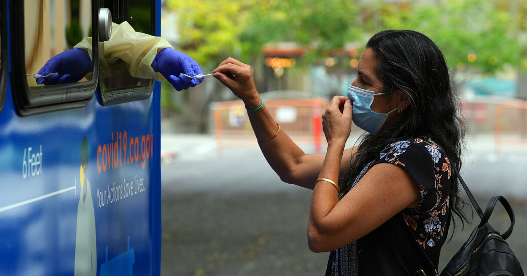
Is XBB.1.5 common?Worldwide, XBB.15 is still fairly rare. As of early January, it accounted for less than 1 percent of coronaviruses being sequenced. For now, it is most common in certain hot spots, like the northeastern United States, where it makes up about three-quarters of new cases.Is XBB.1.5 a cause for serious concern?Yes. Even though it is still rare, it is multiplying much faster than other subvariants of Omicron. However, many experts doubt it will produce a record-breaking wave of infections the way Omicron did in early 2021.

Read more →

'We couldn't get an ambulance and my husband died'

Ambulance bosses have apologised to the family of a man who died after he had a heart attack but no ambulance came.Martin Clark, 68, started suffering with chest pains at his home in East Sussex on 18 November – before any strike action started in the NHS.His family rang three times for an ambulance and after waiting 45 minutes drove him in their car to hospital.He died after arriving at hospital.Average waiting times in England for category 2 999 calls in November, when Martin died, were the worst since records began.

Read more →

Faster knee for better walking

Osaka Metropolitan University scientists delved into the relationship between gait function and knee extension velocity after total knee arthroplasty and compared the effects of various factors on walking. The results reveal that knee extension velocity, measured while seated, on the operated side was the most important determinant of gait function. These findings are expected to contribute to the development of new rehabilitation programs for efficient gait function improvement.
“Speed or strength, which is more important?” may be a critical question for not only athletes but also knee surgery patients. Osaka Metropolitan University scientists have revealed that knee extension velocity while seated is a stronger predictor of walking performance than muscle strength in elderly patients after their total knee arthroplasty (TKA) surgery.
TKA is the most common surgical intervention for knee osteoarthritis, a musculoskeletal disorder that mainly progresses with age. This surgery has been shown to be effective in relieving pain and restoring joint range of motion; however, TKA-enabled improvement in gait function may not be sufficient. Although the strength of the quadriceps — a muscle used to extend the knee — has been deemed to have a significant effect on postoperative gait function in TKA patients, there are many cases in which gait function does not improve even after quadriceps strength is restored. This raises a need to identify other factors influencing gait function.
A research team led by Professor Akira Iwata, from the Graduate School of Rehabilitation Science at Osaka Metropolitan University, hypothesized that the movement velocity of knee extension (i.e., knee extension velocity) is a strong determinant of gait function in TKA patients, and tested this hypothesis on 186 elderly patients who underwent TKA. Measurements were taken before and at 2 and 3 weeks after surgery and included gait function (gait speed and Timed Up and Go test), knee extension velocity, quadriceps strength, knee range of motion, and knee pain. Multiple regression analysis was performed to analyze the relationship between gait function and other variables.
The results show that the knee extension velocity on the operated side was the most important predictor of gait function in the participating patients. The findings were published in PLOS ONE.
“Thus far, rehabilitation programs aimed at recovering gait function after TKA surgery have focused on training to improve quadriceps strength,” explained Professor Iwata. “However, this study’s results suggest that training to enhance knee extension velocity on the operated side could be effective. We will continue to examine the effects of rehabilitation that concentrates on movement velocity.”

Read more →

Screen-printing method can make wearable electronics less expensive

The glittering, serpentine structures that power wearable electronics can be created with the same technology used to print rock concert t-shirts, new research shows.
The study, led by Washington State University researchers, demonstrates that electrodes can be made using just screen printing, creating a stretchable, durable circuit pattern that can be transferred to fabric and worn directly on human skin. Such wearable electronics can be used for health monitoring in hospitals or at home.
“We wanted to make flexible, wearable electronics in a way that is much easier, more convenient and lower cost,” said corresponding author Jong-Hoon Kim, associate professor at the WSU Vancouver’s School of Engineering and Computer Science. “That’s why we focused on screen printing: it’s easy to use. It has a simple setup, and it is suitable for mass production.”
Current commercial manufacturing of wearable electronics requires expensive processes involving clean rooms. While some use screen printing for parts of the process, this new method relies wholly on screen printing, which has advantages for manufacturers and ultimately, consumers.
In the study, published in the ACS Applied Materials and Interfaces journal, Kim and his colleagues detail the electrode screen-printing process and demonstrate how the resulting electrodes can be used for electrocardiogram monitoring, also known as ECG.
They used a multi-step process to layer polymer and metal inks to create snake-like structures of the electrode. While the resulting thin pattern appears delicate, the electrodes are not fragile. The study showed they could be stretched by 30% and bend to 180 degrees.
Multiple electrodes are printed onto a pre-treated glass slide, which allows them to be easily peeled off and transferred onto fabric or other material. After printing the electrodes, the researchers transferred them onto an adhesive fabric that was then worn directly on the skin by volunteers. The wireless electrodes accurately recorded heart and respiratory rates, sending the data to a mobile phone.
While this study focused on ECG monitoring, the screen-printing process can be used to create electrodes for a range of uses, including those that serve similar functions to smart watches or fitness trackers, Kim said.
Kim’s lab is currently working on expanding this technology to print different electrodes as well as entire electronic chips and even potentially whole circuit boards.
In addition to Kim, co-authors on the study includes researchers from the Georgia Institute of Technology and Pukyong National University in South Korea as well as others from WSU Vancouver. This research received support from the National Science Foundation.

Read more →

Fall rate nearly 50% among older Americans with dementia

With falls causing millions of injuries in older adults each year, it is an increasingly important public health concern. Older adults living with dementia have twice the risk of falling and three times the risk of incurring serious fall-related injuries, like fractures, compared to those without dementia. For older adults with dementia, even minor fall-related injuries can lead to hospitalization and nursing home admission. A new study from researchers in Drexel University’s College of Nursing and Health Professions, has shed light on the many and varied fall-risk factors facing older adults in community-living environments.
Recently published in Alzheimer’s & Dementia: The Journal of the Alzheimer’s Association, the research led by Safiyyah Okoye, PhD, an assistant professor at Drexel, and Jennifer L. Wolff, PhD, a professor at Johns Hopkins Bloomberg School of Public Health, examined a comprehensive set of potential fall-risk factors — including environmental factors, in addition to health and function — in older community-living adults in the United States, both with and without dementia.
“Examining the multiple factors, including environmental ones like a person’s home or neighborhood, is necessary to inform fall-risk screening, caregiver education and support, and prevention strategies for this high-risk population of older adults,” said Okoye.
Despite awareness of this elevated risk, there are very few studies that have examined fall-risk factors among people with dementia living in a community setting (not nursing homes or other residential facilities). The studies that do exist, overwhelmingly focus on health and function factors. According to the authors, this is the first nationally representative study to compare a comprehensive set of potential risk factors for falls for older Americans living with dementia to those without dementia.
The research team examined data from the 2015 and 2016 National Health and Aging Trends Study (NHATS), a population-based survey of health and disability trends and trajectories of adults 65 and older in the U.S. They were able to obtain potential sociodemographic, health and function predictors of falls, as well as potential social and physical environmental predictors.
Data from NHATS showed that nearly half (45.5%) of older adults with dementia had experienced one or more falls in 2016, compared to less than one third (30.9%) of older adults without dementia.
Among older adults living with dementia, three characteristics stood out as significantly associated with a greater likelihood of falls: a history of falling the previous year; impaired vision; and living with others (versus alone). For older adults without dementia, financial hardship, a history of falling, fear of falling, poor lower extremity performance, depressive symptoms and home disrepair were strongly associated with increased risk of falls.
While prior history of falling and vision impairment are well-known risk factors for falls among older adults in general; the researchers’ findings indicate that these were strong risk factors for falls among people living with dementia. According to the team, this suggests that people living with dementia should be assessed for presence of these characteristics. If they’re present, the individuals should receive further assessment and treatment, including examining their feet and footwear, assessing their environment and ability to carry out daily living activities, among other items.
The finding that older adults living with dementia who lived with a spouse or with non-spousal others had higher odds of experiencing a fall, compared to those who lived alone, highlights that caregiver support and education are understudied components of fall prevention programs for older adults with dementia who live with family caregivers, and deserve greater attention from clinicians, researchers and policy makers.
“Overall, our findings demonstrate the importance of understanding and addressing fall-risk among older adults living with dementia,” said Okoye. “It confirms that fall-risk is multidimensional and influenced by environmental context in addition to health and function factors.”
The results of the study indicate the need to further investigate and design fall-prevention interventions, specifically for people living with dementia.
“To decrease the high rates of falls among older adults with dementia, additional tailored fall-risk screening and fall-prevention interventions should be developed and tested,” said Okoye.

Read more →