Human body found to predict mealtimes

The human body can predict the timing of regular meals, according to a new study from the University of Surrey. The research team also found that daily blood glucose rhythms may be driven not only by meal timing but by meal size.
In the first study of its kind, researchers from Surrey, led by Professor Jonathan Johnston, investigated if the human circadian system anticipates large meals. Circadian rhythms/systems are physiological changes, including metabolic, that follow a 24-hour cycle and are usually synchronised to environmental signals, such as light and dark cycles.
Previous studies in this field have focussed on animal controls and until now it has been undetermined whether human physiology can predict mealtimes and food availability.
Jonathan Johnston, Professor of Chronobiology, and Integrative Physiology at the University of Surrey said:
“We often get hungry around the same time every day, but the extent to which our biology can anticipate mealtimes is unknown. It is possible that metabolic rhythms align to meal patterns and that regularity of meals will ensure that we eat at the time when our bodies are best adapted to deal with them.”
To learn more, 24 male participants undertook an eight-day laboratory study with strict sleep-wake schedules, exposure to light-dark cycles, and food intake. For six days, 12 participants consumed small meals hourly throughout the waking period, with the remaining participants consuming two large daily meals (7.5 and 14.5 hours after waking).

After six days, all participants were then put on the same feeding schedule for 37 hours and received small meals hourly in a procedure known to reveal internal circadian rhythms. Glucose was measured every 15 minutes during the study, and hunger levels were measured hourly during waking hours on days two four and six in the first stage of the study and then hourly for the final 37 hours.
Analysing results of the first six days of the study, researchers found the glucose concentration of participants in the small meal group increased upon waking and remained elevated throughout the day until declining after their last meal. In the large meal group, there was a similar increase in glucose concentration upon waking however there was a gradual decline leading up to the first meal.
In the final 37 hours, when both groups were fed the same small meals hourly, all participants exhibited an initial rise in glucose concentration upon waking. However, in those who had previously received two large meals, glucose levels began to decline before the anticipated large meal (which they did not receive) whereas for participants who had always consumed small meals hourly, their glucose levels continued to rise as previously seen. In addition, in the large meal group, there was an increase in hunger preceding projected mealtimes which sharply declined after the anticipated mealtime had passed.
Professor Johnston added:
“What we have found is that the human body is rhythmically programmed to anticipate mealtimes particularly when food is not readily accessible. This suggests that there is a physiological drive for some people to eat at certain times as their body has been trained to expect food rather than it just being a psychological habit.”
This study was published in the journal Current Biology.
Also involved in this study are Dr Cheryl Isherwood, Professor Debra Skene, Dr Daan Van Deer Veen and Dr Hana Hassanin.

Read more →

What makes the immune systems of prematurely born babies susceptible to deadly infections?

LMU physician Markus Sperandio has discovered why the immune system of prematurely born babies is not working properly after birth.
Every year, thousands of babies in Germany are born many weeks too early and often have to struggle for months. The earlier the babies are born, the higher the risk of life-threatening complications. Infections can lead to sepsis and are among the most frequent causes of death.
“In the case of very prematurely born infants, a bacterial infection can lead to death within hours,” says LMU physician Prof. Markus Sperandio. The physiologist and former pediatrician and neonatologist researches the causes of this high susceptibility to infection together with his team at LMU’s Biomedical Center Munich. Now the researchers have demonstrated that an immunostimulatory signaling pathway is suppressed in the developing immune system.
Important immune cells in prematurely born infants do not work properly after birth
Sperandio had already shown in earlier studies that in the fetus and in newborns important cells of the innate immune system — so-called neutrophils -do not work as in adults. In contrast to the situation in adults, fetal and neonatal neutrophils do not manage to sufficiently attach to the walls of blood vessels and extravasate into inflamed tissue. This is necessary, however, to trigger an inflammatory response and thus initiate immune defense.
Now the LMU researchers, working in collaboration with the Children and Women’s Clinic at University of Munich Hospital, have investigated which mechanisms are behind this immaturity. By means of a so-called transcriptomic analysis, they compared the gene activity of neutrophils in umbilical cord blood of premature and full-term babies with adult neutrophils. Compared to adults, there is a lot of gene activity in premature and full-term infants that counteracts immune defense. “In this case, these neutrophils act as if they were switched off,” says Sperandio.
Balance shift of immunoregulatory signaling pathways
This particularly affects signals transmitted via the NF-κB signaling pathway, which plays a decisive role in immune and inflammatory responses. It consists of two possible pathways for signals: one that promotes inflammation and one that can suppress it. Therefore, the activity of these two pathways must be finely balanced for proper regulation of the immune response.
“Our experiments have shown that this balance is shifted towards the anti-inflammatory pathway in fetal and neonatal neutrophils,” says Sperandio. “Whereas this regulation of neutrophil function is clearly a requirement for normal fetal growth in utero, it leads to immune defense problems in prematurely born infants who have to adapt ‘too soon’ to the world outside the uterus.” To what extent these findings will be a springboard for new therapeutic approaches in the future remains to be seen. “Due to the complex processes in the growing fetal and neonatal organism, maturation-adapted therapies are conceivable but remain a long way off at this stage,” says Sperandio.

Read more →

Arsenic contaminates private drinking water wells across the western Great Basin

In the arid and drought-stricken western Great Basin, sparse surface water means rural communities often rely on private groundwater wells. Unlike municipal water systems, well water quality in private wells is unregulated, and a new study shows that more than 49 thousand well users across the region may be at risk of exposure to unhealthy levels of arsenic in drinking water.
Led by researchers at DRI and the University of Hawai’i Cancer Center and published February 16th in Environmental Science and Technology, the study used data from groundwater wells across the western Great Basin to build a model to predict the probability of elevated arsenic in groundwater, and the location and number of private well users at risk. According to the study, the Carson Desert basin (including the town of Fallon, Nevada), Carson Valley (Minden and Gardnerville, Nevada), and the Truckee Meadows (Reno), have the highest population of well users at risk. The new study builds on previous research showing that 22% of 174 domestic wells sampled in Northern Nevada had arsenic levels exceeding the EPA guideline.
“What we are finding is that in our region, we have a high probability for elevated arsenic compared to most other regions in the country,” said Daniel Saftner, M.S., a hydrogeologist at DRI and lead author of the study. “And we are seeing that geothermal and tectonic processes that are characteristic of the Great Basin contribute to the high concentrations of naturally occurring arsenic in the region’s groundwater.”
The region’s mountains are also primary sources of arsenic. “As the arsenic-rich volcanic and meta-sedimentary rocks that form the mountains erode, sediment is transported to the valleys below,” says Steve Bacon, Ph.D., DRI geologist and study co-author. Water percolating through the valley floor then carries arsenic into the groundwater. Deeper, older groundwater and geothermal waters tend to have a higher arsenic concentration and can migrate upward along faults and mix with shallow groundwater.
“We really wanted to better understand the unique geologic factors that contribute to high arsenic in this study,” Saftner says. “It’s important for us to think about the role of the environment as it pertains to human health — where we live can influence what our long-term health looks like.”
To train and test the predictive model, the research team used data collected through the Healthy Nevada Project, including water samples from 163 domestic wells primarily located near Reno, Carson City, and Fallon. These data were supplemented with 749 groundwater samples compiled from the USGS National Water Information System. The model uses tectonic, geothermal, geologic, and hydrologic variables to predict the probability of elevated arsenic levels across the region.
Although the U.S. EPA has set an arsenic concentration guideline of 10 µg/L for public drinking water, previous research has shown a range of health effects from long-term exposure to levels above 5 µg/L. Using this concentration as the benchmark, the model and map show that much of the region’s groundwater — particularly in western and central Nevada — is predicted to have more than a 50% probability of elevated arsenic levels.
“Community members can use our arsenic hazard map to see what the risk is at their location, which might motivate them to test their well water,” says Monica Arienzo, Ph.D., associate research professor at DRI and study co-author. “Then, if they have high levels of arsenic or other contaminants, they can take steps to reduce their exposure, such as installing a water treatment system.”
The findings from this study are potentially useful for a range of different applications. “The results can be useful for water utilities or water managers who tap similar shallow aquifers for their water supply,” says Saftner, “as well as irrigation wells that source water from these aquifers.”
The research team plans to use their model to take a closer look at the health impacts of prolonged arsenic exposure. “Through the Healthy Nevada Project, genetic data and health records are paired with environmental data to help determine whether there are associations between the levels of arsenic in a community’s groundwater and specific health outcomes,” stated Joe Grzymski, Ph.D., research professor at DRI and principal investigator of the project.

Read more →

Mpox Often Leads to Severe Illness, Even Death, in People With Advanced H.I.V.

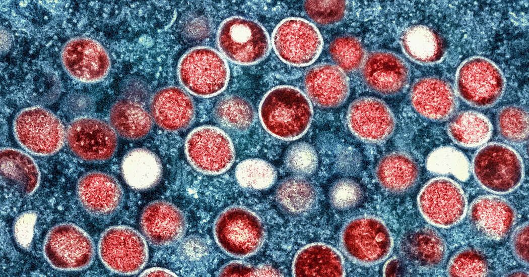
The death rate among these patients is about 15 percent, researchers reported. The virus should be added to the list of opportunistic infections seen in patients with advanced H.I.V., scientists said.In people with advanced H.I.V. disease, the mpox virus — formerly known as monkeypox — often causes severe illness, with a death rate of about 15 percent, researchers reported on Tuesday.The seriousness of the infection warrants the inclusion of mpox among the opportunistic conditions that are particularly dangerous to people with advanced H.I.V., the researchers said at the Conference on Retroviruses and Opportunistic Infections in Seattle.“These findings make it very, very clear that every single person with mpox should have an H.I.V. test,” said Dr. Chloe Orkin, an H.I.V. expert at Queen Mary University of London and the researcher who led the work. She and her colleagues also described the results in the journal The Lancet on Tuesday.The mpox outbreak began unfolding last May. Although the number of cases has slowed to a trickle in most regions, it has so far affected roughly 86,000 people in 110 countries and killed 92. Multiple studies estimated that from 40 percent to 50 percent of infected individuals were living with H.I.V.When antiretroviral drugs keep H.I.V. in check, mpox is about as dangerous as it is to those without H.I.V. infection. But a C.D.C. study last year, along with observations from previous outbreaks in Nigeria, indicated that mpox was more severe, and far more lethal, in those with high levels of H.I.V.In the new study, an international group of clinicians tracked 382 adults in 28 countries who had advanced H.I.V. disease and were infected with mpox. They analyzed the amount of H.I.V. and the number of CD4 cells, a type of immune system cell, in these patients.The typical range for CD4 cells is from 500 to 1,500 per cubic millimeter of blood. All 27 deaths in the study were among people who had fewer than 200 CD4 cells. Mpox killed nearly 30 percent of those with fewer than 100 CD4 cells.The nature of the illness was also strikingly different in patients with weakened immune systems. While most people infected with mpox show lesions only at the site of exposure, those with advanced H.I.V. developed large, ulcerated lesions teeming with virus throughout the body.“It’s on the back, it’s on the feet, it’s in the eyes, it’s everywhere — it’s horrifying,” Dr. Orkin said. “It’s because the immune system is not able to contain the virus at all.”Many patients also had nodules in the lung that caused acute respiratory distress, she added.Adding mpox to the list of opportunistic infections in people with advanced H.I.V. would encourage health care workers to identify and prioritize patients most at risk of severe disease and death.Patients would need antibiotics to prevent other opportunistic infections, and should be offered two doses of vaccine injected under the skin, rather than between layers of the skin as is currently done, Dr. Orkin said.The United States added mpox to the list of possible opportunistic infections in people with H.I.V. in September. The World Health Organization plans to discuss doing the same over the next few months, said Dr. Meg Doherty, the director of global H.I.V., hepatitis and sexual transmitted infections programs at the W.H.O.The new data make a “compelling case” for mpox’s addition to the list of opportunistic conditions, Dr. Doherty said.In parts of the world where people with H.I.V. may not have access to mpox vaccines, or to treatment for mpox and H.I.V., she said, “this should just raise the awareness that we have more to do in those areas than we have.”

Read more →

Rewiring blood cells to give rise to precursors of sperm

Different cell types — say, heart, liver, blood, and sperm cells — possess characteristics that help them carry out their unique jobs in the body. In general, those characteristics are hard-wired. Without intervention, a heart cell won’t spontaneously transform into a liver cell.
Yet researchers from the University of Pennsylvania School of Veterinary Medicine, working with collaborators from the University of Texas at San Antonio and Texas Biomedical Research Institute, have prompted marmoset blood cells to acquire the flexibility of stem cells. Then they directed those stem cells to take on the characteristics of sperm precursors.
In the journal eLife, they report on their step-by-step process of rewiring cells. The findings — the first in the marmoset, a small monkey — open new possibilities for studying primate biology and developing novel assisted reproductive technologies like in vitro gametogenesis, a process of generating germ cells, sperm or eggs, in a laboratory dish, akin to how in vitro fertilization involves the generation of an embryo outside the human body.
“Scientists know how to generate functional sperm and egg from induced pluripotent stem cells in mice, but mouse germ cells are very different from human germ cells,” says Kotaro Sasaki, an assistant professor at Penn Vet. “By studying marmosets, whose biology more closely resembles ours, we can bridge the gap.”
To understand how to generate germ cells, the researchers first studied germ cell precursors from marmoset embryos, which had never been rigorously characterized for the species. They found that these early-stage cells, known as primordial germ cells (PGCs), bore certain molecular markers that could be tracked over time. Performing single-cell RNA sequencing on these cells revealed that PGCs expressed genes characteristic of early-stage germ cells and those related to epigenetic modifications, which regulate gene expression. PGCs did not, however, express genes known to be turned on later in the process of germ cell development, when precursor cells migrate to the ovaries or testes to complete their maturation.
Their findings were “consistent with the notion that marmoset germ cells undergo a reprogramming process,” Sasaki says, that “turns off” certain markers and allows PGCs to proceed through the stages of germ cell development. The patterns the researchers observed in marmoset cells closely resembled what has been found in both humans and other monkey species but were distinct from those of mice, another reason why the marmoset could be a valuable model for reproductive biology studies.

With that information in hand, the team set about trying to reconstitute the process of development artificially, in the lab. The first step: to transform blood cells into induced pluripotent stem cells (iPSCs), cells that retain the ability to give rise to a number of other cell types.
“I have a lot of experience in working with cell culture and induced pluripotent stem cells, but establishing a stable culture for the marmoset cells was a difficult part of the study,” says Yasunari Seita, a postdoctoral researcher in Sasaki’s lab and a lead author.
After much trial and error and applying lessons learned from mouse, human, and other investigations, Seita landed upon a strategy that enabled him to generate and sustain stable cultures of iPSCs. A key to success was the addition of an inhibitor of the signaling pathway governed by the Wnt protein, which is involved in a variety of cellular functions, such as cell differentiation.
The next step was to move from iPSCs to germ cell precursors. Once again, considerable experimentation went into developing the protocol for this transformation. The method that worked best involved adding a cocktail of growth factors to successfully prompt between 15-40% of their culture to take on the characteristics of these germ cell precursors.
“We were excited to see that efficiency and were able to expand our cultures, passaging them multiple times and seeing nice, exponential growth,” Sasaki says. “The cells maintained key germ cell markers but didn’t express other markers that are associated with the migration to the gonad.”
In a final stage of the study, the research team coaxed these lab-grown cells to take on the characteristics of later-stage germ cells. Based on a method Sasaki and colleagues had established earlier in human cells and reported in a 2020 Nature Communications paper, they cultured the cells with mouse testicular cells over the course of a month. The result was a successful growth with some cells beginning to turn on genes associated with later-stage sperm cell precursors.

Developing new approaches to study the marmoset sets up the Penn and University of Texas at San Antonio teams — as well as the scientific community in general — to make use of the species as an important research model. Marmosets, for example, have cognitive functioning that resembles that of humans in many ways and thus could lead to new insights in neuroscience.
For Sasaki’s group, most interested in development of the reproductive system, marmosets represent a new avenue for pursuing studies of normal and abnormal development as well as fertility.
“When you think about the clinical applications of an assisted reproductive technology like in vitro gametogenesis, there are a lot of ethical, legal, and safety concerns that could arise,” Sasaki says. “We definitely need a good preclinical model to explore before we move to human clinical translation.”
Kotaro Sasaki is an assistant professor of biomedical sciences at the University of Pennsylvania School of Veterinary Medicine.
Yasunari Seita is a postdoctoral researcher in the School of Veterinary Medicine at Penn.
Sasaki and Seita’s coauthors are Penn Vet’s Keren Cheng; the University of Texas at San Antonio’s John R. McCarrey, Nomesh Yadu, Isamar Santana Toro, Li-hua Yen, Sean Vargas, Christopher S. Navara, and Brian P. Hermann; and the Texas Biomedical Research Institute’s Ian H. Cheeseman, Alec Bagwell, and Corinna N. Ross. Seita and Cheng were co-first authors, and Sasaki, Navara, and Hermann were co-corresponding authors.
The study was supported by the Open Philanthropy Project (grants 197906 and 10080664), National Institutes of Health (grants DA054170, HD090007, OD011133, and MD007591) and National Science Foundation (grants 1337513 and 2018408).

Read more →

Air pollution speeds bone loss from osteoporosis

Elevated levels of air pollutants are associated with bone damage among postmenopausal women, according to new research led by scientists at Columbia University Mailman School of Public Health. The effects were most evident on the lumbar spine, with nitrous oxides twice as damaging to the area than seen with normal aging.
The research findings appear in the peer-reviewed journal eClinicalMedicine, part of The Lancet Discovery Science suite of open-access journals.
Previous studies on individual pollutants have suggested adverse effects on bone mineral density, osteoporosis risk, and fractures in older individuals. The new study is the first to explore the connection between air pollution and bone mineral density specifically in postmenopausal women, and the first to explore the effects of air pollution mixtures on bone outcomes.
The researchers analyzed data collected through the Women’s Health Initiative study, an ethnically diverse cohort of 161,808 postmenopausal women. They estimated air pollution (PM10, NO, NO2, and SO2) exposures based on participants’ home addresses. They measured bone mineral density (BMD; whole-body, total hip, femoral neck, and lumbar spine) at enrollment at follow-up at year one, year three, and year six using dual-energy X-ray absorptiometry.
The magnitude of the effects of nitrogen oxides on lumbar spine BMD would amount to 1.22 percent annual reductions — nearly double the annual effects of age on any of the anatomical sites evaluated. These effects are believed to happen through bone cell death by way of oxidative damage and other mechanisms.
“Our findings confirm that poor air quality may be a risk factor for bone loss, independent of socioeconomic or demographic factors. For the first time, we have evidence that nitrogen oxides, in particular, are a major contributor to bone damage and that the lumbar spine is one of the most susceptible sites of this damage,” says study first author Diddier Prada, MD, PhD, associate research scientist in the Department of Environmental Health Sciences at Columbia Mailman School of Public Health.
“Improvements in air pollution exposure, particularly nitrogen oxides, will reduce bone damage in postmenopausal women, prevent bone fractures, and reduce the health cost burden associated with osteoporosis among postmenopausal women. Further efforts should focus on detecting those at higher risk of air pollution-related bone damage,” says lead author Andrea Baccarelli, MD, PhD, chair of the Department of Environmental Health Sciences at Columbia Mailman School of Public Health.
Car and truck exhaust is a major source of nitrous oxides, as are the emissions from electrical power generation plants.
Approximately 2.1 million osteoporosis-related bone fractures occur annually, resulting in up to $20.3 billion in annual direct health costs. Osteoporosis impacts women more than men, with 80 percent of the estimated 10 million Americans with osteoporosis being women. Postmenopausal women are at higher risk, with one in two women over 50 experiencing a bone fracture because of osteoporosis.
Previously, Columbia researchers showed that long-term air pollution exposure reduces BMD and increases bone fracture risk in later life. Subsequently, these findings have been confirmed in multiple human studies.
Study co-authors include Carolyn J. Crandall at UCLA; Allison Kupsco, Marianthi-Anna Kioumourtzoglou, Yike Shen, Gary Miller, Iuliana Ionita-Laza at Columbia Mailman; James D. Stewart, Eric A. Whitsel at UNC Chapel Hill; Duanping Liao and Jeff D. Yanosky at Public Health Sciences, Hershey, PA; Andrea Ramirez at National Autonomous University of Mexico; and Jean Wactawski-Wende at SUNY Buffalo.
The research was supported by grants from the National Institutes of Health (ES030163, TR00187, ES020836, ES025225, ES009089, AG069120, ES032242, ES027747, ES031688, AG058704, ES028805, ES030616, ES029943).

Read more →

Pill for skin disease may also curb excessive drinking

Researchers from Oregon Health & Science University and institutions across the country have identified a pill used to treat a common skin disease as an “incredibly promising” treatment for alcohol use disorder.
The study was recently published in the Journal of Clinical Investigation.
On average, the people who received the medication, called apremilast, reduced their alcohol intake by more than half — from five drinks per day to two.
“I’ve never seen anything like that before,” said co-senior author Angela Ozburn, Ph.D., associate professor of behavioral neuroscience in the OHSU School of Medicine and a research biologist with the Portland VA Health Care System.
The lead author is Kolter Grigsby, Ph.D., a postdoctoral fellow in the Ozburn laboratory at OHSU.
Beginning in 2015, Ozburn and collaborators searched a genetic database looking for compounds likely to counteract the expression of genes known to be linked to heavy alcohol use. Apremilast, an FDA-approved anti-inflammatory medication used to treat psoriasis and psoriatic arthritis, appeared to be a promising candidate.

They then tested it in two unique animal models that have a genetic of risk for excessive drinking, as well as in other strains of mice at laboratories across the country. In each case, apremilast reduced drinking among a variety of models predisposed to mild to heavy alcohol use. They found that apremilast triggered an increase in activity in the nucleus accumbens, the region of the brain involved in controlling alcohol intake.
Researchers at the Scripps Research Institute in La Jolla, California, then tested apremilast in people.
The Scripps team conducted a double-blind, placebo-controlled clinical proof-of-concept study involving 51 people who were assessed over 11 days of treatment.
“Apremilast’s large effect size on reducing drinking, combined with its good tolerability in our participants, suggests it is an excellent candidate for further evaluation as a novel treatment for people with alcohol use disorder,” said co-senior author Barbara Mason, Ph.D., Pearson Family professor in the Department of Molecular Medicine at Scripps.
The clinical study involved people with alcohol use disorder who weren’t seeking any form of treatment, and Mason predicts that apremilast may be even more effective among people who are motivated to reduce their alcohol consumption.
“It’s imperative for more clinical trials to be done on people seeking treatment,” Ozburn said. “In this study, we saw that apremilast worked in mice. It worked in different labs, and it worked in people. This is incredibly promising for treatment of addiction in general.”
An estimated 95,000 people in the United States die every year from alcohol-related deaths, according to the National Institute on Alcohol Abuse and Alcoholism.
Currently, there are three medications approved for alcohol use disorder in the United States: Antabuse, which produces an acute sensitivity akin to a hangover when alcohol is consumed; acamprosate, a medication thought to stabilize chemical signaling in the brain that is associated with relapse; and naltrexone, a medication that blocks the euphoric effects of both alcohol and opioids.

Read more →

Paul Berg, Nobel-Winning Pioneer of Genetic Engineering, Is Dead at 96

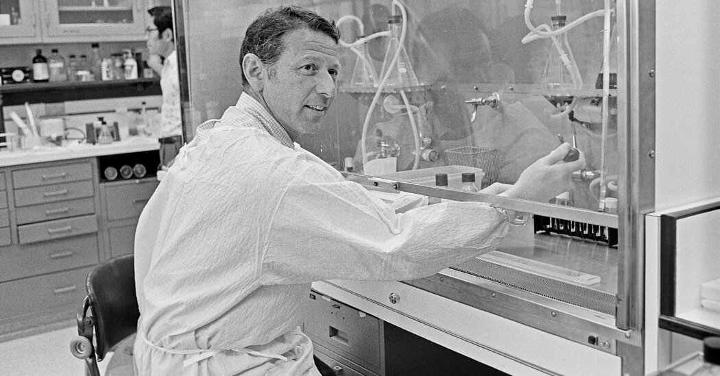
In 1971, he transferred material from one organism to another, creating the first recombinant DNA, or rDNA. He later helped lead a historic effort to write protocols for genetic research.Paul Berg, a Nobel Prize-winning biochemist who ushered in the era of genetic engineering in 1971 by successfully combining DNA from two different organisms, died on Wednesday at his home on the Stanford University campus in California. He was 96.His death was announced by the Stanford School of Medicine.After his breakthrough with DNA, Dr. Berg led a momentous convocation of scientists to establish safeguards against the misuse of genetic research.In 1971, he was already a well-known researcher at Stanford University when he oversaw the artificial introduction of DNA from one virus into another, creating the first recombinant DNA, or rDNA. The achievement was the first link in the chain of advances that has led to the genetic engineering of new therapeutic treatments for diseases and of vaccines, like the messenger RNA versions used to counter the virus that causes Covid-19.Dr. Berg’s work earned him the 1980 Nobel Prize in Chemistry, which he shared with Walter Gilbert and Frederick Sanger, who were cited for their work on genetic sequencing. In remarks at a Nobel banquet, Dr. Berg said that through his research he had “experienced the indescribable exhilaration, the ultimate high, that accompanies discovery, the breaking of new ground, the entering into areas where man had not been before.”Often described as the blueprint for every cell, DNA, or deoxyribonucleic acid, is the spiral-staircase-shaped strand of molecules that carry the code by which cells duplicate themselves. Dr. Berg showed that the blueprint could be altered and cells made to produce new offspring that could ultimately do — or not do — very different things from the original cells.As David A. Jackson, a postdoctoral fellow who was one of Dr. Berg’s trainees, later recalled to Dr. Berg’s biographer, Errol C. Friedberg: “One morning Paul and I got together and he suggested that we attempt to put new genes into SV40 DNA and use the recombinant molecules to introduce foreign DNA into animal cells.”The researchers used the DNA part of a virus (a circular DNA), which can be propagated in the E. coli bacteria, and incorporated it into a simian virus (a circular SV40 DNA genome). Each of the circular DNAs was converted into linear DNAs with an enzyme. Using an existing technique, these linear DNAs were modified so that the modified ends attracted each other. Mixed together, the two DNAs recombined and created a loop of rDNA, which contained the genes from the two different organisms. Dr. Berg and his team began preparing for the next step: introducing the rDNA into E. coli and animal cells. But as word about his work spread among researchers, Dr. Berg was challenged to guarantee that this newly created DNA — which, after all, consisted partly of material from a virus that lived in one of the world’s most common bacteria, E. coli — could not escape the laboratory and cause incalculable harm.Dr. Berg recognized that such an absolute certainty was not then possible, and he halted further experiments, although other researchers quickly moved forward.Dr. Berg, left, received the Nobel Prize in Chemistry in 1980 from King Carl Gustaf of Sweden in Stockholm. Afterward he said that through his research he had “experienced the indescribable exhilaration, the ultimate high, that accompanies discovery.”Reportagebild/Tobbe Gustavsson pool, via Associated PressDr. Berg used the break in his experiments to focus on the larger ethical and public health issues raised by the manipulation of genes, including human genes. As a public figure who had testified before Congress in favor of federal funding for basic scientific research, and who had a wide range of contacts among biochemists, he was well positioned to help organize a conference at Asilomar, Calif., in February 1975.About 150 leading DNA researchers from the United States and 12 other countries — including James Watson, a co-discoverer of the double-helix structure of DNA — discussed and then subscribed to rules to govern their own work. The conference was historic: Never before had scientists gathered to write regulations for their own research.The eventual recommendations were deemed voluntary and drew a few dissents, including from Dr. Watson, but they were adopted by government regulators. In 2017, the event was the template for another Asilomar convention on a technology many consider equally fraught: artificial intelligence.Dr. Berg’s early concerns were highlighted four decades after his experiment when a Chinese scientist claimed in 2018 that he had created the world’s first genetically edited babies. Dr. Berg joined 17 other leading microbiologists in condemning the work and calling for a five-year moratorium on the clinical use of technologies for the editing of heritable genes.Paul Berg was born June 30, 1926, in Brooklyn, a son of Harry and Sarah (Brodsky) Bergsaltz, immigrants from Russia. His father was a furrier.Paul attended Abraham Lincoln High School, in Coney Island, where he developed his interest in science.After serving as an ensign in the Navy during World War II, Dr. Berg graduated from Pennsylvania State University in 1948. He received a doctorate in biochemistry from Western Reserve University (now Case Western Reserve University) in Cleveland in 1952, then did postdoctoral work at the Institute of Cytophysiology in Copenhagen and at Washington University in St. Louis. He joined the university faculty in 1955.Dr. Berg in 2002. His work was the first link in the chain of advances that has led to the engineering of vaccines, like the messenger RNA versions used to counter Covid-19.Linda A. Cicero/Stanford News ServiceDr. Berg, an expert in enzymes, was recruited to Washington University in 1953 by another future Nobel laureate, Arthur Kornberg (also a Lincoln High School alumnus). In 1959, Dr. Kornberg, who that year received the Nobel Prize in Physiology or Medicine, moved to Stanford University to set up a new biochemistry department and brought along his Washington University team, including Dr. Berg.As he became increasingly well known for his basic research, some of it funded by the American Cancer Society, Dr. Berg often received letters from the parents of children with cancer, and despite a crowded schedule, he would respond with personal replies of encouragement.Along with the 1980 Nobel, Dr. Berg was also a recipient of the Eli Lilly Award in Biological Chemistry in 1959, the Albert Lasker Basic Medical Research Award in 1980 and the National Medal of Science in 1983. He was the author, with the molecular biologist Maxine Singer (another organizer of the Asilomar Conference), of “Genes and Genome,” (1991); “Dealing With Genes: The Language of Heredity” (1992); and “George Beadle: An Uncommon Farmer” (2003).He married Mildred Levy in 1947. She died in 2021. His survivors include their son, John.In later years Dr. Berg would hark back to his student days at Abraham Lincoln High School in Brooklyn in tracing his path to a life in science. He credited in particular the keeper of the school science department’s supply room, a woman named Sophie Wolfe.“Her love of young people and interest in science led her to start an after school program of science clubs,” Dr. Berg wrote in an autobiographical sketch for the Nobel committee. “Rather than answering questions we asked, she encouraged us to seek solutions for ourselves, which most often turned into mini research projects. Sometimes that involved experiments in the small lab she kept, but sometimes it meant going to the library to find the answers.“The satisfaction derived from solving a problem with an experiment was a very heady experience, almost addicting,” he continued. “Looking back, I realize that nurturing curiosity and the instinct to seek solutions are perhaps the most important contributions education can make. With time, many of the facts I learned were forgotten, but I never lost the excitement of discovery.”

Read more →