Your Hair Is Going Gray. This Glitch May Explain Why.

Experiments using mice found a malfunction in adult stem cells that offers insights into why we turn into silver foxes and vixens.Many of the signs of aging are invisible, slow, and subtle — changes in cell division capacity, cardiac output and kidney function don’t exactly show up in the mirror. But gray hairs are one of the most obvious clues that the body isn’t working like it used to.Our hair turns gray when melanin-producing stem cells stop functioning properly. A new study in mice, but with implications for people and published Wednesday in the journal Nature, provides a clearer picture of the cellular glitches that turn us into silver foxes and vixens.“This is a really big step toward understanding why we gray,” said Mayumi Ito, an author of the study and a dermatology professor at New York University’s Grossman School of Medicine.Unlike embryonic stem cells, which develop into all sorts of different organs, adult stem cells have a more set path. The melanocyte stem cells in our hair follicles are responsible for producing and maintaining the pigment in our hair.Each hair follicle keeps immature melanocyte stem cells in storage. When they’re needed, those cells travel from one part of the follicle to another, where proteins spur them to mature into pigment-producing cells, giving hair its hue.Scientists assumed that gray hair was the result of that pool of melanocyte stem cells running dry. However, previous studies with mice made Dr. Ito and her co-author, Qi Sun, wonder if hair could lose its pigment even when stem cells are still present.Each hair follicle keeps immature melanocyte stem cells in storage, left. When they’re needed, those cells travel from one part of the follicle to another, where proteins spur them to mature into pigment-producing cells, giving hair its hue, right.NatureTo learn more about stem cell behavior throughout different phases of hair growth, the researchers spent two years tracking and imaging individual cells in mouse fur. To their amazement, the stem cells traveled back and forth within the hair follicle, transitioning into their mature, pigment-producing state and then out of it again.“We were surprised,” said Dr. Sun, who said seeing one group of stem cells switching back and forth between mature and young states did not match up with existing explanations.But as time wore on, the melanocyte cells couldn’t keep up the double act. A hair falling out and growing back takes a toll on the follicle, and eventually, the stem cells stopped making their journey, and thus, stopped receiving protein signals to make pigment. From then on, the new hair growth didn’t get its dose of melanin.The researchers further explored this effect by plucking hairs from mice, simulating a faster hair growth cycle. This “forced aging” led to a buildup of melanocyte stem cells stuck in their storage place, no longer producing melanin. The mice’s fur went from dark brown to salt-and-pepper.While the study was conducted with rodents, the researchers say their findings should be relevant to how human hair gets and loses its color. What’s more, they hope their findings could be a step toward preventing or reversing the graying process.Melissa Harris, a biologist at the University of Alabama at Birmingham who was not involved with the study, said the findings help “clinch” previous evidence she’s seen suggesting that “not all melanocyte stem cells are created equal, and even if you have some left over, they may not be useful.”Dr. Harris said she takes the study’s findings about its “forced aging” of mouse hair “with maybe a little bit of a grain of salt,” as a plucked hair might not behave the same as naturally aged hair. But she found the study valuable, not just because a cure for graying hair might be a hit with the public; the insights into stem cell behavior might help researchers understand things like cancer and cell regeneration.“I think sometimes people take the hair for granted,” she said, “but in a sense, it makes it actually really easy for us to see potential ways in which aging or other perturbations affect our bodies.”

Read more →
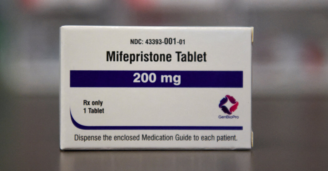
Generic Abortion Pill Maker GenBioPro Sues FDA to Protect Mifepristone

The suit by GenBioPro, the generic maker of mifepristone, is the latest strand in the intense legal battle over abortion pills.The company that makes a majority of the abortion pill mifepristone sold in the United States sued the Food and Drug Administration on Wednesday, a new legal volley in a string of recent court maneuverings over the fate of the drug.The lawsuit adds another strand to the intense legal battle underway over a case filed in federal court in Texas in which a consortium of anti-abortion groups are seeking to overturn the F.D.A.’s 23-year-old approval of mifepristone, the first pill used in a two-drug medication abortion regimen.In the new case, GenBioPro, which makes the generic version of mifepristone, seeks to block the F.D.A. from complying if the courts ultimately order mifepristone off the market. The case was filed in the U.S. District Court of Maryland.Earlier this month, the federal judge in the Texas case issued a preliminary ruling invalidating the approval of mifepristone. Last week a federal appeals court panel said the drug could remain on the market while the case was being litigated, but it ordered a reversal of all regulatory actions the F.D.A. has taken on mifepristone since 2016, which include the approval in 2019 of GenBioPro’s generic version of the exact same drug.That order has been briefly paused by the Supreme Court, which is expected to decide by midnight on Friday whether it will extend the stay until the full case can be heard.The GenBioPro lawsuit claims that the F.D.A. has repeatedly failed to stipulate that it would follow a regulatory process established by Congress and afford the drug company due process rights guaranteed by the Constitution if the agency was ordered to suspend or revoke its approval of GenBioPro’s product.By leaving open the possibility that it would immediately obey such a court order, the lawsuit argues, the F.D.A. has “left GenBioPro at risk of severe civil and criminal penalties if it does not cease shipments of mifepristone.”The F.D.A. issued a statement saying: “F.D.A. doesn’t comment on pending litigation.”In testimony Wednesday before the Senate Appropriations Committee, the F.D.A. commissioner, Dr. Robert M. Califf, fielding questions about the Texas lawsuit, said the agency was concerned about the potential implications of the case, “from the well-being of patients who need access to this drug, the pharmaceutical industry and our ability to implement our statutory authority.” He noted that the F.D.A. was fighting the case in court, adding “I’ll just say the FDA intends to comply with any court orders.”Evan Masingill, GenBioPro’s chief executive, said Wednesday that uncertainty about the outcome of the Texas case has led to fewer orders of mifepristone. “The market disruption is already pervasive, impacting orders that include tens of thousands of units,” he said.The case could also have implications for the broader drug industry. The suit claims that it would be unprecedented for the F.D.A. to follow a court order to immediately revoke the approval of a drug. A drug’s approval can only be revoked if the F.D.A. determines that it presents “an imminent hazard to the public health,” the lawsuit says. The F.D.A. has forcefully argued in court that mifepristone is very safe and cited scores of studies showing that serious complications are rare and that less than 1 percent of patients need hospitalization.“People develop drugs in this country and not in others because we’ve typically had a pretty predictable regulatory structure, and with the Texas lawsuit, is that becoming not the case?” said Skye Perryman, a lawyer for GenBioPro and president of Democracy Forward, a center-left legal advocacy organization. “That has industry wide implications.”GenBioPro says that it supplies about two-thirds of the drug sold in the United States and that it sold more than 850,000 units of the drug between 2017 and 2020. GenBioPro’s lawsuit cites filings the F.D.A. submitted to the Supreme Court, in which the agency said that if the appeals court decision were to take effect, “the generic version of the drug would cease to be approved altogether.”The company said in the lawsuit that such statements amounted to a policy decision by the federal agency and that “the F.D.A. decision is erroneous and unlawful.”The suit says that the F.D.A. has declined to say otherwise in response to three letters GenBioPro sent it in March and April. In those letters, GenBioPro asked the agency to clarify that it would adhere to the congressionally mandated process that typically involves a detailed and lengthy review before any decision about withdrawing a drug is made.The company said that the F.D.A. had responded to only the first letter, sent in March before any decision was announced in the anti-abortion groups’ lawsuit, and that it said only that the “F.D.A. will, of course, need to review the Court’s opinion and order before determining what steps may be necessary to comply with it.”“We are not challenging F.D.A.’s scientific or medical judgment,” Ms. Perryman said, “but F.D.A. has failed to confirm it will respect our clients’ rights and so we are seeking a court order.”Christina Jewett

Read more →

Neuroscientists identify cells especially vulnerable to Alzheimer's

Neurodegeneration, or the gradual loss of neuron function, is one of the key features of Alzheimer’s disease. However, it doesn’t affect all parts of the brain equally.
One of the first brain regions to show neurodegeneration in Alzheimer’s disease is a part of the hypothalamus called the mammillary body. In a new study, MIT researchers have identified a subset of neurons within this body that are most susceptible to neurodegeneration and hyperactivity. They also found that this damage leads to memory impairments.
The findings suggest that this region may contribute to some of the earliest symptoms of Alzheimer’s disease, making it a good target for potential new drugs to treat the disease, the researchers say.
“It is fascinating that only the lateral mammillary body neurons, not those in the medial mammillary body, become hyperactive and undergo neurodegeneration in Alzheimer’s disease,” says Li-Huei Tsai, director of MIT’s Picower Institute for Learning and Memory and the senior author of the study.
In a study of mice, the researchers showed that they could reverse memory impairments caused by hyperactivity and neurodegeneration in mammillary body neurons by treating them with a drug that is now used to treat epilepsy.
Former MIT postdoc Wen-Chin (Brian) Huang and MIT graduate students Zhuyu (Verna) Peng and Mitchell Murdock are the lead authors of the paper, which appears today in Science Translational Medicine.
Predisposed to degeneration

As Alzheimer’s disease progresses, neurodegeneration occurs along with the buildup of amyloid beta plaques and misfolded Tau proteins, which form tangles in the brain. One question that remains unresolved is whether this neurodegeneration strikes indiscriminately, or if certain types of neurons are more susceptible.
“If we could identify specific molecular properties of classes of neurons that are predisposed to dysfunction and degeneration, then we would have a better understanding of neurodegeneration,” Murdock says. “This is clinically important because we could find ways to therapeutically target these vulnerable populations and potentially delay the onset of cognitive decline.”
In a 2019 study using a mouse model of Alzheimer’s disease, Tsai, Huang, and others found that the mammillary bodies — a pair of structures found on the left and right underside of the hypothalamus — had the highest density of amyloid beta. These bodies are known to be involved in memory, but their exact role in normal memory and in Alzheimer’s disease is unknown.
To learn more about the mammillary body’s function, the researchers used single-cell RNA-sequencing, which can reveal the genes that are active within different types of cells in a tissue sample. Using this approach, the researchers identified two major populations of neurons: one in the medial mammillary body and the other in the lateral mammillary body. In the lateral neurons, genes related to synaptic activity were very highly expressed, and the researchers also found that these neurons had higher spiking rates than medial mammillary body neurons.
Based on those differences, the researchers wondered if the lateral neurons might be more susceptible to Alzheimer’s disease. To explore that question, they studied a mouse model with five genetic mutations linked to early-onset Alzheimer’s in humans. The researchers found that these mice showed much more hyperactivity in lateral mammillary body neurons than healthy mice. However, the medial mammillary body neurons in healthy mice and the Alzheimer’s model did not show any such differences.

The researchers found that this hyperactivity emerged very early — around two months of age (the equivalent of a young human adult), before amyloid plaques begin to develop. The lateral neurons became even more hyperactive as the mice aged, and these neurons were also more susceptible to neurodegeneration than the medial neurons.
“We think the hyperactivity is related to dysfunction in memory circuits and is also related to a cellular progression that might lead to neuronal death,” Murdock says.
The Alzheimer’s mouse model showed impairments in forming new memories, but when the researchers treated the mice with a drug that reduces neuronal hyperactivity, their performance on memory tasks was significantly improved. This drug, known as levetiracetam, is used to treat epileptic seizures and is also in clinical trials to treat epileptiform activity — hyperexcitability in the cortex, which increases the risk of seizures — in Alzheimer’s patients.
Comparing mice and humans
The researchers also studied human brain tissue from the Religious Orders Study/Memory and Aging Project (ROSMAP), a longitudinal study that has tracked memory, motor, and other age-related issues in older people since 1994. Using single-cell RNA-sequencing of mammillary body tissue from people with and without Alzheimer’s disease, the researchers found two clusters of neurons that correspond to the lateral and medial mammillary body neurons they found in mice.
Similar to the mouse studies, the researchers also found signatures of hyperactivity in the lateral mammillary bodies from Alzheimer’s tissue samples, including overexpression of genes that encode potassium and sodium channels. In those samples, they also found higher levels of neurodegeneration in the lateral neuron cluster, compared to the medial cluster.
Other studies of Alzheimer’s patients have found a loss of volume of the mammillary body early in the disease, along with deposition of plaques and altered synaptic structure. All of these findings suggest that the mammillary body could make a good target for potential drugs that could slow down the progression of Alzheimer’s disease, the researchers say.
Tsai’s lab is now working on further defining how the lateral neurons of the mammillary body are connected to other parts of the brain, to figure out how it forms memory circuits. The researchers also hope to learn more about what properties of the lateral neurons of the mammillary body make them more vulnerable to neurodegeneration and amyloid deposition.
The research was funded by the JBP Foundation, the Ludwig Family Foundation, and the U.S. National Institutes of Health.

Read more →

New cause identified for metabolic disease that strikes Native Americans

Duke Health researchers have identified the cause of an inherited metabolic disease common among people with Lumbee and other Native American heritage, overturning decades of settled science and pointing to new, more effective therapies.
The finding, publishing online April 19 in the journal Science Translational Medicine, shatters the textbook explanations for how a type of protein breaks down in a child’s brain, becoming toxic and leading to potentially fatal neurological problems.
The inherited condition is called Glutaric Aciduria Type I (GA-1), and current literature describes the toxic substances as being produced in the brain instead of arising elsewhere and crossing the blood-brain barrier.
Treatments for the condition, including a strict, low-protein diet, have limited success. Up to a third of children with the condition suffer long-term neurologic damage and some die.
Because other metabolic disorders have been shown to break down proteins in the liver and then cause brain damage, the Duke researchers reopened the science into GA-1. The work was led by senior author Karl-Dimiter Bissig, M.D., Ph.D., an associate professor in Duke’s departments of Pediatrics, Medicine, Biomedical Engineering and Pharmacology and Cancer Biology.
Bissig and colleagues launched experiments in mice specially bred to have GA-1. They found that catabolites — the residue left by the breakdown of an essential amino acid called lysine — accumulate in the liver and do cross the blood-brain barrier. This leads to a toxic build-up of glutaric acid in the brain, causing nerve damage that impacts motor skills.

The researchers were able to cure the condition in mice with either a liver transplant or CRISPR gene-editing technology. Other liver-targeted gene therapies might also be effective and could be administered once in a lifetime.
“The original experiments led to the interpretation that the toxic catabolites were produced locally in the brain,” Bissig said. “What our work demonstrates is the importance of challenging paradigms, particularly as new technologies and research approaches are available.”
Bissig said inadequate measures to address different mutations in specific populations are leading to health disparities. People with Native American, Amish and Irish heritage have high susceptibility to GA-1, which can be identified during newborn screenings; the genetic variant common in Lumbee populations seems to cause the most damaging disease.
Because states decide what diseases are included in newborn screenings, GA-1 goes undiagnosed if it’s not part of a state’s chosen screening panel. Screenings could also be missed if babies are delivered at home.
While early diagnosis and a low-protein diet have been lifesaving, the benefits are concentrated in Amish- and Irish-heritage children, who have historically had better access to health care services than Native Americans.
“With a better understanding of this disease, we can now work to develop treatments that are more effective and easier to access,” Bissig said. “It’s much easier to treat the liver than the brain. We are now working to advance the more efficient and convenient therapies.”
In addition to Bissig, study authors include Mercedes Barzi, Collin G. Johnson, Tong Chen, Ramona M. Rodriguiz, Madeline Hemmingsen, Trevor J Gonzalez, Alan Rosales, James Beasley, Cheryl K. Peck, Yunhan Ma, Ashlee R. Stiles, Timothy C. Wood, Raquel Maeso-Diaz, Anna Mae Diehl, Sarah P. Young, Jeffrey I. Everitt, William C. Wetsel, William R. Lagor, Beatrice Bissig-Choisat, Aravind Asokan, and Areeg El-Gharbawy.
The study received funding support from The Alice and Y. T. Chen Center for Genetics and Genomics; the National Institute of Diabetes and Digestive and Kidney Disease (DK115461,
DK124477, T32DK060445); the National Heart Lung and Blood Institute (HL132840, R01HL089221); and the National Institute of General Medical Sciences (T32GM088129).

Read more →

Nanoparticles provoke immune response against tumors but avoid side effects

Cancer drugs that stimulate the body’s immune system to attack tumors are a promising way to treat many types of cancer. However, some of these drugs produce too much systemic inflammation when delivered intravenously, making them harmful to use in patients.
MIT researchers have now come up with a possible way to get around that obstacle. In a new study, they showed that when immunostimulatory prodrugs — inactive drugs that require activation in the body — are tuned for optimal activation timing, the drugs provoke the immune system to attack tumors without the side effects that occur when the active form of the drug is given.
The researchers designed prodrugs with bottlebrush-like structures based on a class of compounds called imidazoquinolines (IMDs). Mice treated with these bottlebrush prodrugs designed with optimized activation kinetics showed a significant reduction in tumor growth, with no side effects. The researchers hope that this approach could be used to boost immune system responses in cancer patients, especially when combined with other immunotherapy drugs or cancer vaccines.
“Our bottlebrush prodrug library enabled us to show an immunological effect of controlling immunotherapy kinetics, allowing us to boost immune responses while minimizing the side effects,” says Sachin Bhagchandani, an MIT graduate student who is the lead author of the study. “This kind of approach opens up avenues for scientists who want to decouple toxicity from some promising immunotherapy agents.”
Jeremiah Johnson, an MIT professor of chemistry, and Darrell Irvine, the Underwood-Prescott Professor with appointments in MIT’s departments of Biological Engineering and of Materials Science and Engineering, are the senior authors of the paper, which appears today in Science Advances. Irvine is also an associate director of MIT’s Koch Institute for Integrative Cancer Research and a member of the Ragon Institute of MGH, MIT, and Harvard.
Tailored prodrugs
Organic molecules known as IMDs bind to cell receptors called Toll-like receptors that are found on macrophages and other cells of the innate immune system. When activated, these cells begin producing cytokines and other inflammatory molecules.

In 1997, the FDA approved topical IMD drugs to treat certain types of skin cancer. Since then, many other IMD drugs have been tested in clinical trials for a variety of types of cancer, but none of these were approved, in part because the drugs produced too much systemic inflammation.
The MIT team set out to explore whether prodrugs of IMDs, which are inactivated until turned “on” in the tumor microenvironment, could reduce those side effects. In recent years, Johnson’s lab has developed a novel type of prodrug platform shaped like a bottlebrush. These nanoscale, cylindrical structures consist of chains that extend from a central backbone, giving the molecule a bottlebrush-like structure. Inactivated drugs are bound along the bottlebrush backbone through cleavable linkers that define the rate of active IMD release.
The researchers generated and compared six bottlebrush prodrugs that only differed by their release rate, in order to investigate how prodrug activation kinetics impact antitumor responses. Using these bottlebrush prodrugs, the researchers hoped they could deliver active IMDs to tumors while avoiding release into the bloodstream.
“Our ability to synthesize six bottlebrush prodrugs with identical sizes and shapes uniquely allows us to isolate and study release kinetics as a key variable. Excitingly, we find that it is possible to identify prodrug structures that limit IMD exposure to the whole body, thereby avoiding toxicity, and that activate in tumors to give antitumor efficacy,” Johnson says.
In preliminary studies in cells and mice, the researchers found that the fastest-activating prodrugs did cause immune-related side effects, including weight loss and elevated cytokine levels. However, the medium- and slow-releasing versions did not produce these effects.

The researchers then tested the IMD bottlebrush prodrugs in two different mouse models of colon cancer. Because the prodrugs are so small (approximately10 nanometers), they are able to efficiently accumulate in tumors. Once there, they get taken up by innate immune cells, where their linkers are cleaved. The resulting release of active IMDs causes immune cells to release cytokines and other molecules that create a pro-inflammatory environment. This series of events activates nearby T cells to attack the tumor.
In both models, mice treated with the bottlebrush prodrugs showed significantly slowed tumor growth. When the treatment was combined with a checkpoint blockade inhibitor — another class of immunotherapy drug — tumors were completely eliminated in about 20 percent of the mice.
While mice treated with the IMD used in this study, known as resiquimod, showed weight loss, elevated cytokine levels, and reduction in white blood cell count, as expected, mice given resiquimod bottlebrush prodrugs did not show any of these effects.
“Our molecules were able to safely reduce these effects by controlling how much of the active drug is released in the blood,” Bhagchandani says. “If you minimize release of the active compound there, then you’re able to get anti-tumor effects at the tumor site without the systemic side effects.”
Enhanced response
The findings suggest that the most promising use for IMD bottlebrush prodrugs could be to give them along with another drug that stimulates the immune response. Another possibility is using IMD bottlebrush prodrugs as adjuvants to enhance the immune system’s response to cancer vaccines.
“The ability of the bottlebrush prodrug strategy to change both where the drug accumulates in the body and when it is active is very attractive for activating immune responses against cancer or other disease safely,” Irvine says.

Read more →

Informed by mechanics and computation, flexible bioelectronics can better conform to a curvy body

Today, foldable phones are ubiquitous. Now, using models that predict how well a flexible electronic device will conform to spherical surfaces, University of Wisconsin-Madison and University of Texas at Austin engineers could usher in a new era in which these bendy devices can integrate seamlessly with parts of the human body.
In the future, for example, a flexible bioelectronic artificial retina implanted in a person’s eyeball could help restore vision, or a smart contact lens could continuously sense glucose levels in the body.
“With our powerful simulation model, we can now predict the conformability immediately, which dramatically speeds up the design process for flexible electronics,” says Ying Li, an associate professor of mechanical engineering at UW-Madison, whose research group developed the computational models. “The simulation results give very clear guidance for experimentalists, who can now determine the optimal design without needing to do a lot of extremely time-consuming experiments.”
The researchers detailed their work in a paper published April 19, 2023, in the journal Science Advances.
To perform as expected, bioelectronic devices must make very close contact with living tissue and avoid buckling or creasing. However, researchers have struggled to get flexible electronics to fully conform to so-called “non-developable surfaces” — surfaces such as spheres that can’t be flattened without breaking or creasing — which are all over the human body.
In this study, the research team used a combination of experimental, analytical and numerical approaches to systematically investigate how circular polymer sheets (which mimic the mechanical properties of flexible electronics), as well as partially cut circular sheets, conform on spherical surfaces. Analyzing those results enabled the researchers to derive a ready-to-use formula that reveals the underlying physics and predicts the conformability of flexible electronics.
“The results from our three different methods all pointed to the same physics, which is exciting,” says Nanshu Lu, a professor in the Department of Aerospace Engineering and Engineering Mechanics at the University of Texas at Austin, who led the experimental research. “We formulated a very simple mathematical equation to guide the design of flexible electronics for maximum conformability, and this should have a significant impact in the field.”
In addition, the researchers demonstrated a simple and elegant method for greatly enhancing the ability of flexible sheets to conform on spherical surfaces. Inspired by the Japanese art of kirigami, in which paper is cut and folded, the researchers made the simplest possible radial cuts in the circular sheet, improving its conformability from 40% to more than 90%.
Li says this advance will drive innovation in the field by enabling many other researchers to design improved flexible electronics.
“This is the first work to provide a full picture to understand the complex process of how flexible electronics conform to these complicated surfaces,” Li says. “This advance will pave the way for all the future studies in the area of developing bioelectronics that can better conform to the human body.”

Read more →

Newfound link between Alzheimer's and iron could lead to new medical interventions

There is a growing body of evidence that iron in the brain may play a role in Alzheimer’s disease. Lending weight to that idea, a new imaging probe has for the first time shown that in the same regions of the brain where the amyloid beta plaques associated with Alzheimer’s occur, there is also an increase in iron redox, meaning the iron in these regions is more reactive in the presence of oxygen. Their imaging probe could yield even more details about the causes of Alzheimer’s and help in the search for new drugs to treat it.
A team from The University of Texas at Austin and the University of Illinois at Urbana-Champaign published a study today on the new imaging technique and findings in Science Advances.
“The link between iron redox and Alzheimer’s disease has been a black box,” said Yi Lu, corresponding author and professor of chemistry at UT Austin. “The most exciting part to me is that we now have a way to shine light into this black box so that we can begin to understand this whole process in much more detail.”
About a decade ago, scientists discovered ferroptosis, a process in the body that is dependent on elevated iron levels, leads to cell death and plays a key role in neurodegenerative diseases, such as Alzheimer’s. Using magnetic resonance imaging on living Alzheimer’s patients, scientists have observed that these patients tend to have elevated iron levels in the brain, although that method doesn’t differentiate between different forms of iron. Together, these findings suggested that iron might play a role in destroying brain cells in Alzheimer’s patients.
For the new study, the researchers developed DNA-based fluorescent sensors that can detect two different forms of iron (Fe2+ and Fe3+) at the same time in cell cultures and in brain slices from mice genetically modified to mimic Alzheimer’s. One sensor glows green for Fe2+ and the other glows red for Fe3+. This is the first imaging technique that can simultaneously detect both forms of iron in cells and tissue while also indicating their quantity and spatial distribution.
“The best part about our sensor is that we can now visualize the changes of Fe2+ and Fe3+ and their ratios in each location,” said Yuting Wu, a co-first author of the study and a postdoctoral researcher in Lu’s lab at UT Austin. “We can change one parameter at a time to see if it changes the plaques or the oxidative states of iron.”
That ability could help them better understand why there is an increased ratio of Fe3+ to Fe2+ in the location of amyloid beta plaques and whether increased iron redox is involved in forming the plaques.
Another key question is whether the iron redox is directly involved in cell death in Alzheimer’s, or simply a byproduct. The researchers plan to explore this question in Alzheimer’s mice. If further research determines that iron and its redox changes indeed cause cell death in Alzheimer’s patients, that information could provide a potential new strategy for drug development. In other words, perhaps a drug that change the ratio Fe3+ to Fe2+ could help protect brain cells. The new imaging probe could be used to test how well drug candidates work at changing the ratio.
To develop the sensors, the scientists first hired a commercial lab to produce a library of 100 trillion short DNA strands, through a chemical process called oligonucleotide synthesis. They then conducted a screening process to find those strands that recognize — or in chemistry parlance “bind tightly to and conduct a catalytic reaction with” — a specific form of iron and not any other forms. To complete the sensors, other components were added including molecules called fluorophores that glow in a specific color when the probe recognizes the specific form of iron.
Lu, who moved his lab to UT Austin from the University of Illinois at Urbana-Champaign in the summer of 2021, collaborated with researchers there including professor of chemistry Liviu Mirica.
This work was supported by the National Institutes of Health, the Alzheimer’s Association and the Robert A. Welch Foundation. Lu holds the Richard J.V. Johnson — Welch Regents Chair in Chemistry.

Read more →

Psychologist's death due to AstraZeneca Covid vaccine reaction – inquest

Published4 days agoShareclose panelShare pageCopy linkAbout sharingImage source, Graham Baker PhotographyBy Harry LowBBC NewsThe death of a psychologist after his Oxford-AstraZeneca Covid-19 jab was due to “unintended complications of the vaccine”, an inquest has ruled.Stephen Wright, an NHS employee in south-east London, died 10 days after his first dose in January 2021, senior coroner Andrew Harris found.Dr Wright, 32, suffered a blood clot to the brain after receiving the vaccine.His wife Charlotte has been trying to get the “natural causes” wording on her husband’s death certificate changed.She is pursuing legal action against the pharmaceutical company, along with dozens of other people. At London Inner South Coroner’s Court, Mr Harris described it as a “very unusual and deeply tragic case”.Image source, Charlotte WrightDr Wright suffered from a combination of a brainstem infarction, bleed on the brain and “vaccine-induced thrombosis”, the inquest heard. His condition rapidly worsened, but the nature of the bleed meant he was unfit for surgery.After the inquest, Mrs Wright, from Sevenoaks in Kent, said: “It was made clear that Stephen was [previously] fit and healthy and that his death was by vaccination of AstraZeneca. For us, it allows us to be able to continue our litigation against AstraZeneca. This is the written proof.”Speaking to BBC Radio 4’s World at One, Mrs Wright agreed that some people had not been prepared to listen to her over how her husband had died. She said: “Even with people in my life, there were questions and queries about whether I was actually telling the truth so, two years later, I can finally say it is the truth.”Dr Wright’s mother, Anne Wright, revealed he had been due to start a job at Great Ormond Street Hospital in London the week after he died. She said: “He loved his job, he loved the children he worked with, he loved the young people, and he had a real empathy with them and they really seemed to get on with him.”Speaking about the coroner’s ruling, mother-of-two Charlotte Wright said: “It provides relief but it doesn’t provide closure. I think we’re only going to get that when we have an answer from AstraZeneca and the government.”She added: “I find it very comforting that I have two boys that remind me of him every day. I’m just very thankful that I got to marry such a great man and raise our boys in his honour.”When he outlined the facts of the case, senior coroner Mr Harris told the court it was “very important to record as fact that it is the AstraZeneca vaccine – but that is different from blaming AstraZeneca”. He added: “It seems to me that there is not an action one can take at the moment.”Responding to the coroner’s findings, an AstraZeneca (AZ) spokesman said “the benefits of vaccination outweigh the risks of extremely rare potential side effects”.He added: “We are very saddened by Stephen Wright’s death and extend our deepest sympathies to his family for their loss. Patient safety is our highest priority and regulatory authorities have clear and stringent standards to ensure the safe use of all medicines, including vaccines.”Complex chain reaction Mrs Wright, who was on maternity leave when her husband died, said that before she received £120,000 from the government’s Vaccine Damage Payment Scheme (VDPS) in August, she had used food banks to help support her children, now aged nine and three.Up to 21 March, only 63 out of 4,178 claims received by the VDPS had led to payments, according to NHS figures.From May 2021, the AZ jab was no longer offered to adults under 40 after it became clear the vaccine carried an extremely rare risk of blood clots which could be fatal.Research into why that happens suggests a part of the AZ vaccine can trigger a complex chain reaction involving the immune system which can then result in clots developing in very rare circumstances.Is the Oxford-AstraZeneca vaccine safe?The battle over vaccine-damage compensationCovid jabs: Did nationalism spoil UK’s ‘gift to world’?The UK medicines safety regulator, the MHRA (Medicines and Healthcare products Regulatory Agency), continues to monitor the effects of the AZ vaccine as well as all other Covid vaccines.Side effects of the AZ jab can include changes to the heartbeat, shortness of breath and swelling of the lips, face or throat, according to the UK government. It estimates the vaccine programme prevented more than 100,000 deaths and more than 200,000 hospitalisations from Covid during the first eight months of the rollout in 2021.According to a study in the Lancet, Covid vaccinations – many of which would have been AZ jabs – prevented 14 million deaths in 185 countries between December 2020 and December 2021. Out of more than 50 million first and second doses of the AZ vaccine administered, there have been 1,300 reports to the regulator of suspected deaths after taking the jab. The MHRA has always said that the benefits of any vaccines or medicines must outweigh their risks. Speaking to the BBC last year, Mrs Wright said of her husband: “Being in the profession he was in, I truly believe that if he had been told all of the possible reactions, he would have still taken it [the vaccine] because I am aware it is a rare situation.”A Department of Health and Social Care spokesman said: “More than 144 million Covid vaccines have been given in England, which has helped the country to live with Covid and saved thousands of lives.”All vaccines being used in the UK have undergone robust clinical trials and have met the MHRA’s strict standards of safety, effectiveness and quality.”The vaccine damage payments scheme provides financial support to help ease the burden on individuals who have, in extremely rare circumstances, been severely disabled or died due to receiving a government-recommended vaccine.” Follow BBC London on Facebook, Twitter and Instagram. Send your story ideas to hellobbclondon@bbc.co.ukMore on this storyThe battle over vaccine-damage compensation23 June 2022Scientists find trigger for rare AstraZeneca clots2 December 2021Is the Oxford-AstraZeneca vaccine safe?7 May 2021

Read more →

Simple addition to corn bran could boost grain's nutritional value 15-35%

What if, by adding a couple of cell layers inside a corn kernel, the grain could become significantly richer in essential nutrients like iron, zinc, and protein? Such an improvement could benefit people who rely on corn for a large portion of their diet, as in many parts of the global south.
In a new study, University of Illinois scientists show it’s possible to increase iron up to 35% and zinc up to 15% compared to parent lines simply by adding cell layers in the bran.
“People have been using traditional means to breed corn with higher micronutrients and protein for many, many years. It takes a lot of effort and time. For us to show increases like this with just a single trait, it’s like, why didn’t we do this a long time ago? It’s so simple,” says study co-author Jack Juvik, professor in the Department of Crop Sciences, part of the College of Agricultural, Consumer and Environmental Sciences (ACES) at U of I.
Juvik and co-author Michael Paulsmeyer, now a post-doctoral scientist with the USDA, focused on the aleurone layer, typically a single layer of cells sitting just inside the outer coating of a corn kernel. Although it only makes up about 2% of the total volume of the kernel, the aleurone is rich in proteins and micronutrients.
A few rare corn varieties produce multiple aleurone layers (MAL) naturally, but until now, no one had looked at how these extra layers could be manipulated to affect the nutritional quality of the grain. Juvik and Paulsmeyer sourced two MAL lines — a yellow variety, with five to six aleurone layers; and a blue variety, with three aleurone layers — from the Maize Genetics Cooperation Stock Center. They quickly started making crosses with normal corn varieties to learn how the MAL trait is inherited and how it can change the grain’s nutritional value.
By looking at how MAL was expressed in offspring of those crosses, the team traced MAL to a small section on corn chromosome 8, but also found other gene regions that contributed to the trait. The researchers then developed molecular markers to identify MAL genes quickly for future breeding programs.

“Using molecular markers, we can take a little sample of the seed, do a DNA analysis, and identify whether the seedling will have the trait we want,” Juvik explains. “It saves a great deal of time and energy compared to traditional breeding where you have to plant all the seeds you have and wait until they mature to see if the trait is there.”
The researchers also tested the nutritional quality of MAL offspring compared to the single-aleurone-layer parents. In addition to higher iron and zinc, offspring from the blue MAL parents produced 20-30% more anthocyanin, a red to purple pigment prized in the food manufacturing industry as a natural alternative to artificial colorants.
Juvik has been working to increase anthocyanin content in corn for years, but he had mainly focused on the pericarp, the outer layer of the kernel. When he realized some corn varieties also carry anthocyanin in their aleurone layers, a light bulb went off.
“In some cases, the aleurone will have genes that can create anthocyanins. We thought if we can increase the number of layers of aleurone as well as the pericarp, we could increase the amount of color we can extract from corn kernels. That was actually our original intent for this project,” Juvik says. “But when we sent our samples to be analyzed for micronutrients, lo and behold, there was a very significant increase in iron and zinc.”
Juvik says MAL is a simple and promising trait to increase nutrition and anthocyanin content in corn but notes it’s not quite ready for prime time. In the study, the team crossed MAL corn lines with corn with low iron and zinc values. If they introduced the MAL trait into hybrids with higher levels of those micronutrients, would the increase seem less dramatic or more? Juvik isn’t sure, but he’s working to find an answer.
He is currently using genetically identical corn hybrids to further isolate the effect of MAL on nutritional quality and anthocyanin content. After that, he plans to introduce the trait into hybrids that are locally adapted to areas of the global south where a nutritional boost would be most beneficial.
“We hope we can improve zinc and iron content to a level where staple diets, which can be upwards of 50-70% maize, can provide enough micronutrients to overcome nutritional problems, particularly in pregnant women and very young children. That’s the target. It’s a big if, but it looks promising enough to continue this work,” Juvik says.

Read more →

Wonder drug-capsule may one day replace insulin injection for diabetics

Scientists in Melbourne have designed a new type of oral capsule that could mean pain-free delivery of insulin and other protein drugs.
Co-lead researcher Professor Charlotte Conn, a biophysical chemist from RMIT University, said protein drugs had proven challenging to deliver orally as the drugs degrade very quickly in the stomach — until now.
“These types of drugs are typically administered with an injection — thousands of diabetics in Australia need insulin injections up to several times a day, which can be unpleasant for the patient and results in high healthcare costs,” said Conn, from RMIT’s School of Science.
She said the new technology could also be used to deliver other protein drugs orally — including a new type of oral antibiotic developed by the RMIT team that can avoid resistance by dangerous superbugs.
“Other protein drugs such as monoclonal antibodies have been developed to treat inflammatory conditions, cancer and other diseases with a projected market value of about $400 billion by 2030,” Conn said.
An international patent application has been filed for RMIT’s technology.

Strong pre-clinical results provide optimism for a new way to deliver insulin
The team has tested the new oral capsule with insulin in a pre-clinical study and the results have been published in the international journal Biomaterials Advances.
“We think the results are really exciting, and we’re doing a suite of pre-clinical testing so we can move to clinical trials as soon as possible,” Conn said.
The research paper assessed the performance of the oral capsules with both fast-acting and slow-acting insulin.
“When controlling the blood-sugar, you need a very fast response if you’re eating a meal. That’s known as fast-acting insulin,” Conn said.

A slow-acting form acts over a much longer timeframe — up to a day or so — to keep the insulin in the body steady. Most diabetics take a combination of both types of insulin.
“We had excellent absorption results for the slow-acting form — about 50% better than injection delivery for the same quantity of insulin,” Conn said.
The capsule achieved good absorption results for fast-acting insulin, but the significant lag in the insulin taking effect compared with injection delivery would likely make it less practical.
“Our results show there is real promise for using these oral capsules for slow-acting insulin, which diabetics could one day take in addition to having fast-acting insulin injections,” Conn said.
“The oral capsules could potentially be designed to allow dosing over specific time periods, similar to injection delivery. We need to investigate this further, develop a way of doing so and undergo rigorous testing as part of future human trials.”
How does the team’s drug capsule work?
Dr Jamie Strachan, the first author on the paper, said the capsule protected the drug inside so that it passed safely through the stomach to the small intestine.
“The capsule has a special coating designed to not breakdown in the low pH environment of the stomach, before the higher pH levels in the small intestine trigger the capsule to dissolve,” said Strachan, from RMIT’s School of Science.
“We package the insulin inside a fatty nanomaterial within the capsule that helps camouflage the insulin so that it can cross the intestinal walls.
“It’s actually similar to how the Pfizer and the Moderna COVID vaccines work where the mRNA in those vaccines is also packaged within fats, helping to keep the drugs active and safe during delivery in the body.”
These vaccines contain mRNA, which is similar to DNA, to safely carry the instructions for making a viral protein within the body, activating our immune system.
A cheaper and more efficient way to deliver protein drugs
Dr Céline Valéry, a pharmaceutical scientist from RMIT and study co-author, said they used the same amount of insulin in the oral capsules and in the injection delivery.
“For many pre-clinical trials the oral formulations by necessity contain much higher levels of insulin to achieve the same response as the injection delivery. This is not a very cost-effective way to deliver protein drugs which tend to be expensive,” said Valéry, from RMIT’s School of Health and Biomedical Sciences.
“It’s a great starting point but we need to do further trials to develop an alternative, pain-free method for the delivery of insulin and other protein drugs.”

Read more →