Study finds hallmarks of T cell exhaustion within hours of tumor exposure

Immune system T cells that should be able to kill cancer cells become dysfunctional or “exhausted” within hours of encountering a tumor, according to a study reported Aug. 3 in Nature Immunology.
The surprising findings have implications for cancer immunotherapies that aim to harness the tumor-killing power of T cells, and they challenge existing ideas about how T cells become exhausted, said Mary Philip, MD, PhD, assistant professor of Medicine in the Division of Hematology and Oncology at Vanderbilt University Medical Center.
“The idea has been that T cells that are exposed to an antigen (such as a tumor or pathogen) for a long time are working and working, and then at some point they sort of peter out. That’s where the term exhaustion comes from,” said Philip, senior author of the study.
“I don’t think anyone expected that within six to 12 hours, T cells would look dysfunctional or exhausted; that’s a very fast time window.”
To explore how T cells become exhausted and identify targets that could prevent or reverse it, Philip and her colleagues used their previously established genetic mouse model, in which the mice develop liver tumors as they age in a manner analogous to human patients. The researchers can track immune responses as tumors develop, and they can introduce trackable T cells and study how the cells respond to established tumors.
“In patients who are diagnosed with cancer, we can’t go back in time to find out how the immune system responded,” Philip said. “The mouse model allows us to do this, to say, ‘What is happening when T cells first see tumors; when and how do the T cells get exhausted?”
In parallel studies, the researchers are able to compare how T cells respond to tumors versus infection. In the context of acute infection, T cells become functional, whereas in the context of tumors, they become dysfunctional, Philip explains.

Read more →

Ear today, gone tomorrow? A new discovery in a cause of inner-ear bone loss

Chronic inflammation of the middle ear can cause several problems and complications that can affect a person’s hearing and balance. One such problem is the formation of a cholesteatoma, which is an abnormal collection of cells in the ear that can cause bone erosion if left untreated. In turn, this can cause symptoms such as hearing loss, dizziness, facial paralysis, and even a brain infection.
In a study published recently in Nature Communications, researchers from Osaka University have revealed the cause of cholesteatomas, which may help in developing new therapies for patients who are suffering from this disease.
Cholesteatomas are made up of cysts or bumps in the ear that consist of skin, collagen fibers, skin cells, fibroblasts, keratin, and dead tissue. There are many theories on how these cholesteatomas can cause bone erosion, including the activation of cells responsible for the breakdown of the minerals and matrix of the bone, the presence of inflammatory markers and enzymes, and the accumulation and pressure from dead cells and tissues in the ear; however, the exact mechanism for the creation of cholesteatomas remains unknown. “A cholesteatoma can still return or happen again even after its surgical removal, so it is important to know what is actually causing it,” says lead author Kotaro Shimizu.
To investigate this, researchers looked at human cholesteatoma tissues that were surgically removed from patients. A process called single-cell RNA sequencing analysis was employed to identify cells responsible for triggering bone erosion; these were called osteoclastogenic fibroblasts. This study demonstrated how these fibroblasts expressed an abundant amount of activin A, a molecule that regulates different physiologic functions of the body. The presence of activin A is said to cause bone erosion through a process in which specialized cells initiate bone resorption through a process wherein the minerals and matrix of the bones are broken down and absorbed by the body.
The researchers were successful in showing the relationship between activin A and bone erosion in cholesteatoma. “Our study showed that targeting activin A is a potential treatment in the management of cholesteatomas,” states senior author Masaru Ishii.
Currently in clinical settings, the only effective treatment for cholesteatomas is complete surgical removal. However, the discovery of how a cholesteatoma can cause bone erosion in this study offers new hope for developing novel medical treatments as first-line management for cholesteatomas.

Read more →

Enhanced tumor modeling using Laponite bioinks for 3D bioprinting

Scientists from the Terasaki Institute for Biomedical Innovation (TIBI) have developed a nanoengineered bioink with improved bonding and cross-linking capabilities for 3D bioprinting of tumor models. A key component of this bioink is Laponite, highly charged, disk-shaped, crystalline nanoparticles. As explained in their recent paper in Biofabrication, these nanoparticles were shown to enhance the biological signaling that occurs in the tumor microenvironment so that more accurate tumor models can be created for study and anti-tumor drug development.
Tumor microenvironments are complex, with a supportive, connective tissue matrix containing multiple cellular components such as tumor cells, immune cells, organ specific cells, and collagen-producing cancer-associated fibroblasts. In addition, there is extensive cell-to-cell physical interaction and interactive signaling via a variety of biological factors that are difficult to model in an accurate and representative manner.
The use of 3D bioprinting offers a versatile approach for creating in vitro tumor models where precise 3D tissue structures can be created. To create an in vitro model, the printers use a cell laden biomaterial solution termed bioink custom made for the intended tissues. The bioinks in this study were comprised of cancer and normal cells from the tumor microenvironment, which were embedded in a biocompatible gel, typically a type of polymer. This cellular/gel mixture must be optimally formulated to exhibit mechanical and biofunctional properties that accurately recapitulate the tumor microenvironment.
In order to achieve these properties, the TIBI team chose gelatin methacyloyl (GelMA) as the polymer base for their bioink, a biocompatible material with tunable properties for structural stability and porosity, with binding sites conducive to cellular adhesion and survival. Adding Laponite to the GelMA not only created a reinforced network within the bioink, but also improved printability by conferring shear-thinning properties — the ability to deform under stress and then quickly self-recover. While using higher concentrations of GelMA and Laponite, the team was able to create bioinks with significantly improved shear-thinning properties.
In initial tests of their new composite as a biomaterial for the fabrication of a tumor model, the scientists made different GelMA/Laponite formulations and used them to encapsulate human pancreatic cancer cells. The formulations which gave optimum cell viability were identified and used in creating 3D bioprinted multicellular tumor models.
These multicellular models were made by adding fibroblasts, cells which are pivotal to pancreatic cancer tumor progression, to the GelMA/Laponite/pancreatic cell bioink. Tests of these models showed that the cells had good viability, particularly with higher Laponite concentrations. Furthermore, higher concentrations of Laponite were also observed to increase the size and distribution of co-aggregations of the two cell types; this more fully depicts the native pancreatic tumor structure and its promotion of cell-to-cell interactions.
The team went on to study the effects of Laponite on the gene expression of various biomarkers which are promoters or indicators of tumor progression in pancreatic cancer. They found that increased Laponite concentrations upregulated production of tumor cell growth factors, as well as remodeling and cell differentiation genes, by 10-20-fold.
The effect of Laponite on gene expression was even more pronounced on the fibroblasts, with overall upregulation of their gene pools, especially for genes related to growth factors, which were increased up to 60-fold. As fibroblasts play an important role in pancreatic tumor progression via signaling by these growth factors, this is a significant demonstration of Laponite’s influence.
The improved signaling between cancer cells and fibroblasts by released biological factors also increased the expression of genes that are related to the cell cycle — specifically, genes that indicate a decrease in the proliferation of cancer cells. The decrease in the proliferation of cancer cells is known as cancer dormancy, which is one of the factors affecting resistance to chemotherapy and cancer recurrence after treatment.
“Our study of the effects of Laponite on 3D printed tumor models has shown that not only does Laponite improve the mechanical characteristics of the model, but it can also selectively influence the biological signaling that are a part of tumor progression,” said Ali Khademhosseini, Ph.D., TIBI’s Director and CEO. “This gives us the versatility to re-create more accurate tumor models of different types so that targets for effective therapeutic drugs can be identified.”

Read more →

Window into mechanisms of rare disease

A University of Ottawa-led research team has published rigorous new research that advances a quest to understand a puzzling — and heartbreaking — ultra-rare disease that’s found almost exclusively in boys.
XLP-2 is a genetic X-linked lymphoproliferative disease first described in 2006. It typically has severe complications among patients who become infected with the Epstein-Barr virus, an exceedingly common virus that infects most people without problems in their teenage years or young adulthood. But when the few individuals with XLP-2 encounter the Epstein-Barr virus the experience is often fatal because it results in immunodeficiency — a derailing of the immune system.
The team aimed to examine how the inactivation of a protein-coding gene called XIAP triggers this immunodeficiency, according to senior author Dr. Subash Sad, whose uOttawa Faculty of Medicine lab studies the mechanisms that maintain a healthy immune system and prevent the development of inflammatory diseases.
“How inactivation of XIAP results in immunodeficiency was not clear. It was speculated that an inactivation of XIAP impacts T cells directly. However, our work demonstrates that this is not entirely true. We showed inactivation of XIAP impacts the survival of T cells through direct and indirect effects which comprehensively result in a state of immunodeficiency,” says Dr. Sad, a cellular immunologist and professor in the Faculty’s Department of Biochemistry, Microbiology and Immunology.
First author Parva Thakkar and the other researchers used recombinant bacteria and T cell transgenic cells to monitor the impact of XIAP on the differentiation and memory development of T cells following infection. Most of the experiments were done in vivo with a mouse model. Other mechanistic experiments were done in various cells of the immune system.
Ultimately, the team’s efforts revealed two underlying mechanisms: 1) Inactivation of XIAP results in poor expression of Interleukin-6 (IL-6) which compromises the proliferation of activated T cells, and 2) inactivation of XIAP compromises the ability of activated T cells to survive long-term.
The consequence of this phenomenon is that memory T cells, which patrol the body and are induced during prior infections or vaccinations, survive poorly if there is an inactivating mutation in XIAP. This explains the reasons behind immunodeficiency in XLP-2 patients, Dr. Sad says.

Read more →

New research casts doubt on role of fungus in driving pancreatic cancer

Four years ago, a report that a common species of fungus might fuel pancreatic cancer offered a promising new view of the deadly disease.
But in working to validate the finding, Duke Health researchers have found no such association. In a study appearing online Aug. 3 in the journal Nature, the Duke researchers conducted a multi-pronged analysis of data from the earlier study and found no link between the pancreatic microbiome and the development of pancreatic cancer.
“We were intrigued by the original finding, as were many research teams,” said senior author Peter Allen, M.D., professor in the Department of Surgery and chief of the Division of Surgical Oncology at Duke University School of Medicine.
“There is a growing body of literature connecting the human microbiome to disease, and this was particularly compelling for pancreatic cancer,” Allen said. “But our findings did not support an association between fungi and the development of pancreatic cancer in humans.”
Allen and colleagues worked to recreate the 2019 findings published in Nature by a different research team. The original study raised hopes that there might be a possible method of preventing pancreatic cancer with the use of antifungals or some other approach to protect from infection.
Focusing on the research team’s original raw sequencing data, the Duke researchers were unable to reproduce the findings. Additional studies, using pancreatic cancer tissue in Duke repositories, also failed to produce the original results.
“We believe our findings highlight the challenges of using low biomass samples for microbiome sequencing studies,” Allen said. “The inclusion of appropriate negative controls and efforts to identify and remove sequencing contaminants is critical to the interpretation of microbiome data.”
In addition to Allen, study authors include Ashley A. Fletcher, Matthew S. Kelly, and Austin M. Eckhoff.
The work was funded by the Duke University School of Medicine through a grant from the Duke Microbiome Center. Kelly and Eckhoff receive funding from the National Institutes of Health (K23-AI135090, T32-CA093245).

Read more →

Study reveals unexpected importance of the thymus in adults

The thymus gland — which produces immune T cells before birth and during childhood — is often regarded as nonfunctional in adults, and it’s sometimes removed during cardiac surgery for easier access to the heart and major blood vessels. New research led by investigators at Massachusetts General Hospital (MGH) and published in the New England Journal of Medicine has uncovered evidence that the thymus is in fact critical for adult health generally and for preventing cancer and perhaps autoimmune disease.
To determine whether the thymus provides health benefits to adults, the team evaluated the risk of death, cancer, and autoimmune disease among 1,146 adults who had their thymus removed during surgery and among 1,146 demographically matched patients who underwent similar cardiothoracic surgery without thymectomy. The scientists also measured T cell production and blood levels of immune-related molecules in a subgroup of patients.
Five years after surgery, 8.1% of patients who had a thymectomy died compared with 2.8% of those who did not have their thymus removed, equating to a 2.9-times higher risk of death. Also during that time, 7.4% of patients in the thymectomy group developed cancer compared with 3.7% of patients in the control group, for a 2.0-times higher risk.
“By studying people who had their thymus removed, we discovered that the thymus is absolutely required for health. If it isn’t there, people’s risk of dying and risk of cancer is at least double,” says senior author David T. Scadden, MD, director of the Center for Regenerative Medicine at MGH and co-director of the Harvard Stem Cell Institute. “This indicates that the consequences of thymus removal should be carefully considered when contemplating thymectomy.”
In an additional analysis involving all patients in the thymectomy group with more than five years of follow-up, the overall mortality rate was higher in the thymectomy group than in the general U.S. population (9.0% vs. 5.2%), as was mortality due to cancer (2.3% vs. 1.5%).
Although Scadden and his colleagues found that the risk of autoimmune disease did not differ substantially between the thymectomy and control groups as a whole in their study, they observed a difference when patients who had infection, cancer, or autoimmune disease before surgery were excluded from the analysis. After excluding these individuals, 12.3% of patients in the thymectomy group developed autoimmune disease compared with 7.9% in the control group, for a 1.5-times higher risk.
In the subgroup of patients in whom T cell production and immune-related molecules were measured (22 in the thymectomy group and 19 in the control group, with an average follow-up of 14.2 postoperative years), those who had undergone thymectomy had consistently lower production of new T cells than controls and higher levels of pro-inflammatory molecules in the blood.
Scadden and his team now plan to assess how different levels of thymus function in adults affect individuals’ health. “We can test the relative vigor of the thymus and define whether the level of thymus activity, rather than just whether it is present, is associated with better health,” he says.
Additional co-authors include Kameron A. Kooshesh, MD, Brody H. Foy, DPhil, David B. Sykes, MD, PhD, and Karin Gustafsson, PhD.
This work was supported by the Tracey and Craig A. Huff Harvard Stem Cell Institute Research Support Fund, the Gerald and Darlene Jordan Professorship of Medicine, and a grant (U19AI149676, to Dr. Scadden) from the National Institutes of Health. Dr. Kooshesh received support from the American Society of Hematology. Dr. Gustafsson received support from the Swedish Research Council and the John S. Macdougall Jr. and Olive R. Macdougall Fund.

Read more →

Modern antidepressants may reduce risk of relapse for patients with bipolar depression

Treatment with modern antidepressants may help prevent patients with bipolar disorder from relapsing into a depressive episode, according to an international clinical trial led by researchers at the University of British Columbia.
The findings, published today in the New England Journal of Medicine, challenge current clinical practice guidelines and could change how bipolar depression is managed globally.
“Treating depression in bipolar disorder is challenging and the depressive episodes can be quite devastating for patients and their families,” said Dr. Lakshmi Yatham, professor and head of the department of psychiatry at UBC, and the study’s lead author. “Reducing the risk of relapse is important because it can provide patients with a great deal of stability that ultimately lets them get back to the activities they enjoy and can greatly improve their quality of life.”
Patients with bipolar disorder experience extreme changes in their emotional state that cycle through periods of intense highs (mania or hypomania) and lows (depression). During depressive episodes, patients can experience feelings of sadness, hopelessness and loss of interest or pleasure in activities, in addition to trouble sleeping, changes in appetite and suicidal thoughts.
Antidepressant adjunctive therapy — in which antidepressants are prescribed alongside mood stabilizers and/or second-generation antipsychotic medications — is a commonly used strategy by clinicians to treat depressive episodes. However, the duration of this therapy is hotly debated due to a lack of evidence and concerns that antidepressants may induce mania, mixed states or rapid cycling between mania and depression.
Practice guidelines for the management of bipolar disorder published by the Canadian Network for Mood and Anxiety Treatments (CANMAT) and International Society for Bipolar Disorders (ISBD) currently recommend discontinuing antidepressant treatment eight weeks after remission of depression.
“It’s an area that hasn’t been widely studied and there is not a lot of consensus among experts,” said Dr. Yatham. “Some studies have shown that up to 80 per cent of patients continue receiving antidepressants for six months or longer.”
Now, results from the world’s first randomized clinical trial assessing the duration of adjunctive antidepressant therapy suggest that extending the treatment period beyond current guidelines may help prevent depressive relapses.

Read more →

Researchers find drugs that reduce infant death may lead to long-term health issues

Steroids commonly offered to pregnant people with increased risk of preterm birth may be unnecessary and may leadto long-term health issues for the infants, according to new research led by McMaster University.
The research, published in The BMJ on Aug. 2, analyzed data from 1.6 million infants and found approximately 40 per cent of infants with early exposure to corticosteroids — defined as exposure at 34 weeks gestation or earlier — were born at term. The full-term infants had an increased risk of both short and long-term health issues, including neonatal intensive care admission, respiratory and growth issues, and adverse neurodevelopmental outcome, researchers found.
Corticosteroids are used to increase very preterm infant survival rates and reduce health issues, however the effects on the infant’s long-term health have not been well understood, particularly in infants who exceed expectations and are born at term. The research suggests that many babies exposed to steroids avoid preterm birth, but new risks for other future health complications are introduced.
“Preterm birth is very challenging to predict; we need better prediction models to avoid over-exposure to interventions like steroids as there may be a potential risk,” said Sarah McDonald, senior author of the study and a professor in the Department of Obstetrics and Gynecology at McMaster University.
To conduct the study, researchers conducted a systematic review and meta-analysis of data from seven randomised controlled trials and 10 population-based studies involving 1.6 million infants born since 2000.
More than half of infants with early exposure to corticosteroids were born at term (37 weeks or more) and late preterm (34-36 weeks) combined, and researchers found similar outcomes among this combined group. For infants born very prematurely, antenatal steroids potentially save lives and reduce severe morbidity but as pregnancy progresses to term, the benefits shift to risks.
“Antenatal steroids are a double-edged sword: very beneficial for babies born very early, and potentially harmful for babies born at term,” said McDonald, who is a Canada Research Chair in Maternal and Child Disease Prevention and Intervention.
The authors say more research with long-term follow-up in randomized controlled trials is critical. They also caution for a less liberal approach to the use of steroids during pregnancy.
The study was supported by the Canada Research Chairs program.

Read more →

New insights on pelvic floor damage after vaginal birth, and new directions for treatment

University of California San Diego researchers are leading a team reporting new insights in Science Translational Medicine on pelvic floor muscle (PFM) dysfunction, which is one of the key risk factors for pelvic floor disorders which impact close to a quarter of women in the U.S. Pelvic floor disorders are a set of conditions that include pelvic organ prolapse and urinary and fecal incontinence that have a strong association with vaginal childbirth.
The UC San Diego team reports new direct evidence of both atrophy and fibrosis in the skeletal muscles of the pelvic floor of women with symptoms of pelvic organ prolapse. In addition, the team showed that in a well-established rat model for vaginal birth injury in humans, the pelvic floor muscles of the female rats sustained the same kinds of negative muscle transformations – atrophy and fibrosis – seen in the pelvic floor muscle biopsies of women. Finally, the UC San Diego team treated rats subjected to simulated birth injuries with an acellular injectable skeletal muscle extracellular matrix (ECM) hydrogel either at the time of or four weeks after simulated birth injury. They found that the administration of the hydrogel reduced the negative impact of the simulated birth injury on the rat pelvic floor muscles.
The work, published in the August 02, 2023 issue of Science Translational Medicine, is part of a larger effort from this team of bioengineers, physician-scientists and basic scientists who are working together to advance understanding, treatment and prevention of pelvic floor muscle dysfunction in humans.
Pelvic floor muscle damage
“Understanding the natural pelvic floor muscle response after birth injury is crucial for developing and applying regenerative medicine approaches,” said Pamela Duran, PhD, the first author on the paper. Duran recently completed her PhD in bioengineering at the University of California San Diego Jacobs School of Engineering. Duran is currently a postdoctoral researcher at the University of Michigan. “In this new work, we first investigated the pelvic skeletal muscles’ short- and long-term responses after birth injury using a rat preclinical model. With these findings, we rationalized applying a cell-free biomaterial to prevent and treat the pathological changes of the pelvic floor muscle after simulated birth injury. The use of a low-cost and minimally invasive biomaterial is crucial for the clinical translation of this regenerative medicine approach to counteract the negative alterations of the pelvic floor muscles.”
In the new paper, the team presents new tissue-level research demonstrating that for cis-women who have given birth vaginally and have symptoms of pelvic organ prolapse, their pelvic floor muscles show the damage signs of atrophy and fibrosis – which includes the excess buildup of collagen. This new direct evidence of both atrophy and fibrosis in the skeletal muscles of the pelvic floor of women with symptoms of pelvic organ prolapse is an important step toward developing strategies to either prevent damage or aid recovery after damage occurs. This tissue-level research included samples from age-matched human cadavers as well as women undergoing surgery for pelvic organ prolapse.
Insights from a preclinical model of birth injury
“Current clinical and preclinical strategies for treating damaged pelvic floor muscles have focused on late stage treatments that are suboptimal for patients and do not address the underlying causes of muscle damage. In this preclinical research, we have shown that injecting a hydrogel directly into muscle tissue of the pelvic floor offers a potential method for encouraging and accelerating the natural healing process. In particular, we see the possibility of muscle fiber restoration rather than the unhealthy buildup of collagen,” said Karen L. Christman, a professor in the Shu Chien-Gene Lay Department of Bioengineering at the UC San Diego Jacobs School of Engineering.

Read more →

Leprosy May Be Endemic in Central Florida, Scientists Report

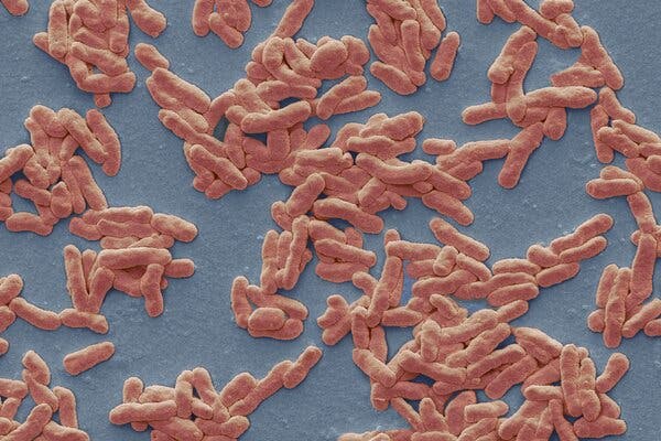
The NewsLeprosy, a fearsome scourge of ancient civilizations, may have become a permanent fixture in Florida, according to a new study.The authors described a 54-year-old man who was diagnosed with the illness but had no known risk factors and had never traveled outside Florida. Other people have similarly become infected without obvious explanation, suggesting that leprosy is now endemic in the state, the researchers said.Their report appeared in the journal Emerging Infectious Diseases.Still, there is no rising tide of leprosy in Florida. In the United States, the number of infections plummeted after peaking in 1983 but began a slow rise again about 20 years ago. The number of cases in the United States is fewer than 200 each year, and it is not rising.“It’s a drop in the bucket, especially when you view it through a global lens,” said Dr. Charles Dunn, a dermatologist and an author of the study.“Our paper simply highlights that there appears to be this really intriguingly strong geographic predilection for this illness that’s very uncommon,” he added.The Centers for Disease Control and Prevention “does not believe there is a great concern to the American public,” a spokeswoman said in an email. The number of cases “is very small.”Leprosy, also known as Hansen’s disease, is caused by slow-growing bacteria called Mycobacterium leprae. About 95 percent of people are genetically resistant to the bacteria.Steve Gschmeissner/Science SourceWhy It Matters: The disease can be treated if doctors recognize it.Leprosy, also known as Hansen’s disease, is caused by slow-growing bacteria called Mycobacterium leprae. About 95 percent of people are genetically resistant to the bacteria.There were 159 new cases in 2020, the most recent year for which national data are available. New cases are reported most commonly in Florida, California, Louisiana, Hawaii, New York and Texas. Central Florida accounts for 81 percent of the cases reported in that state.The bacteria are thought to be transmitted by droplets from the nose and mouth of an infected patient, but only after close, sustained contact. Armadillos famously carry the bacteria, and people may become infected through contact with the animals.Caught early enough, leprosy can be cured with standard antibiotic drugs taken over a year. Treatment can make patients noninfectious within a week.But, left untreated, the bacteria can damage nerves and lead to permanent disabilities including paralysis and blindness. The physical changes associated with the disease can also lead to the enduring stigmatization and isolation of infected people.“The fact that this patient had never traveled outside of the state of Florida was something that we just wanted to bring to light to those clinicians and physicians that are in the area,” said Dr. Rajiv Nathoo, a dermatologist and senior author on the study.What It Looks Like: Striking symptoms can take decades to develop.M. leprae may damage skin, peripheral nerves, the upper respiratory tract and the eyes.The disease starts with either discolored, numb patches on the skin or with tiny nodules under it. Early symptoms can easily be mistaken for other skin conditions like psoriasis or eczema. Symptoms can develop as many as 20 years after exposure, making it even more challenging to diagnose the disease.Left untreated, the bacteria slowly destroy nerves and muscles, leading to striking deformities in the hands and feet, sometimes referred to as claw hands and hammer toes.The disease was first described thousands of years ago. While it may seem like a thing of the past, roughly 200,000 new infections continue to crop up all over the world each year, with a majority in Southeast Asia and India, according to the World Health Organization.Eliminating the disease in some countries like India has proved to be much more challenging than public health officials expected.What Scientists Don’t Know: How some cases are acquired. Researchers have identified a second type of bacteria that leads to leprosy. Both pathogens are close cousins of the bacteria that cause tuberculosis.None of these bacterial species can easily be cultured in the lab, leaving many questions unanswered about the disease’s transmission and progression.New cases of leprosy were often diagnosed in people who had traveled to other parts of the world. But since 2015, more than one-third of the cases in the United States have been locally acquired.Many new patients report no travel or contact with armadillos that would explain their infection, according to the researchers.

Read more →