
Extreme Heat and Your Body: What Happens When It Gets Too Hot?
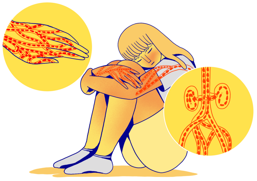
Heat is the leading cause of weather-related deaths in the United States. While the human body is able to withstand […]
Read more →
Heat is the leading cause of weather-related deaths in the United States. While the human body is able to withstand […]
Read more →
Research from experts at Michigan Medicine, the Children’s Hospital of Philadelphia and Penn Medicine is breaking ground on new ways of treating blood disorders, such as sickle cell anemia, through gene therapy.
To cure blood disorders, patients must undergo high dose chemotherapy and bone marrow transplantation. This requires a match between the recipient and donor immune system, but ~30% of patients do not have a match. Even when they do the donor immune system can attack the patient, graft versus host disease.
Gene therapy corrects the mutation in a patient’s own cells but still requires chemotherapy and transplantation of one’s own corrected cells. The new research shows that blood stem cells can be genetically engineered while still in the bone marrow, in a single treatment.
Co-first author Michael Triebwasser, M.D., Ph.D., clinical instructor in Pediatric Hematology and Oncology reported, “This is the first time the blood stem cells that create the blood and immune system over our lifetime can be genetically engineered while still in the bone marrow.
“This technology can be used to correct disease cause mutations such as the single mutation that causes sickle cell anemia in ~7.5 million people worldwide, and it can be used to control stem cells using messenger RNA (mRNA). To do this we utilized a type of nanoparticle similar to the Pfizer COVID mRNA vaccine but designed it to find these stem cells specifically.”
The risks patients undergo for gene therapy highlights the need for improved treatments. In addition, eliminating the need for stem cell collection and treatment outside the body can cut costs for patients and improve access to critical gene therapies for many patients.
The recently approved gene therapy for another blood disorder, beta-thalassemia, costs $2.8 million dollars.
“This approach is highly flexible and has reduced toxicity when treating stem cells outside the blood compared to current methods. It will hopefully lead to improved methods for correcting stem cells.
“The ultimate goal would be to do these same gene corrections while the stem cells remain in the body. This would open the door for cures in resource limited countries where the infrastructure for bone marrow transplantation is not present, and the cost is prohibitive.”
This research was supported by the National Institutes of Health (NIH grants 5T32HL007150 and 5T32HL007622), The Thomas B. and Jeannette E. Laws McCabe Fund at the University of Pennsylvania.
A scientific journey decades in the making at Duke University has found a new antibiotic strategy to defeat gram-negative bacteria like Salmonella, Pseudomonas and E. coli, the culprits in many urinary tract infections (UTIs). The synthetic molecule works fast and is durable in animal tests.
It works by interfering with a bacterium’s ability to make its outer lipid layer, its skin, so to speak.
“If you disrupt the synthesis of the bacterial outer membrane, the bacteria cannot survive without it,” said lead investigator Pei Zhou, a professor of biochemistry in the Duke School of Medicine. “Our compound is very good and very potent.”
The compound, called LPC-233, is a small molecule that has proven effective at wrecking the outer membrane lipid biosynthesis in every gram-negative bacterium it was tested against. Co-authors at the University of Lille in France tested it against a collection of 285 bacterial strains, including some that were highly resistant to commercial antibiotics, and it killed them all.
And it works fast. “LPC-233 can reduce bacterial viability by 100,000-fold within four hours,” Zhou said.
The compound is also tenacious enough to survive all the way to the urinary tract after oral administration, which may make it a vital tool against stubborn urinary tract infections (UTIs).
Tests run at high concentrations of the compound showed “exceedingly low rates of spontaneous resistance mutations in these bacteria,” according to a paper describing the findings, which appears Aug. 9 in Science Translational Medicine.
People who take acid reflux medications called proton pump inhibitors for four-and-a-half years or more may have a higher risk of dementia compared to people who do not take these medications, according to new research published in the August 9, 2023, online issue of Neurology®, the medical journal of the American Academy of Neurology. This study does not prove that acid reflux drugs cause dementia; it only shows an association.
Acid reflux is when stomach acid flows into the esophagus, usually after a meal or when lying down. People with acid reflux may experience heartburn and ulcers. People with frequent acid reflux may develop gastroesophageal reflux disease, or GERD, which can lead to cancer of the esophagus.
Proton pump inhibitors reduce stomach acid by targeting the enzymes in the stomach lining that produce that acid.
“Proton pump inhibitors are a useful tool to help control acid reflux, however long-term use has been linked in previous studies to a higher risk of stroke, bone fractures and chronic kidney disease,” said study author Kamakshi Lakshminarayan, MBBS, PhD, of the University of Minnesota School of Public Health in Minneapolis, and a member of the American Academy of Neurology. “Still, some people take these drugs regularly, so we examined if they are linked to a higher risk of dementia. While we did not find a link with short-term use, we did find a higher risk of dementia associated with long-term use of these drugs.”
The study included 5,712 people, age 45 and older, who did not have dementia at the start of the study. They had an average age of 75.
Researchers determined if participants took acid reflux drugs by reviewing their medications during study visits and during yearly phone calls. Of the participants, 1,490 people, or 26%, had taken the drugs. Participants were then divided into four groups based on whether they had taken the drugs and for how long, as follows: people who did not take the drugs; those who took the drugs for up to 2.8 years; those who took them for 2.8 to 4.4 years; and people who took them for more than 4.4 years.
Participants were then followed for a median duration of 5.5 years. During this time, 585 people, or 10%, developed dementia.
In the ongoing search for a cure for Alzheimer’s disease, a burgeoning branch of medicine is bringing new hope. Stem cell therapies are already being used to treat various cancers and disorders of the blood and immune system. In a new proof-of-concept study, scientists at University of California San Diego show stem cell transplants may also be a promising therapeutic against Alzheimer’s.
In the study, publishing this month in Cell Reports, the researchers demonstrate that transplanting hematopoietic stem and progenitor cells was effective in rescuing multiple signs and symptoms of Alzheimer’s in a mouse model of the disease. Mice that received healthy hematopoietic stem cells showed preserved memory and cognition, reduced neuroinflammation and significantly less β-amyloid build-up compared to other Alzheimer’s mice.
“Alzheimer’s is a very complex disease, so any potential treatment has to be able to target multiple biological pathways,” said senior study author Stephanie Cherqui, PhD, professor at UC San Diego School of Medicine. “Our work shows that hematopoietic stem and progenitor cell transplantation has the potential to prevent complications from Alzheimer’s and could be a promising therapeutic avenue for this disease.”
The success of the therapy stems from its effects on microglia, a type of immune cell in the brain. Microglia have been implicated in the initiation and progression of Alzheimer’s disease in various ways. It’s known that sustained microglia inflammation can contribute to Alzheimer’s, as the release of inflammatory cytokines, chemokines and complement proteins leads to increased β-amyloid production. In healthy conditions, microglia also play a major role in clearing β-amyloid plaques, but this function is impaired in Alzheimer’s. The resulting β-amyloid build-up also puts stress on other brain cells, including endothelial cells that affect blood flow to the brain.
Postdoctoral researcher and first author Priyanka Mishra, PhD, set out to test whether transplanting stem cells could lead to the generation of new, healthy microglia that might reduce the progression of Alzheimer’s disease. The Cherqui lab had already found success using similar stem cell transplants to treat mouse models of cystinosis, a lysosomal storage disease, and Friedreich’s ataxia, a neurodegenerative disease.
Mishra and her colleagues performed systemic transplantations of healthy wild-type hematopoietic stem and progenitor cells into Alzheimer’s mice and found that the transplanted cells did differentiate into microglia-like cells in the brain.
The researchers then evaluated the animals’ behavior and found that memory loss and neurocognitive impairment were completely prevented in mice that received the stem cell transplant. These mice showed better object recognition and risk perception, as well as normal anxiety levels and locomotor activity, compared to non-treated Alzheimer’s mice.
In Parkinson’s disease, growing evidence targets neuroinflammation as essential for brain pathogenesis. But which group of immune cells that reside in the brain direct this inflammatory response?
In a study published in Nature Communications, Ashley Harms, Ph.D., and colleagues at the University of Alabama at Birmingham, used a mouse model of Parkinson’s disease to show that border-associated macrophages — not microglia — mediate the neuroinflammatory response in the brain. Neuroinflammation has previously been shown to exacerbate neurodegradation in the mouse model of Parkinson’s.
In humans, abnormal accumulation of the protein alpha-synuclein results in the loss of brain neurons that produce the neurotransmitter dopamine. The mouse model — where human alpha-synuclein is overproduced in dopaminergic neurons — replicates many of the inflammatory features seen in human disease.
Microglia and border-associated macrophages, or BAMs, are two specific subsets of macrophage immune cells that reside in the brain. They are also known as central nervous system-resident macrophages, or CRMs. Macrophages are a type of white blood cell that engulf and digest “foreign” pathogens like cancer cells and bacteria. They then play a key role to stimulate the adaptive immune response for large-scale defense by presenting pieces of protein from the pathogen, known as antigens, on the surface of the macrophage. The T cells that are essential to the escalation of the immune response become activated when they interact with those presented antigens. The antigens are presented on the macrophage by a carrier protein called MHCII.
Previous studies had shown that MHCII has a pivotal role in Parkinson’s neuroinflammation and neurodegradation. However, identification of the specific cells presenting the antigen remained elusive. The UAB researchers showed that deleting MHCII from CRMs — which includes both microglia and BAMs — was neuroprotective against dopaminergic cell loss in the mouse model, meaning that antigen presentation specifically by CRMs is essential for the inflammation and subsequent neurodegradation induced by alpha-synuclein.
However, Harms and colleagues were surprised to find that deletion of MHCII specifically in microglia caused no reduction in neuroinflammation. Thus, they reasoned that some other non-microglial subset of CRMs has to be presenting the alpha-synuclein antigen to CD4+ T cells, thus producing neuroinflammation and the infiltration of peripheral immune cells into the brain from outside the central nervous system.
In contrast to the microglia experiment, they found that specific depletion of BAMs reduced multiple measures of neuroinflammation. “Together, these findings indicate that BAMs, not microglia, are required for peripheral immune cell recruitment into the brain parenchyma and antigen re-stimulation, a process responsible for alpha-synuclein-induced neurodegeneration,” said Harms, an associate professor in the UAB Department of Neurology.
What do superheroes Deadpool and Elastigirl have in common? Each was used in a college anatomy class to add relevance to course discussions — Deadpool to illustrate tissue repair and Elastigirl, aka Mrs. Incredible, as an example of hyperflexibility.
Instructors at The Ohio State University College of Medicine created a “SuperAnatomy” course in an attempt to improve the experience of undergraduate students learning the notoriously difficult — and for some, scary or gross — subject matter of human anatomy.
Surveys showed that most students who took the class found the use of superheroes increased their motivation to learn, fostered deeper understanding of the material, and made the content more approachable and enjoyable.
A few of the many content examples also included considering how Wolverine’s claws would affect his musculoskeletal system and citing Groot in a discussion of skin disorders.
The effort was aimed at bringing creativity to the classroom — in the form of outside-the-box instruction and as a way to inspire students’ imagination and keep them engaged, said Melissa Quinn, associate professor of anatomy at Ohio State and senior author of a study on the course’s effectiveness.
“In these introductory courses, it’s a little tougher to talk about clinical relevance because students don’t fully understand a lot of the mechanics,” Quinn said. “But if you bring in pop culture, which everybody is inundated with in some way, shape or form, and tie it to the foundational sciences, then that becomes a way to apply it a little bit more.”
The study was published recently in the journal Anatomical Sciences Education.
In a small, exploratory study, levels of certain types of microbes in babies’ guts were shown to be associated with performance in tests of early cognitive development. Sebastian Hunter of the University of British Columbia, Canada, and colleagues present these findings in the open-access journal PLOS ONE on August 9, 2023.
Mounting evidence has highlighted numerous ways in which the community of diverse microbes that naturally reside in the human gut — the microbiome — is connected to human health, including brain health. Several studies in animals and humans have hinted at connections between the microbiome and early-life brain development, but few have examined how differences in infants’ microbiomes might be associated with differences in their emerging cognitive abilities.
To help deepen understanding of these potential connections, Hunter and colleagues analyzed data from 56 infants aged four to six months. The infants had each completed at least one of three evaluations of various cognitive abilities, and the researchers evaluated their gut microbiomes using fecal samples.
They found that infants who succeeded at a test of social attention known as “point and gaze” — which measures the ability to share focus on an object with another person — tended to have higher amounts of microbes in the Actinobacteria phylum, the genus Bifidobacterium, and the genus Eggerthella, and lower amounts of microbes in the Firmicutes phylum, the Hungatella genus, and the Strepcococcus genus.
Meanwhile, electroencephalogram measurements of infants’ brain activity in response to hearing a steady beat showed that certain patterns of activity linked to better rhythmic processing were associated with higher or lower levels of certain microbe types, as well as with levels of certain metabolic chemical reactions involving microbes that prior studies have linked to brain and spinal cord development.
No links were found between the microbiome and measurements of blood flow in the infants’ brains in response to hearing recordings of both forward and backward human speech.
Overall, these findings are in line with the idea that the microbiome might influence early cognitive development, but more research is needed to confirm and clarify this role.
The authors add: “In our small pilot study, we observed interesting associations between the microbiome and brain function in early infancy. Further replication and research could be fruitful for understanding the role of the microbiome in early cognitive development.”
While engaging in cell division research, Silke Hauf and members of her lab made a surprisingly quiet discovery. When cells express RNA, there is always some fluctuation, or noise, in how much RNA is produced. Hauf’s group found several genes whose noise dips below a previously established threshold, known as the noise floor, during expression.
“We have solid data for this phenomenon,” said Hauf, associate professor in the Department of Biological Sciences at Virginia Tech. “There are some genes that are different and can have super low noise.”
Often upstaged by the more striking, well-publicized high-noise genes, Hauf and her team were intrigued by these ultra-low noise genes as they provide a window into the understanding of gene expression and gene expression noise.
This discovery, published in the journal Science Advances on Aug. 9, includes contributions from co-authors Abhyudai Singh, professor of electrical and computer engineering at the University of Delaware, and Ramon Grima, professor of computational biology at the University of Edinburgh. Both Singh and Grima are also mathematical biologists.
Cells will be cells
Hauf said the discovery’s importance lies in helping gain a basic understanding of how these cells do what they do. Cells can’t avoid making noise, but for them to function well, the noise needs to be minimized. She compared it with airports attempting to keep their flights on time in order to gain maximum functionality.”So it’s exciting to see that there are genes that operate with a minimum level of noise,” said Hauf. “Imagine there was a flight that always left within five minutes of the scheduled departure time. Wouldn’t you want to know how the airline does it?”
Opens the door to more discoveries
Hauf is excited about understanding how these cells express in such a quiet manner and learning more about the mechanisms behind it. She also would like to find other genes in this category.
“We saw these minimal fluctuations in one particular organism and cell type, but we really need to check other cells to determine if it is universal,” Hauf said.
This research has been funded by grants from the National Institute of General Medical Sciences, a unit within the National Institutes of Health, and Virginia Tech’s College of Science Lay Nam Chang Dean’s Discovery Fund.
Whether it’s work or play that prevents us from getting enough shut-eye during the week, assuming we can make up for it by sleeping in over the weekend is a mistake. New research led by Penn State reveals that cardiovascular health measures, including heart rate and blood pressure, worsen over the course of the week when sleep is restricted to five hours per night, and attempting to catch up on sleep over the weekend is insufficient to return these measures to normal.
“Only 65% of adults in the U.S. regularly sleep the recommended seven hours per night, and there’s a lot of evidence suggesting that this lack of sleep is associated with cardiovascular disease in the long term,” said Anne-Marie Chang, associate professor of biobehavioral health and co-author of the work, published in the journal Psychosomatic Medicine. “Our research reveals a potential mechanism for this longitudinal relationship, where enough successive hits to your cardiovascular health while you’re young could make your heart more prone to cardiovascular disease in the future.”
The team recruited 15 healthy men between the ages of 20 and 35 to participate in an 11-day inpatient sleep study. For the first three nights, the participants were allowed to sleep up to 10 hours per night to achieve a baseline sleep level. For the next five nights, the participants’ sleep was restricted to five hours per night, followed by two recovery nights, in which they were again allowed to sleep up to 10 hours per night. To evaluate the effects of this sleep regime on cardiovascular health, the researchers measured the participants’ resting heart rates and blood pressure every two hours during the day.
Chang explained that the team’s study is unique because it measured heart rate and blood pressure multiple times throughout the day for the duration of the study, which enabled them to account for any effects that time of day might have on heart rate and blood pressure. For example, heart rate is naturally lower upon waking than later in the day, so measuring heart rate multiple times throughout the day can account for this difference.
The team, which included David Reichenberger, lead author and graduate student in biobehavioral health, Penn State, found that heart rate increased nearly one beat per minute (BPM) with each successive day of the study. Specifically, the average baseline heart rate was 69 BPM, while the average heart rate by the end of the study on the second day of recovery was nearly 78 BPM. Systolic blood pressure also increased by about 0.5 millimeters of mercury (mmHg) per day. The average baseline systolic blood pressure was 116 mmHg and was nearly 119.5 mmHg by the end of the recovery period.
“Both heart rate and systolic blood pressure increased with each successive day and did not return to baseline levels by the end of the recovery period,” Reichenberger said. “So, despite having additional opportunity to rest, by the end of the weekend of the study, their cardiovascular systems still had not recovered.”
Chang noted that longer periods of sleep recovery may be necessary to recover from multiple, consecutive nights of sleep loss.
“Sleep is a biological process, but it’s also a behavioral one and one that we often have a lot of control over,” Chang said. “Not only does sleep affect our cardiovascular health, but it also affects our weight, our mental health, our ability to focus and our ability to maintain healthy relationships with others, among many other things. As we learn more and more about the importance of sleep, and how it impacts everything in our lives, my hope is that it will become more of a focus for improving one’s health.”
Other Penn State authors on the paper include Stephen Strayer, former graduate student in neuroscience; Margeaux Schade, assistant research professor of biobehavioral health; and Orfeu Buxton, Elizabeth Fenton Susman Professor of Biobehavioral Health. Kelly Ness, postdoctoral fellow, University of Washington, and Gina Marie Mathew, postdoctoral associate, Stony Brook University, also are authors.