Childhood plastic exposure could be fueling obesity, infertility, and asthma

Childhood exposure to chemicals used to make plastic household items presents growing health risks that can extend long into adulthood, experts from NYU Langone Health report.
This is the main conclusion after a review of hundreds of the latest studies on the topic, publishing online Sept. 21 in the journal The Lancet Child & Adolescent Health.
The article is being released to coincide with a gathering of experts the same week in New York City to discuss the global impact of plastics on human health.
In their report, the authors outline decades of evidence that substances often added to industrial and household goods may contribute to disease and disability, particularly when they are encountered early in life. The review focuses on three classes of chemical — phthalates used to make plastic flexible, bisphenols, which provide rigidity, and perfluoroalkyl substances (PFAS), which help materials resist heat and repel water.
The results of the studies, which together assessed thousands of pregnant mothers, fetuses, and children, tied these toxins to a wide range of long-term health concerns, including heart disease, obesity, infertility, and asthma.
“Our findings point to plastic’s role in the early origins of many chronic diseases that reverberate into adolescence and adulthood,” said study lead author and pediatrician Leonardo Trasande, MD, MPP. “If we want kids to stay healthy and live longer, then we need to get serious about limiting the use of these materials,” added Trasande, the Jim G. Hendrick, MD, Professor of Pediatrics at NYU Grossman School of Medicine.
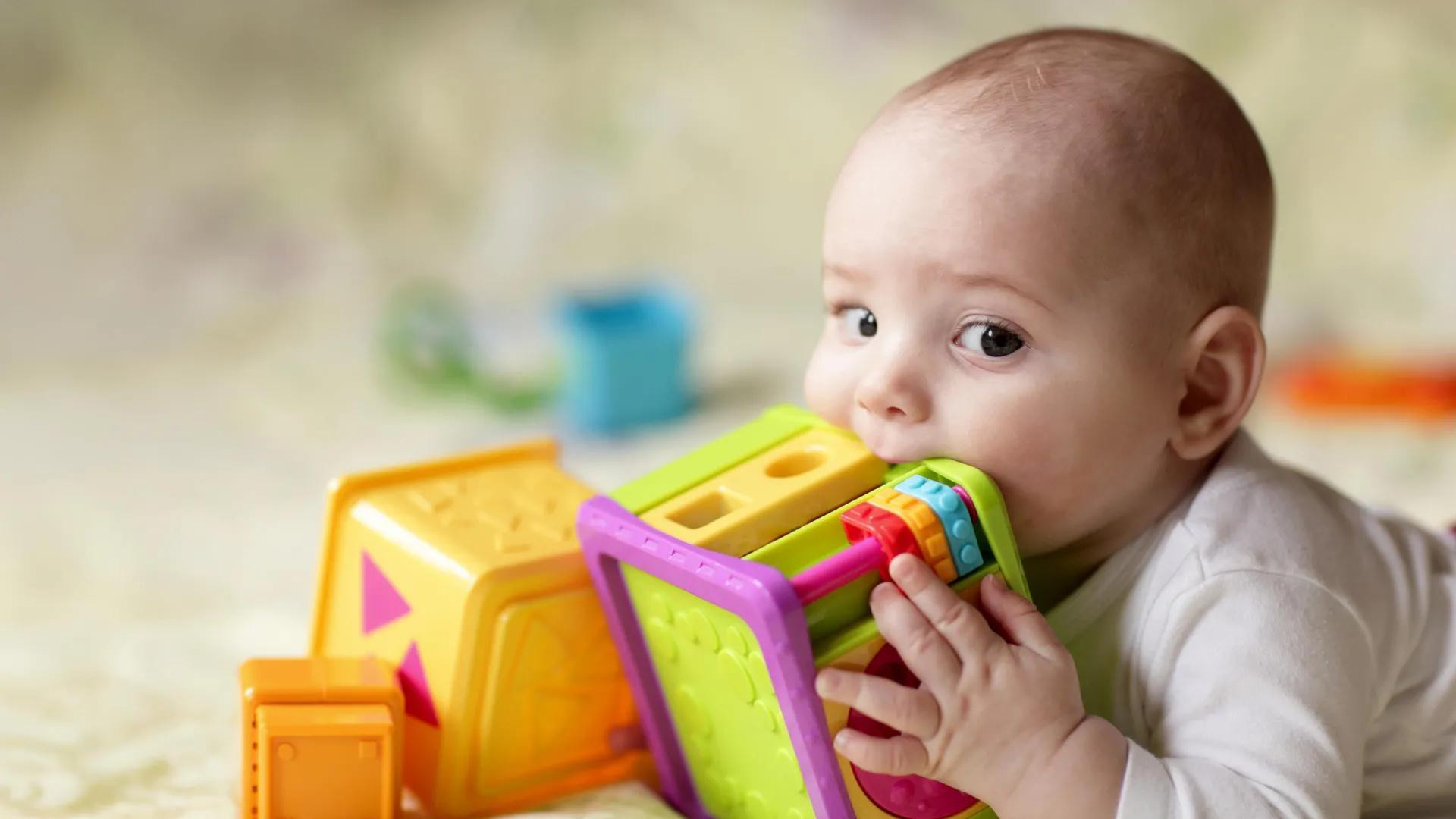
The chemicals are found in a range of items, such as food packaging, cosmetics, and paper receipts, notes Trasande, who is also a professor in the Department of Population Health. Experts have found that as plastics are used, heated, or chemically treated, microplastic and nanoparticles are released and become ingested.

Chemicals used in plastic materials have been shown to prompt an overactive immune response (inflammation) throughout the body’s tissues as well as disrupt the function of hormones that influence many bodily processes. The substances are also believed to affect brain development, with numerous studies linking early-life exposure to IQ loss and neurodevelopmental issues such as autism and attention deficit hyperactivity disorder.
The Lancet review also explored strategies to reduce the use of plastic and to help safeguard human health.
“There are safe, simple steps that parents can take to limit their children’s plastic exposure without breaking the bank,” said Trasande, who serves as director of NYU Grossman School of Medicine’s Division of Environmental Pediatrics and NYU Langone Health’s Center for the Investigation of Environmental Hazards.
Replacing plastic containers with glass or stainless steel, and avoiding microwaving and dishwashing plastic, have proved helpful, he says.
Trasande adds that by offering clear guidance, health care providers can empower parents to make informed decisions about the products they use and steer them toward safer options. He also suggests that clinicians partner with schools and community organizations to engage younger generations about the health risks of plastic exposure.
At the policy level, the researchers call for stricter regulatory measures to reduce the use of nonessential plastic items, particularly in low-income communities with profound heath disparities.

Their review comes on the heels of the most recent round of negations for the United Nations’ Global Plastics Treaty, which took place in Geneva last month. The developing treaty represents an international effort to tackle plastic pollution, with more than 100 countries calling for legally binding caps on production.
According to Trasande, the findings in the article support the urgent need for a strong agreement to help protect not only the environment but human health as well.
He notes that while the economic value of the plastics industry is commonly raised as a barrier to enacting regulations, the resulting health care costs from exposure are enormous, with his research estimates reaching roughly $250 billion per year in the United States alone.
The Global Plastics Treaty will be part of the discussion during NYU Langone Health’s 2025 Plastics, Human Health, and Solutions Symposium. At the event, experts will discuss the latest research on the health impact of microplastics, recent policy developments, and the critical role of regulations in addressing this public health crisis.
Despite its health risks, plastic can play an essential role in pediatric medicine, such as its use in ventilators and feeding tubes for premature infants, nebulizers for children with asthma, and masks that help prevent the spread of infection. The findings, the researchers say, do not challenge the need for the material in health care but instead highlight the dangers of its unnecessary use elsewhere.
The symposium will be held at NYU Langone Health on Sept. 22. The event will also be live streamed on YouTube for registered attendees.
Funding for the study was provided by National Institutes of Health grants R01ES022972, R01ES029779, R01ES032214, R01ES034793, and P2CES033423. Further funding was provided by several Argentinian foundations as well as the Science Fund of the Republic of Serbia.
Along with Trasande, Marina Olga Fernandez, PhD, at the National Council for Scientific and Technical Research in Buenos Aires, Argentina, serves as study senior author.
Another study co-investigator is Aleksandra Buha Đorđević, PhD, at the University of Belgrade in Serbia.

Read more →

Sneezing from cats or dust? Safe UV light may neutralize allergens in minutes

Cats. Dust mites. Mold. Trees.
For people with allergies, even a brief whiff of the airborne allergens these organisms produce can lead to swollen eyes, itchy skin and impaired breathing.
Such allergens can persist indoors for months after the original source is gone, and repeated exposure can exacerbate, and even lead to, asthma.
What if you could just flip a switch and disable them? You can, according to new University of Colorado Boulder research.
“We have found that we can use a passive, generally safe ultraviolet light treatment to quickly inactivate airborne allergens,” said study author Tess Eidem, a senior research associate in the Department of Civil, Environmental and Architectural Engineering.
“We believe this could be another tool for helping people fight allergens in their home, schools or other places where allergens accumulate indoors.”
The findings were published in August in the journal ACS ES&T Air.

Why you can’t kill an allergen
Walk into a room with a cat and, if you sneeze, it’s not actually the cat you are reacting to. It’s likely airborne flecks of a protein called Fel d1 produced in their saliva. The protein spreads when they lick themselves and ends up in microscopic flakes of dead skin floating in the air, a.k.a. dander. When we inhale these particles, our immune system produces antibodies that bind to the protein’s unique 3D structure, kicking off an allergic reaction.
Dogs, mice, dust mites, mold and plants all emit their own unique proteins, with their own unique structure. Unlike bacteria and viruses, these allergens can’t be killed because they were never alive.
“After those dust mites are long gone, the allergen is still there,” said Eidem. “That’s why, if you shake out a rug, you can have a reaction years later.”
Standard methods of reducing allergens — like vacuuming, washing walls, using an air filter and regularly bathing pets — can work OK but are hard to maintain long-term studies show.
Eidem and co-authors Mark Hernandez, a professor of Civil, Environmental and Architectural Engineering, and Kristin Rugh, a microbiologist in the lab, sought a simpler way.

Instead of eliminating the proteins that cause allergies, they sought to change their structure — much like unfolding an origami animal — so the immune system wouldn’t recognize them.
“If your immune system is used to a swan and you unfold the protein so it no longer looks like a swan, you won’t mount an allergic response,” explained Eidem.
UV light, their study suggests, can do that.
Let there be light
Previous research has shown that UV light can kill airborne microorganisms, including the virus that causes COVID-19.
It’s already used widely to disinfect equipment in hospitals, airports and elsewhere, but the bandwidth is typically so strong (a wavelength of 254 nanometers) that users must wear protective equipment to prevent damage to skin and eyes.
Eidem used 222-nanometer-wavelength lights, a less-intense alternative considered safe for occupied spaces because it doesn’t penetrate deep into cells. (It does not come entirely without risks, including ozone production, she notes, so exposure should be limited.)
The team pumped microscopic aerosolized allergens from mites, pet dander, mold and pollen into an unoccupied and sealed 350-cubic-foot chamber. Then they switched on four lunchbox-sized UV222 lamps on the ceiling and floor.
When they sampled the air at 10-minute intervals and compared it to untreated, allergen-filled air via laboratory tests, they saw significant differences. In the treated samples, immunorecognition was reduced, meaning the antibodies no longer recognized many of the proteins and stuck to them.
After just 30 minutes, airborne allergen levels effectively decreased by about 20% to 25% on average, the study showed.
“Those are pretty rapid reductions when you compare them to months and months of cleaning, ripping up carpet, and bathing your cat,” said Eidem.
A portable allergy buster?
UV222 lights are already commercially available, mostly for industrial antimicrobial uses.
But Eidem envisions a day when companies could engineer portable versions for people to switch on when they visit a friend with a pet or clean out a dusty basement.
UV222 systems could also potentially protect workers frequently exposed to allergens, such as those who work around live animals or in cannabis grow houses where, her own research shows, allergic reactions can be deadly.
One-in-three adults and children in the United States have allergies, according to the Centers for Disease Control. Eidem hopes her research, and more to come, can provide them with some relief — or even save lives.
“Asthma attacks kill about 10 people every day in the United States, and they are often triggered by airborne allergies,” she said. “Trying to develop new ways to prevent that exposure is really important.”

Read more →

A hormone that silences the immune system may unlock new cancer treatments

Researchers at UT Southwestern Medical Center have discovered how a hormone interacts with a receptor on the surface of immune cells to shield cancer cells from the body’s natural defenses. The findings, published in Nature Immunology, could lead to new immunotherapy approaches for treating cancer as well as potential treatments for inflammatory disorders and neurologic diseases.
“Myeloid cells are among the first group of immune cells recruited to tumors, but very quickly these tumor-fighting cells turn into tumor-supporting cells. Our study suggests that receptors on these myeloid cells get stimulated by this hormone and end up suppressing the immune system,” said Cheng Cheng “Alec” Zhang, Ph.D., Professor of Physiology and a member of the Harold C. Simmons Comprehensive Cancer Center at UT Southwestern. Dr. Zhang co-led the study with first author Xing Yang, Ph.D., a postdoctoral researcher in the Zhang Lab.
Current immunotherapies, such as immune checkpoint inhibitors, are effective for only about 20%-30% of cancer patients, Dr. Zhang said, suggesting that there are multiple ways that cancers evade attack from the immune system.
Several years ago, researchers in the Zhang Lab studying cancer-fighting immune cells called myeloid cells identified an inhibitory receptor called LILRB4. Stimulating this receptor blocked the myeloid cells’ ability to attack tumors.
Dr. Zhang, Dr. Yang, and their colleagues then did a genome-wide screen of all proteins that might interact with LILRB4. A promising hit was a hormone called SCG2. Although researchers have suggested that SCG2 plays a role in immune response, its function and receptor were unknown. Laboratory experiments confirmed that SCG2 binds to LILRB4, kicking off a signaling cascade that turned off the cancer-fighting abilities of myeloid cells and inhibited their ability to recruit cancer-fighting T cells to tumors.
In mice genetically altered to express the human form of LILRB4, injected cancer cells that produced SCG2 grew rapidly as tumors. Treating these mice with an antibody that blocks LILRB4 significantly slowed cancer growth, as did artificially ridding the animals’ bodies of SCG2.
Together, these experiments suggest that interactions between LILRB4 and SCG2 allow cancer to grow unchecked by myeloid cells, T cells, and potentially other immune cell types. Dr. Zhang suggested that disrupting this interaction could someday offer a new immunotherapy option to treat cancer. Conversely, because this interaction neutralizes myeloid cells’ immune activity, delivering extra SCG2 could be a promising treatment for autoimmune or inflammatory disorders spurred by myeloid cells. Dr. Zhang and his colleagues plan to investigate both ideas in future studies.

Other UTSW researchers who contributed to this study include Xuewu Zhang, Ph.D., Professor of Pharmacology and Biophysics; Cheryl Lewis, Ph.D., Associate Professor in the Simmons Cancer Center and of Pathology; Lin Xu, Ph.D., Assistant Professor in the Peter O’Donnell Jr. School of Public Health and of Pediatrics; Jingjing Xie, Ph.D., Instructor of Physiology; Qi Lou, Ph.D., Assistant Instructor of Physiology; Lei Guo, Ph.D., Computational Biologist; and Meng Fang, Ph.D., Chengcheng Zhang, Ph.D., Ankit Gupta, Ph.D., and Lianqi Chen, Ph.D., postdoctoral researchers.
Dr. Alec Zhang holds the Hortense L. and Morton H. Sanger Professorship in Oncology and is a Michael L. Rosenberg Scholar in Medical Research. Dr. Xuewu Zhang and Dr. Xu are members of the Simmons Cancer Center.
This study was funded by grants from the National Cancer Institute (NCI) (R01CA248736, R01CA263079, and Lung Cancer 779 SPORE Development Research Program), the Cancer Prevention and Research Institute of Texas (RP220032, RP15150551, RP190561), The Welch Foundation (AU-0042-20030616, I-1702), Immune-Onc Therapeutics Inc. (Sponsored Research Grant No. 111077), the National Institutes of Health (R35GM130289), and NCI Cancer Center Support Grant (P30CA142543).
The University of Texas has a financial interest in Immune-Onc in the form of equity and licensing. Dr. Alec Zhang holds equity in and had sponsored research agreements with Immune-Onc.

Read more →

When cancer cells feel squeezed, they become more dangerous

Cancer cells are notoriously flexible, taking on new features as they move around the body. Many of these changes are due to epigenetic modifications, which influence how DNA is packaged, and not due to mutations in the DNA itself. Such modifications are difficult to target for cancer therapy because they are reversible and can flip on and off.
Epigenetic changes have traditionally been thought to arise from internal cellular processes that result in the chemical tagging of DNA and its histone protein packaging — such as histone methylation or DNA acetylation. But now a new study led by Ludwig Oxford’s Richard White and Miranda Hunter of the Memorial Sloan Kettering Cancer Center and reported in the current issue of Nature shows that the physical environment in which these cells land is also a key instigator of epigenetic transformation.
Using a zebrafish model of melanoma, White, Hunter and their colleagues show that when tumor cells are tightly confined by surrounding tissues, they undergo structural and functional changes. Rather than continuing to divide rapidly, the cells activate a program of ‘neuronal invasion’, enabling them to migrate and spread into the surrounding tissue.
At the center of this transformation is HMGB2: a DNA-bending protein. The study demonstrates that HMGB2 responds to the mechanical stress of confinement by binding to chromatin, altering how genetic material is packaged. This exposes regions of the genome linked to invasiveness, making them newly available for gene expression. As a result, cells with high levels of HMGB2 become less proliferative but more invasive and resistant to treatment.
The team also found that melanoma cells adapt to this external pressure by remodeling their internal skeleton, forming a cage-like structure around the nucleus. This protective shield involves the LINC complex, a molecular bridge that connects the cell’s skeleton to the nuclear envelope, helping to protect the nucleus from rupture and DNA damage caused by confinement-induced stress.
“Cancer cells can rapidly switch between different states, depending on cues within their environment,” White explained. “Our study has shown that this switch can be triggered by mechanical forces within the tumor microenvironment. This flexibility poses a major challenge for treatment, as therapies targeting rapidly dividing cells may miss those that have transitioned to an invasive, drug-resistant phenotype. By identifying the factors that are involved in this switch, we hope to able to develop therapies that prevent or even reverse the invasive transformation.”
The findings highlight the role of the tumor microenvironment in shaping cancer cell behavior, showing how physical cues can drive cells to reorganize their cytoskeleton, nucleus and the architecture of their genomic packaging to shift between states of growth and invasion.
Most notably, however, the study also demonstrates how physical stress can act as a potent — and underappreciated — driver of epigenetic change.
This study was supported by the Ludwig Institute for Cancer Research, National Cancer Institute, the Cancer Research Society, the Canadian Institutes of Health Research, the U.S. National Institutes of Health, the Melanoma Research Alliance, The Debra and Leon Black Family Foundation, the Pershing Square Sohn Foundation, The Mark Foundation, The Alan and Sandra Gerry Metastasis Research Initiative at MSKCC, The Harry J. Lloyd Foundation, Consano, the Starr Cancer Consortium and the American Cancer Society.
Richard White is a member of the Oxford Branch of the Ludwig Institute for Cancer Research and a professor of genetics at the University of Oxford, Nuffield Department of Medicine.

Read more →

Trump will reportedly link autism to pain reliever Tylenol – but many experts are sceptical

Trump officials are expected to link the use of pain reliever Tylenol in pregnant women to autism, according to US media reports. At an Oval Office event on Monday, the US president will reportedly advise pregnant women in the US to only take Tylenol, known as paracetamol elsewhere, to relieve high fevers.At the Charlie Kirk memorial service on Sunday, Trump said he had an “amazing” announcement coming on autism, saying it was “out of control” but they might now have a reason why.Some studies have shown a link between pregnant women taking Tylenol and autism, but these findings are inconsistent and do not prove the drug causes autism. Tylenol is a popular brand of pain relief medication sold in the United States, Canada and some other countries. Its active ingredient is acetaminophen, which is called paracetamol outside North America.Tylenol maker Kenvue has defended the use of the drug in pregnant women.In a statement to the BBC, it said: “We believe independent, sound science clearly shows that taking acetaminophen does not cause autism. We strongly disagree with any suggestion otherwise and are deeply concerned with the health risk this poses for expecting mothers.”Acetaminophen is the safest pain reliever option for pregnant women, it added, and without it, women face a dangerous choice between suffering through conditions like fever or use riskier alternatives.The BBC has contacted the Department of Health and Human Services (HHS) for comment. In April, the leader of HHS, Robert F Kennedy Jr, pledged “a massive testing and research effort” to determine the cause of autism in five months.But experts have cautioned that finding the causes of autism – a complex syndrome that has been researched for decades – would not be simple. The widely held view of researchers is that there is no single cause of autism, which is thought to be the result of a complex mix of genetic and environmental factors.The American College of Obstetrics and Gynecology said doctors across the country have consistently identified Tylenol as one of the only safe pain relievers for pregnant women.”[S]tudies that have been conducted in the past, show no clear evidence that proves a direct relationship between the prudent use of acetaminophen during any trimester and fetal developmental issues,” the group has said. The drug is recommended by other major medical groups as well as other governments around the world. In August, a review of research led by the dean of Harvard University’s Chan School of Public Health found that children may be more likely to develop autism and other neurodevelopmental disorders when exposed to Tylenol during pregnancy. The researchers argued some steps should be taken to limit use of the drug, but said the pain reliever was still important for treating maternal fever and pain, which can also have negative effects for children. But another study, published in 2024, found no relationship between exposure to Tylenol and autism. “There is no robust evidence or convincing studies to suggest there is any causal relationship,” said Monique Botha, a professor in social and developmental psychology at Durham University. Dr Botha added that pain relief for pregnant women was “woefully lacking”, with Tylenol being one of the only safe options for the population. Autism diagnoses have increased sharply since 2000, and by 2020 the rate among 8-year-olds reached 2.77%, according to the US Centers for Disease Control and Prevention (CDC).Scientists attribute at least part of the rise to increased awareness of autism and an expanding definition of the disorder. Researchers have also been investigating environmental factors.In the past, Kennedy has offered debunked theories about the rising rates of autism, blaming vaccines despite a lack of evidence.

Read more →

Want a younger brain? Harvard researchers say eat like this

Following a green-Mediterranean diet — which includes green tea and the aquatic plant Mankai — is associated with slower brain aging, according to a study.
The study, published recently in the journal Clinical Nutrition, was co-authored by researchers at Ben-Gurion University, Harvard T.H. Chan School of Public Health, and the University of Leipzig.
Neurological conditions, including mild cognitive impairment and Alzheimer’s disease, have been associated with a higher brain age gap — a brain that’s “older” than would be predicted given a person’s chronological age. To evaluate the impact of diet on brain age, researchers analyzed data from around 300 participants in the DIRECT PLUS trial, one of the longest-running studies on the link between brain and diet. Over the course of 18 months, the participants consumed one of three diets: a standard healthy diet; a traditional calorie-restricted Mediterranean diet, which was low in simple carbohydrates, rich in vegetables, and replaced red meat with poultry and fish; and the green-Mediterranean diet, which additionally included green tea and Mankai.
When the researchers measured protein levels in the participants’ blood, they found that higher levels of certain proteins were associated with accelerated brain aging. Further, they found that those protein levels decreased in participants who followed the green-Mediterranean diet. The researchers hypothesized that the protective effect of the diet could be a result of the anti-inflammatory molecules contained in green tea and Mankai.
“Studying the circulating proteins in blood allows us to observe, in a real-life setting, how the brain’s aging processes are influenced by lifestyle and dietary changes,” said Anat Meir, postdoctoral research fellow at Harvard Chan School, who co-led the study. “This approach gives us a dynamic window into brain health, helping to reveal biological changes long before symptoms may appear. By mapping these protein signatures, we gain powerful new insight into how interventions, such as diet, may help preserve cognitive function as we age.”
Senior author of the study was Iris Shai, adjunct professor of nutrition at Harvard Chan School. Other Harvard Chan School co-authors included Dong Wang, Frank Hu, and Meir Stampfer.

Read more →

Lib Dems want health warnings on teens’ social media

1 hour agoShareSavePaul SeddonPolitical reporterShareSaveGetty ImagesThe Liberal Democrats say children using social media apps should be shown cigarette-style health warnings to help protect their mental health.The party says the move would protect youngsters from “addictive algorithms”, citing concerns over excessive phone usage.It also wants a two-hour “doomscrolling cap” limiting how long under-18s can access TikTok-style video clips.The party says its own research has shown excessive social media use can lead to anxiety, sleep disruption and shortened attention spans.Announcing the plan at the party’s autumn conference in Bournemouth, technology spokesperson Victoria Collins said excessive social media use brings well-documented risks, “just like cigarettes or alcohol”.Adding that the issue was a public heath “crisis,” she said social media apps had become “more and more addictive” for children.She added that mandatory health alerts would not prevent teenagers using social media or connecting with friends online, but would “ensure they go into the experience with their eyes open to the risks”.”When we pick up a pack of cigarettes or a bottle of wine, we expect to be told about the harm those products will pose to our health,” she argued.”So why is social media, the key driver of a crisis in young people’s mental health, any different?”Graphic pictures to illustrate the dangers of smoking have been mandatory on cigarette packets in the UK since 2008.Alcoholic drinks must display the volume of alcohol, whilst the units and warnings against drinking while pregnant are often displayed under a series of voluntary industry schemes.The government is considering making these mandatory as part of its 10-year NHS plan, along with dedicated alcohol “health warning messages”.The Liberal Democrats are pointing to proposals for social media suggested last year by Vivek Murthy, who was the most senior health adviser in the Biden administration.He had suggested that under-18s accessing social media platforms should be referred to research on the mental health impact of using the apps. Smartphone banChildren aged eight to 17 spend between two and five hours online per day, according to Ofcom research.Children have to be at least 13 to create their own social media profile, with stricter rules on age checks coming into force in July. The new regulations also introduced new rules requiring platforms to change the algorithms which determine what is shown in children’s feeds.The Liberal Democrats have previously pushed unsuccessfully to raise the age at which tech companies can collect children’s data from 13 to 16. The Labour government is currently considering a two-hour cap on the use of individual social media apps for children, along with a 22:00 curfew.But in her speech, Collins said that ministers “make noise” about online safety but had yet failed to deliver on promises of stronger time curbs.Ministers have also previously faced pressure from the Conservatives to legislate for an England-wide ban on smartphones in schools.Labour has so far stopped short of legislating for such a ban, although it has said it is reviewing guidance that allows individual headteachers to ban the devices.

Read more →

Why alcohol blocks the liver from healing, even after you quit

Excessive alcohol consumption can disrupt the liver’s unique regenerative abilities by trapping cells in limbo between their functional and regenerative states, even after a patient stops drinking, researchers at University of Illinois Urbana-Champaign and collaborators at Duke University and the Chan Zuckerberg Biohub Chicago describe in a new study.
This in-between state is a result of inflammation disrupting how RNA is spliced during the protein-making process, the researchers found, providing scientists with new treatment pathways to explore for the deadly disease. The researchers published their findings in the journal Nature Communications.
The liver has a remarkable ability to regenerate itself after damage or partial removal. However, it loses that ability in patients with alcohol-associated liver disease — the leading cause of liver-related mortality worldwide, resulting in roughly 3 million deaths annually.
“We knew that the liver stops functioning and stops regenerating in patients with alcohol-related hepatitis and cirrhosis, even when a patient has discontinued consuming alcohol, but we didn’t know why,” said U. of I. biochemistry professor Auinash Kalsotra, who co-led the study with Duke University School of Medicine professor Anna Mae Diehl. “The only real life-saving treatment option once a patient reaches the liver failure stage in those diseases is transplantation. But if we understood why these livers were failing, maybe we could intervene.”
Both the Kalsotra and Diehl labs havestudied the molecular and cellular underpinnings of liver regeneration. Over the last five years, they found that in order to regenerate, liver cells reprogram their gene expression to revert to fetal-like progenitor cells, multiply and then reverse the process back to become mature functioning cells again. Armed with this knowledge, the group turned to the question of how those mechanisms were disrupted in alcohol-associated liver disease.
The researchers compared samples of healthy livers and samples of livers with alcohol-associated hepatitis or cirrhosis obtained from Johns Hopkins University Hospital through an initiative supported by the National Institute for Alcohol Abuse and Alcoholism, part of the National Institutes of Health.
The first thing the researchers noticed in diseased livers was that, although damaged cells had begun the process of reverting to the regenerative state, they did not complete the process and instead remained in transitional limbo.

“They are neither functional adult cells nor proliferative progenitor cells. Since they are not functioning, more pressure builds on the remaining cells. So they try to regenerate, and they’re all ending up in this unproductive quasi-progenitor state, and that’s what is causing liver failure,” said U. of I. graduate students Ullas Chembazhi and Sushant Bangru, the co-first authors of the study.
To figure out why the cells were getting stuck in this state, the team investigated which proteins were being made by the liver cells and, in turn, the RNA molecules carrying the instructions for those proteins from the DNA to the cell’s protein-building machinery.
While most studies focus only on the total amounts of RNA or protein in a cell, Kalsotra’s team used deep RNA sequencing technology and computational analyses to zoom in on the splicing of RNA fragments, a key step in stitching together different parts of genetic instructions to make proteins.
“In comparing the samples, we saw RNA was getting misspliced broadly in alcohol-related liver disease, across thousands of genes, and it was affecting major functions of proteins,” said Kalsotra, who also is affiliated with the Carl R. Woese Institute for Genomic Biology at Illinois.
The researchers found a possible driver of the RNA missplicing: Alcohol-damaged liver cells had a deficiency of the protein ESRP2, which binds to RNA to splice it properly.
“Proteins function at a very specific place in the cell, and that is directed by sequences within the protein that take the protein to that particular spot. We found that, in many cases, the sequence that dictates where the protein localizes within a cell was misspliced. That’s why it was important that we did the multiple analyses we did,” said Kalsotra, also a member of the Chan Zuckerberg Biohub Chicago. “There was the same amount of RNA and protein, but the protein was not at the right place to function. Due to missplicing, key proteins that are required for productive liver regeneration were getting stuck in the cytoplasm, when they needed to be in the nucleus.”
To verify that ESRP2 deficiency was a likely culprit, the researchers studied mice without the gene that produces ESRP2. They displayed similar liver damage and regeneration failure to that seen in patients with advanced alcohol-related hepatitis.

But why was ESRP2 missing from liver cells from patients with alcohol-related hepatitis? Upon investigation, the researchers found that liver support cells and immune cells, drawn to the liver tissue damaged by alcohol processing, released high amounts of inflammatory and growth factors. Those factors suppress ESRP2 production and activity.
To verify this finding, the researchers treated liver cell cultures with a molecule that inhibits the receptor for one of the inflammation-promoting factors. ESRP2 levels recovered and splicing activity was corrected, pointing to the pathway as a possible treatment target.
“I’m hopeful these findings will become a launching pad for future clinical studies. We can use these misspliced RNAs as diagnostic markers or develop treatments that can curb the inflammation. And if we can correct the splicing defects, then maybe we can improve recovery and restore damaged livers,” Kalsotra said.
The research team also included U. of I. biochemistry graduate students Diptatanu Das and Subhashis Natua; U. of I. undergraduate students Katelyn Toohill, Ishita Purwar, and Anuprova Bhowmik; Brandon Peiffer and Zhaoli Sun from Johns Hopkins University School of Medicine; Aurelia Leona and Yogesh Goyal from Northwestern University and Rajesh Dutta from Duke University School of Medicine.
The National Institutes of Health, the Chan-Zuckerberg Biohub Chicago, the Duke Endowment and the Muscular Dystrophy Association supported this work.
The National Institutes of Health supported this work through grants R01-AA010154, R01-HL126845, R21-HD104039, R01-AA010154, 5R01-DK077794, 1R56-DK1343340 and R24 AA025017.

Read more →

‘My girl can’t walk or talk’: Searching for answers in Wales’ rare disease cluster

9 hours agoShareSaveOscar EdwardsBBC WalesShareSaveRachel Cowley’s daughter Jessica was like any other 10-year-old until she started having seizures.Now aged 20, Jessica is unable to walk or talk, and Ms Cowley fears she will die in the next few years.”I hope when the time comes it is quick,” said Ms Cowley. “But I just don’t think it’s going to be.”Jessica is one of a “cluster” of people in south Wales affected by Dentatorubral-pallidoluysian atrophy, or DRPLA, an inherited and progressive neurological disorder for which there is no known treatment or cure.DRPLA was believed to mainly affect people of Japanese origin and considered rare in the UK, until a 2007 research study found a relatively high prevalence of the condition in Wales.The symptoms of DRPLA can vary significantly, but generally include progressive memory loss and personality change, impaired control of body movements, muscle spasms, seizures and psychiatric disturbances.It is caused by a gene defect, and a person with the condition has a 50% chance of passing it on to a child of their own.Dr Mark Wardle, a consultant neurologist who authored the 2007 study, said there were a number of “common founders” with DRPLA in south Wales which had resulted in the current “cluster of patients in their 20s or 30s”.”It doesn’t take many generations to have very many more cases just from the most common founders,” he added.Rachel CowleyMs Cowley said Jessica was initially diagnosed with epilepsy aged 10, when she began having seizures.”We could never control it,” said Ms Cowley, 45, from Pontypridd, Rhondda Cynon Taf.But when Jessica turned 13, Ms Cowley said her daughter’s legs began to “give way”.The teenager, who previously loved doing gymnastics and horse riding, was now struggling to walk.”I noticed that she could hardly walk up the stairs,” said Ms Cowley.”When she lost her voice, I just thought, these ain’t signs of epilepsy, there’s got to be something else.”After “years of wondering” why her health was deteriorating, Jessica was diagnosed with DRPLA in May.Rachel CowleyJessica now uses a wheelchair and is completely non-verbal.”It’s quite scary because she’s been going through this for the last five to seven years,” said Ms Cowley.”I would speak to the consultant and they just did not know why she was deteriorating like she was.”I think it’s because of this disease being so rare.”Ms Cowley is part of a group called DRPLA in south Wales on Facebook, which currently has more than 100 members.Ms Cowley said it was a “massive surprise” to find other people in her area were facing the same challenges.”We’re all connected in a way,” she said. “It’s nice to talk to some of them and have advice off them, because if I phone the GP, they don’t know nothing about it.”Zoe Hoes adopted her eight-year-old daughter Gabriella when she was a baby.Last year, she was contacted by social services who said that a member of Gabriella’s birth family had tested positive for DRPLA.Ms Hoes, 47, from Cwmbran, Torfaen, noticed her daughter was twitching at night and thought she might have symptoms.”I managed to get a private test done and that confirmed that she did have it,” Ms Hoes added.She said she was left “scrambling to find out information” about the condition.”It’s terrible because at the moment there is no treatment and no cure. “You love them so much and you don’t want to lose them.”Ms Hoes said the private test cost her “in the region of £2,000″ which she acknowledged was unaffordable for many.”A lot of families haven’t got that money to spend,” she said.”So they just carry on with their lives, and anyone’s child could meet someone with [DRPLA] and end up being a carer for their partner and children.”Zoe HoesMs Hoes would like to see more people being tested for the condition at a younger age. “[Parents] need to be able to make plans for those children’s futures because, ultimately, they’re going to be caring for them,” she said.Dr Wardle began his research study into the DRPLA prevalence in south Wales after finding “several families” with the condition.He said the DRPLA gene, also known as the ATN1 gene, was crucial in understanding the disorder.If the gene expands in a large way “over multiple generations” a person will “probably have a more aggressive disease than [their] parent”, explained Dr Wardle.He said the current generation of DRPLA patients had, in this way, experienced “more severe disease”.”That’s why it feels as if we’ve suddenly got more patients,” he said.”When I first found patients with this disease in south Wales, they were in their 50s or 60s.”Now we’ve got a cluster of patients in their 20s or 30s and that’s why we’re seeing that difference.”Dr Wardle said it was “very difficult” to diagnose DRPLA because it was “astonishingly rare” compared to similar conditions like epilepsy.”Unless you’ve got a family history, people aren’t even going to consider it,” he added.He said raising awareness about DRPLA was a “really good thing” as it would make people think about it earlier.”Getting good access to neurological services and genetic testing is really important to push this forward,” he added.But Dr Wardle said there were “moral and ethical issues” with testing people under the age of 18.He said people needed to be mature enough to “understand the consequences” of being tested for the “life-changing” condition.”You can’t do that if you’re six or 12-years-old.”I know of one patient who has been devastated by being screened for this disease,” he added.Clinical trial ‘hope’Dr Wardle said it would be “very different” if there was treatment available.”I think we’d change our approach because we want to identify those patients early.”But that would be on the basis of early access to treatment, which we don’t have at the moment.”The families affected in Wales said that clinical trials in the US for a potential treatment were providing “hope”.But Dr Wardle said these trials could “potentially” cause more harm to families by “raising expectations”.”There’s a pipeline where you’ve got to prove safety and then prove efficacy, and you’ve got to do that right,” he said.Silvia PradesDr Silvia Prades is a research manager at the charity Ataxia UK and CureDRPLA.She is currently working with a team based in the US who are trialling a treatment on two people living with DRPLA.She said the early results had been positive with one of the people on the trial, a teenage boy, showing signs of improvement.”He’s now able to walk a few steps without assistance. He has regained some small movement in his hands and his speech is a bit clearer,” she said.The second patient, a woman in her 30s, was more advanced in the condition and Dr Prades said any improvement had been “less obvious”.She said the treatment effects could “vary” but felt the results were “encouraging”.Dr Prades said she understood why people wanted testing at an earlier age but stressed that it was a “personal choice”.”Some people will feel empowered by having this knowledge. Others feel a huge burden,” she said.Dr Prades said a “combination of many different factors” might explain why DRPLA prevalence appeared to be higher in south Wales than the rest of the UK.”Perhaps it is the access to genetic testing has improved. It could also be that people are more aware of the condition,” she said.”It could also be that the stigma on genetic conditions is being removed.”She said there had been an “increase” in the number of families “engaging” with her team in recent times, which could be because people are experiencing the symptoms of DRPLA at an earlier age.Related internal links

Read more →

New crystal camera lets doctors see inside the body like never before

Physicians rely on nuclear medicine scans, like SPECT scans, to watch the heart pump, track blood flow and detect diseases hidden deep inside the body. But today’s scanners depend on expensive detectors that are difficult to make.
Now, scientists led by Northwestern University and Soochow University in China have built the first perovskite-based detector that can capture individual gamma rays for SPECT imaging with record-breaking precision. The new tool could make common types of nuclear medicine imaging sharper, faster, cheaper and safer.
For patients, that could mean shorter scan times, clearer results and lower doses of radiation.
The study was published on Aug. 30 in the journal Nature Communications.
“Perovskites are a family of crystals best known for transforming the field of solar energy,” said Northwestern’s Mercouri Kanatzidis, the study’s senior author. “Now, they are poised to do the same for nuclear medicine. This is the first clear proof that perovskite detectors can produce the kind of sharp, reliable images that doctors need to provide the best care for their patients.”
“Our approach not only improves the performance of detectors but also could lower costs,” said co-corresponding author Yihui He, a professor at Soochow University. “That means more hospitals and clinics eventually could have access to the best imaging technologies.”
Kanatzidis is a Charles E. and Emma H. Morrison Professor of Chemistry at Northwestern’s Weinberg College of Arts and Sciences and a senior scientist at Argonne National Laboratory. Yihui He is a former postdoctoral fellow from Kanatzidis’ laboratory.

Nuclear medicine, like SPECT (single-photon emission computing tomography) imaging, works like an invisible camera. Physicians implant a tiny, safe, short-lived radiotracer in a specific part of a patient’s body. The tracer emits gamma rays, which pass outward through tissues and eventually hit a detector outside of the body. Each gamma ray is like a pixel of light. After collecting millions of these pixels, computers can construct a 3D image of working organs.
Today’s detectors, which are either made from cadmium zinc telluride (CZT) or sodium iodide (NaI), have several disadvantages. CZT detectors are incredibly expensive, sometimes reaching into the price range of hundreds of thousands to millions of dollars for a whole camera. Because CZT crystals are brittle and prone to cracking, these detectors also are difficult to manufacture. While cheaper than CZT detectors, NaI detectors are bulky and produce blurrier images — like taking a photo through a foggy window.
To overcome these issues, the scientists turned to perovskite crystals, a material that Kanatzidis has studied for more than a decade. In 2012, his group built the first solid-film solar cells made from perovskites. Then, in 2013, Kanatzidis discovered that single perovskite crystals were highly promising for detecting X-rays and gamma rays. This breakthrough, enabled by his group’s growth of high-quality single crystals, sparked a worldwide surge of research and effectively launched a new field in hard radiation detection materials.
“This work demonstrates how far we can push perovskite detectors beyond the laboratory,” Kanatzidis said. “When we first discovered in 2013 that perovskite single crystals could detect X-rays and gamma rays, we could only imagine their potential. Now, we’re showing that perovskite-based detectors can deliver the resolution and sensitivity needed for demanding applications like nuclear medicine imaging. It’s exciting to see this technology moving closer to real-world impact.”
Building on this foundation, Kanatzidis and He led the crystal growth, surface engineering and device design for the new study. By carefully growing and shaping these crystals, the researchers created a pixelated sensor — just like the pixels in a smartphone camera — that delivers record-breaking clarity and stability.
Leading the design and development of the prototype gamma-ray detector, He developed the camera’s pixelated architecture, optimized the multi-channel readout electronics and carried out the high-resolution imaging experiments that validated the device’s capabilities. He, Kanatzidis and their team demonstrated that perovskite-based detectors can achieve record energy resolutions and unprecedented single-photon imaging performance, paving the way for practical integration into next-generation nuclear medicine imaging systems.

“Designing this gamma-ray camera and demonstrating its performance has been incredibly rewarding,” He said. “By combining high-quality perovskite crystals with a carefully optimized pixelated detector and multi-channel readout system, we were able to achieve record-breaking energy resolution and imaging capabilities. This work shows the real potential of perovskite-based detectors to transform nuclear medicine imaging.”
In experiments, the detector was able to differentiate among gamma rays of different energies with the best resolution reported thus far. It also sensed extremely faint signals from a medical radiotracer (technetium-99m) commonly used in clinical practice and distinguished incredibly fine features, producing crisp images that could separate tiny radioactive sources spaced just a few millimeters apart. The detector also remained highly stable, collecting nearly all the tracer’s signal without loss or distortion. Because these new detectors are more sensitive, patients potentially could require shorter scan times or smaller doses of radiation.
Northwestern spinout company Actinia Inc. is commercializing this technology — working with partners in the medical device field to bring it out of the lab and into hospitals. Because they are easier to grow and use simpler components, perovskites offer a far less expensive alternative to CZT and NaI detectors without sacrificing quality. Perovskite-based detectors also offer a realistic pathway to imaging using a lower dose of a radiotracer than can be used with a NaI detector but at a price that ensures widespread patient access.
“Demonstrating that perovskites can deliver single-photon gamma-ray imaging is a milestone,” He said. “It shows these materials are ready to move beyond the laboratory and into technologies that directly benefit human health. From here, we see opportunities to refine the detectors further, scale up production and explore entirely new directions in medical imaging.”
“High-quality nuclear medicine shouldn’t be limited to hospitals that can afford the most expensive equipment,” Kanatzidis said. “With perovskites, we can open the door to clearer, faster, safer scans for many more patients around the world. The ultimate goal is better scans, better diagnoses and better care for patients.”
The study, “Single photon γ-ray imaging with high energy and spatial resolution perovskite semiconductor for nuclear medicine,” was supported by the Defense Threat Reduction Agency (award number HDTRA12020002), the Consortium for Interaction of Ionizing Radiation with Matter University Research Alliance, the National Key R&D Program of China (award number 2021YFF0502600), the National Natural Science Foundation of China (award number U2267211) and the Jiangsu Natural Science Foundation (award number BK20240822).

Read more →