Chatbot outperformed physicians in clinical reasoning in head-to-head study

ChatGPT-4, an artificial intelligence program designed to understand and generate human-like text, outperformed internal medicine residents and attending physicians at two academic medical centers at processing medical data and demonstrating clinical reasoning. In a research letter published in JAMA Internal Medicine, physician-scientists at Beth Israel Deaconess Medical Center (BIDMC) compared a large language model’s (LLM) reasoning abilities directly against human performance using standards developed to assess physicians.
“It became clear very early on that LLMs can make diagnoses, but anybody who practices medicine knows there’s a lot more to medicine than that,” said Adam Rodman MD, an internal medicine physician and investigator in the department of medicine at BIDMC. “There are multiple steps behind a diagnosis, so we wanted to evaluate whether LLMs are as good as physicians at doing that kind of clinical reasoning. It’s a surprising finding that these things are capable of showing the equivalent or better reasoning than people throughout the evolution of clinical case.”
Rodman and colleagues used a previously validated tool developed to assess physicians’ clinical reasoning called the revised-IDEA (r-IDEA) score. The investigators recruited 21 attending physicians and 18 residents who each worked through one of 20 selected clinical cases comprised of four sequential stages of diagnostic reasoning. The authors instructed physicians to write out and justify their differential diagnoses at each stage. The chatbot GPT-4 was given a prompt with identical instructions and ran all 20 clinical cases. Their answers were then scored for clinical reasoning (r-IDEA score) and several other measures of reasoning.
“The first stage is the triage data, when the patient tells you what’s bothering them and you obtain vital signs,” said lead author Stephanie Cabral, MD, a third-year internal medicine resident at BIDMC. “The second stage is the system review, when you obtain additional information from the patient. The third stage is the physical exam, and the fourth is diagnostic testing and imaging.”
Rodman, Cabral and their colleagues found that the chatbot earned the highest r-IDEA scores, with a median score of 10 out of 10 for the LLM, 9 for attending physicians and 8 for residents. It was more of a draw between the humans and the bot when it came to diagnostic accuracy — how high up the correct diagnosis was on the list of diagnosis they provided — and correct clinical reasoning. But the bots were also “just plain wrong” — had more instances of incorrect reasoning in their answers — significantly more often than residents, the researchers found. The finding underscores the notion that AI will likely be most useful as a tool to augment, not replace, the human reasoning process.
“Further studies are needed to determine how LLMs can best be integrated into clinical practice, but even now, they could be useful as a checkpoint, helping us make sure we don’t miss something,” Cabral said. “My ultimate hope is that AI will improve the patient-physician interaction by reducing some of the inefficiencies we currently have and allow us to focus more on the conversation we’re having with our patients.
“Early studies suggested AI could makes diagnoses, if all the information was handed to it,” Rodman said. “What our study shows is that AI demonstrates real reasoning — maybe better reasoning than people through multiple steps of the process. We have a unique chance to improve the quality and experience of healthcare for patients.”
Co-authors included Zahir Kanjee, MD, Philip Wilson, MD, and Byron Crowe, MD, of BIDMC; Daniel Restrepo, MD, of Massachusetts General Hospital; and Raja-Elie Abdulnour, MD, of Brigham and Women’s Hospital.
This work was conducted with support from Harvard Catalyst | The Harvard Clinical and Translational Science Center (National Center for Advancing Translational Sciences, National Institutes of Health) (award UM1TR004408) and financial contributions from Harvard University and its affiliated academic healthcare centers.
Potential Conflicts of Interest: Rodman reports grant funding from the Gordon and Betty Moore Foundation. Crowe reports employment and equity in Solera Health. Kanjee reports receipt of royalties for books edited and membership on a paid advisory board for medical education products not related to AI from Wolters Kluwer, as well as honoraria for continuing medical education delivered from Oakstone Publishing. Abdulnour reports employment by the Massachusetts Medical Society (MMS), a not-for-profit organization that owns NEJM Healer. Abdulnour does not receive royalty from sales of NEJM Healer and does not have equity in NEJM Healer. No funding was provided by the MMS for this study. Abdulnour reports grant funding from the Gordan and Betty Moore Foundation via the National Academy of Medicine Scholars in Diagnostic Excellence.

Read more →

Mosquito detectives track malaria’s history

A group of researchers is calling on colleagues around the world to join them in what they call “pathogen prospecting” by tracking down archival specimens of mosquitoes in museums and other collections to examine them for pathogens that would have infected people with malaria while feeding on their blood.
Researchers from McMaster University, Public Health Ontario and Leiden University in the Netherlands present their case for the deeper study of archival mosquitoes in a paper published in the journal Proceedings of the National Academy of Sciences.
Individual mosquitoes that transmitted malaria to people, housed in museums around the world, can be very useful to scientists using modern tools and methods to study how the often-deadly and still prevalent disease moved through human populations — and how it continues to move today.
“Historic entomology collections around the world have huge potential to answer multiple research questions,” says McMaster paleopathologist Megan Brickley, who holds the Tier 1 Canada Research Chair in the Bioarchaeology of Human Disease. “Our focus was on malaria, but there’s great potential to extend beyond that.”
Mosquitoes housed in public and private collections — some featuring samples collected during colonial expeditions as far back as the 1700s — represent an untapped trove of new information about species of Plasmodium, the single-celled parasites that cause malaria by catching a ride into the flesh by using feeding mosquitoes as carriers, or vectors.
Modern science can extract information about the evolution and movement of malaria through DNA analysis and other forms of testing, the authors say. That can accelerate the effort to protect vulnerable human populations from the continuing threat of malaria. The World Health Organization recorded 249 million cases of malaria worldwide in 2022, including 608,000 deaths. Three quarters of those who died were children under the age of 5 years.
The scientific study of historical malaria has been challenging because the infection typically leaves little trace, particularly in the remains of patients who have survived the fever-inducing disease and died later from other causes.

In recent decades, malaria has been associated primarily with regions with hot climates with robust mosquito populations, including countries in Africa and South Asia. Poverty is a significant influence on the prevalence of malaria today because many people in developing countries are less able to protect themselves from mosquitoes.
As recently as the early 1900s, malaria was also endemic to cooler areas, such as Canada and the United States, where it thrived in the Great Lakes region in particular.
“People tend to associate malaria with tropical and subtropical regions, but in the past, there was also malaria in the Netherlands, the UK, and even in Finland above the Arctic Circle,” says co-author Amanda Cooke, a PhD student whose research focuses on historical malaria in Canada.
Factors including the drainage of wetlands for agriculture and development, the widespread use of insecticides following the Second World War and access to protective equipment such as bed nets ultimately pushed malaria’s endemic reach much farther south.
Today, though, malaria may be on the move again as climate change alters temperature and weather patterns, making the work of understanding malaria’s history more urgent.
“As the deadliest vector-borne disease, malaria continues to present a challenge to those battling the disease and underscores an urgent need for the development of novel insecticides or vaccines,” says co-author Mark Nelder, a medical and veterinary entomologist with Public Health Ontario who specializes in vector-borne diseases. “Pathogen prospecting provides not only an opportunity to reconstruct malaria’s historical epidemiology, but it also informs pathogen/vector evolution and climate-based predictive modelling of malaria distribution.”
Nelder, Cooke and Brickley worked with co-authors Hendrik Poinar, a McMaster University professor who holds the Michael G. DeGroote Chair in Genetic Anthropology, and Rachel Schats of Leiden University’s Laboratory for Human Osteoarchaeology.

Read more →

Person Infected With Bird Flu in Texas After Contact With Cattle

The case adds another worrying wrinkle to a global outbreak that has devastated bird and marine mammal populations, and recently appeared in cattle herds.At least one person in Texas has been diagnosed with bird flu following contact with dairy cows presumed to be infected, state officials said on Monday.The announcement adds a worrying dimension to an outbreak that has affected millions of birds and sea mammals worldwide and, most recently, cows in the United States.So far, there are no signs that the virus has evolved in ways that would help it spread more easily among people, federal officials have said.The patient’s primary symptom was conjunctivitis; the individual is being treated with an antiviral drug and is recovering, according to the Centers for Disease Control and Prevention.The Department of Agriculture announced the first cases in dairy herds in Texas and Kansas last week, and then, a few days later, in an additional herd in Michigan. Preliminary testing suggests that cows in New Mexico and Idaho may also be infected.The virus has been identified as the same version of H5N1, an influenza subtype, that is circulating in North American birds.We are having trouble retrieving the article content.Please enable JavaScript in your browser settings.Thank you for your patience while we verify access. If you are in Reader mode please exit and log into your Times account, or subscribe for all of The Times.Thank you for your patience while we verify access.Already a subscriber? Log in.Want all of The Times? Subscribe.

Read more →

Patients dying needlessly due to A&E delay – study

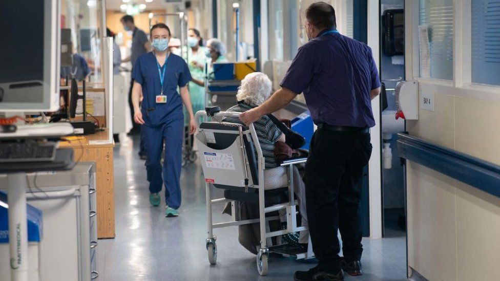
Published39 minutes agoShareclose panelShare pageCopy linkAbout sharingImage source, PA MediaBy Dominic HughesHealth correspondent, BBC NewsMore than 250 patients a week could be dying unnecessarily, due to long waits in A&E in England, according to analysis of NHS data.The Royal College of Emergency Medicine analysed the 1.5 million who waited 12 hours or more to be admitted in 2023. A previous data study had calculated the level of risk of people dying after long waits to start treatment and found it got worse after five hours.The government says the number seen within a four-hour target is improving.This is despite February seeing the highest number of attendances to A&E on record, it adds.The Royal College of Emergency Medicine (RCEM) carried out a similar analysis in 2022, which at that time resulted in an estimate of 300-500 excess deaths – more deaths than would be expected – each week.The analysis uses a statistical model based on a large study of more than five million NHS patients that was published in 2021. At the time NHS England disputed the figures, but it’s roughly what you get if you multiply the number of people waiting long periods in A&E, with the extra risk of dying estimated to come with those long waits (of between five and 12 hours).Postcode check: How’s the NHS coping in your area?12-hour A&E waits in winter ‘becoming normalised’Why did so many people die in 2022?This latest analysis is more detailed following a Freedom of Information request, this time asking for a breakdown of the number of people who waited for more than 12 hours and were subsequently admitted for treatment.That does not include the time that many people spent waiting outside hospitals in ambulances before they were transferred into A&E – meaning the full figures will be even higher, experts say. NHS data for England shows more than 1.5 million patients waited 12 hours or more in major emergency departments in 2023, of which around one million were waiting to be admitted to a hospital bed as a decision had already been made to treat them but a bed was not available at that stage.The RCEM has calculated that, when looking solely at patients awaiting admission, an average of 268 excess deaths are likely to have occurred each week in 2023.RCEM president Dr Adrian Boyle said long waits were continuing to put patients at risk of serious harm. “In 2023, more than 1.5 million patients waited 12 hours or more in major emergency departments, with 65% of those awaiting admission,” he said.”Lack of hospital capacity means that patients are staying in longer than necessary and continue to be cared for by emergency department staff, often in clinically inappropriate areas such as corridors or ambulances.”The direct correlation between delays and mortality rates is clear. Patients are being subjected to avoidable harm.”Sir David Spiegelhalter, emeritus professor of statistics in the Centre for Mathematical Sciences, University of Cambridge, told BBC Radio 4’s Today programme: “It sort of seems intuitive that if you wait a long time in a corridor or somewhere else waiting to be admitted, an average of six hours now, that that’s going to harm people.”He added: “They are not identifiable people, you can’t count people who died because of this. It’s an estimate based on past analysis, that this does increase your risk, but it does seem plausible.”An NHS England statement said : “We have seen significant increases in demand for A&E services, with attendances in February up 8.6% on last year and emergency admissions up 7.7%.”The latest published data shows our urgent and emergency care recovery plan – backed by extra funding with more beds, capacity and greater use of measures like same day emergency care – is delivering improvements.”The Department of Health said it was making progress in reducing waiting times, including adding an extra 5,000 permanent staffed beds this winter to increase capacity.”Thanks to our £200m urgent care recovery plan, A&E four-hour performance improved in February compared to January, despite the highest number of February A&E attendances on record and the impact of industrial action,” a statement said.”We are determined to continue improving experiences for patients and making access to care faster, simpler and fairer.”More on this storyPostcode check: How’s the NHS coping in your area?Published14 March12-hour A&E waits in winter ‘becoming normalised’Published15 FebruaryWhy did so many people die in 2022?Published10 January 2023

Read more →

Hospitals Must Get Written Patient Consent for Pelvic Exams, H.H.S. Says

In a letter to teaching hospitals, the federal health agency said that institutions could lose Medicare funding if they didn’t comply.The Department of Health and Human Services said on Monday that hospitals must obtain written informed consent from patients before they undergo sensitive examinations — like pelvis and prostate exams — especially if the patients will be under anesthesia.A New York Times investigation in 2020 found that hospitals, doctors and doctors in training sometimes conducted pelvic exams on women who were under anesthesia, even when those exams were not medically necessary and when the patient had not authorized them. Sometimes these exams were done only for the educational benefit of medical trainees.On Monday, the secretary of Health and Human Services, along with top officials from the department’s Centers for Medicare and Medicaid Services and Office for Civil Rights, sent a letter to the country’s teaching hospitals and medical schools denouncing the practice of doctors and students conducting the exams without explicit consent. “The Department is aware of media reports as well as medical and scientific literature highlighting instances where, as part of medical students’ courses of study and training, patients have been subjected to sensitive and intimate examinations,” the letter said. “It is critically important that hospitals set clear guidelines to ensure providers and trainees performing these examinations first obtain and document informed consent.”The department issued a set of guidelines clarifying a longstanding requirement that hospitals must obtain written informed consent as a condition for participating in Medicare and Medicaid programs.“Patients who are participating in future clinicians’ education should be aware, should have the opportunity to consent, should be given the same opportunity to participate in that education that they would be given if they were awake and fully clothed,” said Ashley Weitz, who underwent an unauthorized pelvic exam while she was under sedation in an emergency room. “We can only expect to have better trust in medicine when both patients and providers can expect a standard of care that prioritizes patient consent.”

Read more →

Researchers produce grafts that replicate the human ear

Using state-of-the-art tissue engineering techniques and a 3D printer, researchers at Weill Cornell Medicine and Cornell Engineering have assembled a replica of an adult human ear that looks and feels natural. The study, published online in Acta Biomaterialia on March 16, offers the promise of grafts with well-defined anatomy and the correct biomechanical properties for those who are born with a congenital malformation or who lose an ear later in life.
“Ear reconstruction requires multiple surgeries and an incredible amount of artistry and finesse,” said, Dr. Jason Spector, chief of the Division of Plastic and Reconstructive Surgery at NewYork-Presbyterian/Weill Cornell Medical Center and a professor of surgery (plastic surgery) at Weill Cornell Medicine. “This new technology may eventually provide an option that feels real for thousands needing surgery to correct outer ear deformities.”
Many surgeons build a replacement ear using cartilage removed from a child’s ribs, an operation that can be painful and scarring. And though the resulting graft can be crafted to resemble the recipient’s other ear, it generally does not have the same flexibility.
Adding Texture to Structure
One way to produce a more natural replacement ear is to enlist the aid of chondrocytes, the cells that build cartilage. In earlier studies, Dr. Spector and his colleagues used animal-derived chondrocytes to seed a scaffold made of collagen, a key component of cartilage. Though these grafts developed successfully at first, over time the well-defined topography of the ear — its familiar ridges, curves, and whorls — were lost. “Because the cells tug on the woven matrix of proteins as they labor, the ear contracted and shrank by half,” said Dr. Spector.
To address this problem in this study, Dr. Spector and his team used sterilized animal-derived cartilage treated to remove anything that could trigger immune rejection. This was loaded into intricate, ear-shaped plastic scaffolds that were created on a 3D printer based on data from a person’s ear. The small pieces of cartilage act as internal reinforcements to induce new tissue formation within the scaffold. Much like rebar, it strengthens the graft and prevents contraction.
Over the next three to six months, the structure developed into cartilage containing tissue that closely replicated the ear’s anatomical features, including the helical rim, the “anti-helix” rim-inside-the-rim and the central, conchal bowl. “That’s something that we had not achieved before,” said Dr. Spector.
To test the feel of the ear, biomechanical studies were performed in conjunction with Dr. Spector’s long time engineering collaborator Dr. Larry Bonassar, the Daljit S. and Elaine Sarkaria Professor in Biomedical Engineering at the Meinig School of Biomedical Engineering on Cornell’s Ithaca campus. This confirmed that the replicas had flexibility and elasticity similar to human ear cartilage. However, the engineered material was not as strong as natural cartilage and could tear.
To remedy this issue, Dr. Spector plans to add chondrocytes to the mix, ideally ones derived from a small piece of cartilage removed from the recipient’s other ear. Those cells would lay down the elastic proteins that make ear cartilage so robust, producing a graft that would be biomechanically much more similar to the native ear, he said.
This work was supported in part by the National Center for Advancing Translational Sciences of the National Institutes of Health, grant TL1- TR-002386.

Read more →

Aphantasia: Why I cannot see my children in my mind

Published11 minutes agoShareclose panelShare pageCopy linkAbout sharingImage source, Mary WathenBy Philippa RoxbyHealth reporterMost people can picture images in their heads – the look of an apple, the appearance of their kitchen or the smile of their best friend – but not everyone can.Those who cannot visualise anything in their mind’s eye are probably among 1% of people with extreme aphantasia, according to a review of studies on the phenomenon.They are also less likely to recognise faces, remember the sound of a piece of music or the feel of sandpaper, and more likely to work in science, maths or computing.And up to 6% of people may experience some degree of aphantasia. It is not a disorder and does not imply a lack of imagination but can have subtle effects on everyday life, says Prof Adam Zeman, honorary professor of neurology at the University of Exeter, who came up with the term nearly 10 years ago.Image source, Mary WathenMary Wathen, 43, from Cheltenham, Gloucs, finds it “totally mind-blowing” other people can create images in their head.”I just cannot understand what they really mean – where is this image and what does it look like?” she says.”To me, unless you can see something with your eyes, it’s not there.” Mary cannot picture major events in her life such as her wedding day. And unless they are with her, she cannot even picture her two young boys.”I don’t bring up an image – I have all the memories, I just recall it very differently,” Mary says.”As someone once described it, all the hardware is working – but the monitor is not switched on.”‘Gut instinct’Mary discovered she was unlike most other people only when chatting with friends, and was astonished to find her husband could easily visualise past events as if watching a film.On the plus side, she says, she is a very good verbal communicator, because she assumes nothing – it is all about the words. She also feels things deeply.”I’m a very emotional person who is led by gut instincts – so when recalling something, it’s a feeling rather than an image,” Mary says.Mind maps have never been a useful tool for exams, for example, and fantasy fiction is a no-no because she simply cannot escape to that world in her mind.”I only see what is real and in front of me – doesn’t matter whether I saw it a minute or an hour ago,” Mary says.Opposite extremeProf Zeman discovered this way of experiencing the world when he saw a patient who had lost the ability to visualise. When he wrote up the patient’s case, others contacted him to say they had always been that way.And Prof Zeman has since discovered there is an opposite extreme, hyperphantasia, in which people see images so vividly in their heads they cannot tell if they are real or imagined. An estimated 3% of us see the world this way.”One term gave birth to the other,” he says, after borrowing Aristotle’s word for the mind’s eye – “phantasia”.Prof Zeman says 17,000 people have contacted him in the past decade, with experiences of aphantasia and hyperphantasia. Many said they knew they processed information differently to others but had unable to describe how. In your dreamsDifferences in connectivity between regions of the brain may explain why, Prof Zeman says.Asked to picture an apple, for example, most people go through a succession of steps, including nudging the brain to remember what an apple looks like and activating the brain create an image of it.But in those with aphantasia, that process can break down at any point.”Thoughts remain thoughts,” Prof Zeman says, “whereas for others, thought translates into sensory terms.”While aphantasics think about memories, other people are able to recollect and live those memories.But intriguingly, many aphantasics can visualise images while dreaming – probably because it is a more spontaneous task beginning deep down in the brain, Prof Zeman says.And aphantasia can have benefits. It can have a protective effect on someone’s mental health, because they are more likely to live in the moment and less likely to imagine frightening or stressful events, for example.But for Prof Zeman, “the big surprise” was aphantasic artists, who told him their struggle to visualise imagery gave them an extra incentive to make art, by using the canvas as their mind’s eye.Normally, it’s hyperphantasics who are more likely to be creative, like Geraldine van Heemstra.Image source, Paul Bokslag Geraldine, an artist, always had an “enormous imagination” as a child, building entire villages in her mind.And she has always seen the alphabet in colour, as well as numbers and the days of the week.At school, Geraldine used to change answers to maths problems because the colours of the numbers in her head looked wrong sitting next to each other.But she discovered she saw the world differently to most others only when collaborating with musicians and dancers, painting swirls and shapes in response to their rhythms.Feels transported”I remember asking musicians how they saw music – but they didn’t understand what I meant,” Geraldine says.”I thought all musicians saw notes in colour.”Geraldine has a similarly intense experience when painting. “I can walk, sketch, take in the landscape and relive the experience later,” she says.Even when making plans to do something, Geraldine feels transported to the future.Image source, Geraldine van Heemstra”I can be going down another path and it will feel like deja vu,” she says.But constant visualising can also be tiring. And Geraldine’s brain can feel overloaded sometimes, making it hard to sleep.Lots of question remain about aphantasia and hyperphantasia, such as what are the different sub-types and why it might be a genetic thing.Data from large biobanks may provide the answer.Inner livesThe review, in Trends in Cognitive Sciences, found aphantasia ran in families, with the aphantasics’ siblings 10 times more likely to be affected themselves. It has also been suggested aphantasics are more likely to have autism.Prof Zeman says research suggests “conscious sensory imagery is not a prerequisite for human cognition” – or creative imagination.And everyone pictures images in their mind differently.”Our experience is not the norm and other people may have different inner lives,” he adds.Image source, Jamie Mitchell PhotographyRelated Internet LinksTrends in Cognitives SciencesThe BBC is not responsible for the content of external sites.

Read more →

Why Are Older Americans Drinking So Much?

The pandemic played a role in increased consumption, but alcohol use among people 65 and older was climbing even before 2020.The phone awakened Doug Nordman at 3 a.m. A surgeon was calling from a hospital in Grand Junction, Colo., where Mr. Nordman’s father had arrived at the emergency room, incoherent and in pain, and then lost consciousness.At first, the staff had thought he was suffering a heart attack, but a CT scan found that part of his small intestine had been perforated. A surgical team repaired the hole, saving his life, but the surgeon had some questions.“Was your father an alcoholic?” he asked. The doctors had found Dean Nordman malnourished, his peritoneal cavity “awash with alcohol.”The younger Mr. Nordman, a military personal finance author living in Oahu, Hawaii, explained that his 77-year-old dad had long been a classic social drinker: a Scotch and water with his wife before dinner, which got topped off during dinner, then another after dinner, and perhaps a nightcap.Having three to four drinks daily exceeds current dietary guidelines, which define moderate consumption as two drinks a day for men and one for women, or less. But “that was the normal drinking culture of the time,” said Doug Nordman, now 63.At the time of his hospitalization, though, Dean Nordman, a retired electrical engineer, was widowed, living alone and developing symptoms of dementia. He got lost while driving, struggled with household chores and complained of a “slipping memory.”We are having trouble retrieving the article content.Please enable JavaScript in your browser settings.Thank you for your patience while we verify access. If you are in Reader mode please exit and log into your Times account, or subscribe for all of The Times.Thank you for your patience while we verify access.Already a subscriber? Log in.Want all of The Times? Subscribe.

Read more →

Bacteria That Cause Meningitis Are Spreading Again, C.D.C. Warns

The NewsFederal officials are warning health care providers to be on the lookout for invasive meningococcal disease, a rare but potentially deadly illness that has increased in prevalence in recent years and requires prompt treatment with antibiotics to prevent long-term disability or death.Colonies of Neisseria meningitidis bacteria, which cause meningococcal disease.Brodsky/CDC, via Associated PressBy the Numbers: A rising fatality rate.The illness is caused by infection with a bacterium called Neisseria meningitidis. Last year, 422 cases of invasive meningococcal disease were reported in the United States, the highest number since 2014, according to the Centers for Disease Control and Prevention.But as of Monday, 143 cases have been reported to the C.D.C. so far this year, 62 more than the number of cases reported last year during the same period.The illness is extremely dangerous. Even with appropriate treatment, 10 to 15 percent of patients who develop meningococcal disease will die. Many recent cases were caused by an unusual strain of N. meningitidis called ST-1466. This strain caused 17 deaths among 94 patients whose outcomes are known, a fatality rate of 18 percent.Survivors of meningococcal disease may be left with long-term disability, deafness, amputations or brain damage.The Mystery: What’s causing the outbreaks?A majority of people affected in the recent outbreaks were Black people and adults ages 30 to 60.Others who are susceptible to the infection include people living with H.I.V., who account for 15 percent of patients; individuals who have had their spleens removed; people with sickle cell disease; and patients with certain rare immune conditions.A meningitis vaccine that protects against four of six N. meningitidis types — including group Y, which includes ST-1466 — is recommended for adolescents as well as those with medical conditions like H.I.V. Most older adults have not received the vaccine.We are having trouble retrieving the article content.Please enable JavaScript in your browser settings.Thank you for your patience while we verify access. If you are in Reader mode please exit and log into your Times account, or subscribe for all of The Times.Thank you for your patience while we verify access.Already a subscriber? Log in.Want all of The Times? Subscribe.

Read more →

4 Things You Need to Know About Health Care Cyberattacks

Despite the explosion in ransomware hacks like the one against Change Healthcare, regulation is spotty and few new safeguards have been proposed to protect patient data, vulnerable hospitals and medical groups.The recent cyberattack on the billing and payment colossus Change Healthcare revealed just how serious the vulnerabilities are throughout the U.S. health care system, and alerted industry leaders and policymakers to the urgent need for better digital security.Hospitals, health insurers, physician clinics and others in the industry have increasingly been the targets of significant hacks, culminating in the assault on Change, a unit of the giant UnitedHealth Group, on Feb. 21.The ransomware attack on the nation’s largest clearinghouse, which handles a third of all patient records, had widespread effects. Fixes and workarounds have alleviated some distress, but providers are still unable to collect billions of dollars in payments. Many smaller hospitals and medical offices are still having trouble getting paid more than a month after Change was first forced to shut down many of its systems.Even now, very little information about the exact nature and scope of the attack has been disclosed. UnitedHealth said that it had advanced more than $3 billion to struggling providers, and that it expected more of Change’s services to be available in the coming weeks as it brought the systems back online.The F.B.I. and the Department of Health and Human Services are investigating the Change hack, including whether patients’ records and personal information have been compromised. Because Change’s network acts as a digital switchboard that connects information from a patient’s first doctor visit to a diagnosis like cancer or depression and then subsequent treatment to a health insurer for benefits and payments, there is a risk that people’s medical history could be exposed for years.The attack on Change is just the most far-reaching example of what has become nearly commonplace in the health care industry. Ransomware attacks, in which criminals shut down computer systems unless the owners pay the hackers, affected 46 hospital systems last year, up from 25 in 2022, according to the data security firm Emsisoft. Hackers have also taken down companies that provide services such as medical transcription and billing in recent years.We are having trouble retrieving the article content.Please enable JavaScript in your browser settings.Thank you for your patience while we verify access. If you are in Reader mode please exit and log into your Times account, or subscribe for all of The Times.Thank you for your patience while we verify access.Already a subscriber? Log in.Want all of The Times? Subscribe.

Read more →