Simulations reveal mechanism behind protein build-up in Parkinson’s disease

Researchers have used computational models to understand what drives the accumulation of alpha-synuclein protein, a key culprit in the development of Parkinson’s disease.
The study, published today as a Reviewed Preprint in eLife, is described by the editors as providing important biophysical insights into the molecular mechanism underlying the association of alpha-synuclein chains, which is essential for understanding the development of Parkinson’s disease. The data analysis is solid, and the methodology can help investigate other molecular processes involving intrinsically disordered proteins (IDPs).
IDPs play important roles in the human body. These proteins lack a well-defined 3D structure, which allows them to function in a flexible way — adopting different roles as needed. However, this also makes them susceptible to irreversible aggregation, especially if mutated. These aggregates are known to be associated with various diseases, such as neurodegenerative diseases, cancer, diabetes and heart disease. For example, Alzheimer’s disease is characterised by the aggregation of amyloid-beta protein, whereas Parkinson’s disease is linked with the build-up of alpha-synuclein.
“A growing body of evidence has established a connection between intrinsically disordered proteins and liquid-liquid phase separation, or LLPS, the phenomenon you see if you mix oil and water,” says lead author Abdul Wasim, a PhD student at the Tata Institute of Fundamental Research, Hyderabad, India. “This is of interest because LLPS is itself known to form subcellular compartments that can lead to incurable diseases.”
It is known that alpha-synuclein can undergo LLPS, and that the aggregation of alpha-synuclein is influenced by crowding from nearby molecules and surrounding pH. But characterising the precise interactions and dynamics of these miniscule aggregate proteins is challenging.
“Previous attempts have simulated individual IDPs, but these simulations can be extremely time-consuming and resource-intensive, making the study of protein aggregation impractical even with cutting-edge software and hardware,” explains senior author Jagannath Mondal, Associate Professor at the Tata Institute of Fundamental Research. “We used coarse-grained molecular dynamic simulations, which although offering lower resolution allowed us to study the aggregation of multiple IDPs in a mixture.”
Using this model, the authors simulated the collective interaction of many alpha-synuclein chains within droplets under different conditions. First, by studying the protein chains mixed only with water, they found that around 60% of the protein chains remained free and did not show a strong and spontaneous tendency to aggregate together.

Next, they added in some ‘crowder’ molecules — large biological molecules that make the environment a highly crowded space for proteins. Previous studies in Alzheimer’s disease have shown increased aggregation of proteins in a crowded environment. As expected, the addition of crowders led to enhanced alpha-synuclein aggregation and the number of free proteins decreased.
Similarly, the team found that changing the ionic environment by adding salt also promoted aggregation. However, further exploration revealed that these two environmental factors — crowding and salt — caused aggregation by different mechanisms. Adding salt into the mix increased the surface tension of the droplets, but adding in crowder molecules had no surface tension effects. This is important to know, because the larger the surface tension, the higher the tendency of proteins to aggregate. Moreover, merging of droplets to alleviate surface tension is often seen in liquid-liquid phase separated (LLPS) droplets characteristic of diseases involving disordered proteins.
A characteristic of LLPS is that the protein molecules within droplets adopt an extended shape and all orient themselves in a consistent direction. So, the team next set out to see if this was true within their simulations. They found that proteins in the dense (highly concentrated) phase of the liquid-liquid separation indeed had an extended shape, irrespective of whether crowder molecules or salt were present — all protein molecules had similar orientations — suggesting that alpha-synuclein IDPs display the hallmarks of the LLPS phenomenon.
Next, the team wanted to find out how different alpha-synuclein proteins interact with each other to achieve these effects. By studying the position and features of different amino acids within the protein, they could work out the chances of them coming into contact under different conditions. This revealed that certain amino acids in the protein probably exist to prevent aggregation — and that proteins orient themselves to minimise interactions between these residues.
The editors note that there are limitations to the study to be addressed. Namely, they say that benchmarking of the simulations against other methods could be improved to give the reader greater confidence in the conclusions presented.
“Together, these results suggest that both crowder molecules and salt enhance the aggregation of alpha-synuclein, while also stabilising the resulting aggregates,” says Wasim. “Irrespective of the factors causing the aggregation, the interactions that drive the formation of droplets remain the same.”
“Our study focused on normal alpha-synuclein and identified key sites within the protein that are crucial for aggregation,” concludes Mondal. “Inherited mutations in alpha-synuclein are thought to significantly increase the likelihood of aggregation. These mutations, involving minor alterations to protein sequence, highlight the importance of understanding the molecular basis of this process.”


Read more →

Investigators develop novel treatment for T-cell leukemias and lymphomas

A novel treatment for leukemias and lymphomas that arise from immune system T cells, developed by investigators at the Johns Hopkins Kimmel Cancer Center and its Ludwig Center and Lustgarten Laboratory, was found to be effective at killing these cancers in mice bearing human T-cell tumors.
The therapy, an antibody-drug conjugate (ADC), combines an antibody that targets a protein called TRBC1 expressed on the surface of T-cell cancers with an anti-cancer drug, called SG3249. The ADC works by using the antibody to seek out the cancer cells that express TRBC1. Then, those cancer cells ingest the ADC, where SG3249 is released and kills the cancer cells. A description of the work was published March 27 in Nature.
Each year, about 100,000 patients worldwide are affected by T-cell leukemias and lymphomas. Adults with relapsed T-cell cancers have limited therapeutic options and five-year survival rates of 7-38%.
“Developing treatments for T-cell leukemias and lymphomas is much more difficult than for leukemias and lymphomas arising from immune system B cells,” explains senior study author Suman Paul, M.B.B.S., Ph.D., an assistant professor of oncology at the Johns Hopkins University School of Medicine. Effective therapies for B-cell cancers wipe out both cancerous and noncancerous B cells, but patients still do well even without the immune system B cells that help fight infections. However, if similar approaches are used and a treatment wipes out both normal and cancerous T cells, it would leave patients without a functioning immune system and at high risk of dying from infections.
“Not much drug development has happened in this space of T-cell leukemias and lymphomas,” Paul says. “We need new therapies for these cancers, but whatever therapies we develop in the space have to be cancer-specific. We have to preserve some of the normal T cells and wipe out cancerous T cells at the same time.”
T-cell cancers express either TRBC1 or TRBC2, while normal T cells express a mix of TRBC1 and TRBC2. Therefore, selective targeting of TRBC1 can potentially eradicate the normal and cancerous T cells expressing TRBC1 while preserving normal T cells expressing TRBC2. A recent clinical trial conducted elsewhere attempted to target TRBC1 cancers using chimeric antigen receptor (CAR) T-cell therapy. These CAR T cells are genetically engineered T cells that bind to and kill TRBC1 cells. CAR T-cell therapies are FDA-approved treatment options used in several B-cell cancers. However, after administering the TRBC1-targeting CAR T cell therapy in human patients, trial investigators reported that the CAR T cells were not persisting inside the patients. Such persistence is required for effective cancer cell-killing. Interested to understand why, Paul and colleagues found that the CAR T cells targeting TRBC1 could be killed by normal T cells, limiting their persistence.
This lack of CAR T-cell persistence led the team to try TRBC1 targeting with the use of antibody-drug conjugates. Paul and colleagues tried two different formulations of ADCs in mouse models of T-cell cancers. After a single injection of one formulation of the treatment, the cancers initially regressed but then recurred. After a single treatment with the anti-TRBC1-SG3249 ADC combination, investigators observed signs of cancer elimination within seven days and the cancers were eventually undetectable, with no recurrences. “The tumors didn’t come back, and we followed the mice for more than 200 days,” Paul explains.

The treatment was able to eliminate the cancer while preserving half of the remaining normal T cells. “The residual normal T cells should be sufficient to maintain some immune system protection against infectious diseases,” Paul says.
“Witnessing the successful elimination of T-cell cancers while sparing normal T cells in preclinical models was truly gratifying,” adds Jiaxin Ge, a co-author of the study and third-year Ph.D. student in the Ludwig Center. “We believe this approach has the potential to address a critical unmet need in oncology, and we’re committed to advancing it through further research.”
Tushar Nichakawade, first author on the study and a fourth-year Ph.D. student at the Ludwig Center, says, “There are so many lessons to learn from the clinic and it has been exciting to be a part of the iterative process of drug discovery. Every therapy has its pros and cons, but the preclinical efficacy of our ADC gives me hope that it can make a difference for patients suffering from these terrible cancers.”
Investigators are now working with an industry partner to conduct early-phase safety and efficacy trials in human patients.
The study’s co-authors were Brian J. Mog, Bum Seok Lee, Alexander H. Pearlman, Michael S. Hwang, Sarah R. DiNapoli, Nicholas Wyhs, Nikita Marcou, Stephanie Glavaris, Maximilian F. Konig, Sandra B. Gabelli, Evangeline Watson, Cole Sterling, Nina Wagner-Johnston, Sima Rozati, Lode Swinnen, Ephraim Fuchs, Drew M. Pardoll, Kathy Gabrielson, Nickolas Papadopoulos, Chetan Bettegowda, Kenneth W. Kinzler, Shibin Zhou, Surojit Sur and Bert Vogelstein of Johns Hopkins.
The work was supported in part by The Virginia and D.K. Ludwig Fund for Cancer Research, Lustgarten Foundation for Pancreatic Cancer Research, Commonwealth Fund, Bloomberg~Kimmel Institute for Cancer Immunotherapy, Bloomberg Philanthropies and the National Institutes of Health Cancer Center Support Grant P30 CA006973. Paul was supported by the National Cancer Institute (grant K08CA270403), the Leukemia Lymphoma Society Translation Research Program Award, the American Society of Hematology Scholar Award and the Swim Across America Translational Cancer Research Award.

Read more →

Wound treatment gel fights the battle against antibacterial resistance

Hydrogels are popular for use in skin ailments and tissue engineering. These polymer-based biocompatible materials are useful for their abilities to retain water, deliver drugs into wounds, and biodegrade. However, they are complicated to manufacture and not very resilient to external forces like rubbing against clothing, sheets, or wound dressings. They are also not inherently able to battle bacterial infections, so they are often infused with antimicrobial drugs or metal ions, which can cause antibiotic resistance and negative effects on cell growth.
In a paper published this week in APL Materials, by AIP Publishing, researchers created a hydrogel that is easier to synthesize, contains natural antibiotic properties, and promotes cell growth.
“A diabetic patient may have skin wounds that do not heal easily due to metabolic disease,” author Jing Sun said. “The patient may try to treat the wounds with topical medicines such as erythromycin, and it may be effective at first, but over a long period of time, it may fail to relieve symptoms. This could be due to antibiotic resistance.”
Using the common hydrogel Gel-MA, they added the amino acid polylysine and platelet-rich blood plasma to create properties that are well-suited to wound care. The result is a hydrogel that is stronger, expands in the wound, lasts longer, kills bacteria, and creates a healthy environment for new cells to grow.
“The hydrogel continuously releases polylysine on the wound surface and continuously inhibits bacterial growth,” Sun said. “We chose ε-polylysine because it can inhibit the growth of bacteria and solve the problem of antibiotic abuse, drug resistance, and does not affect the proliferation and development of cells. It can also conjugate with gelatin methacrylate, which plays an antimicrobial role and enhances the mechanical strength of the hydrogel.”
In tests with E. coli and S. aureus, the bacterium that causes staph infection, the hydrogel damaged bacteria cell membranes and led to bacterial cell death. For healthy cells, the inclusion of platelet-rich blood plasma resulted in a release of growth factors and an increase of viable cells.
“The most interesting and exciting moment for me was when we mixed the polylysine and platelet-rich plasma solutions to see if they could form a hydrogel under UV irradiation,” Sun said.
The experiment worked, and the hydrogel can be cured under a UV lamp for 30 seconds instead of curing by repeatedly freezing and thawing for up to 8 hours.
“As a clinician and researcher in dermatology, I have the obligation to provide better treatments for patients,” Sun said. “Patients with chronically infected wounds combined with metabolic diseases, such as diabetes, malnutrition, and other diseases, as well as long-term bedridden patients will be helped by this solution.”

Read more →

Scientists link certain gut bacteria to lower heart disease risk

Changes in the gut microbiome have been implicated in a range of diseases including type 2 diabetes, obesity, and inflammatory bowel disease. Now, a team of researchers at the Broad Institute of MIT and Harvard along with Massachusetts General Hospital has found that microbes in the gut may affect cardiovascular disease as well. In a study published in Cell, the team has identified specific species of bacteria that consume cholesterol in the gut and may help lower cholesterol and heart disease risk in people.
Members of Ramnik Xavier’s lab, Broad’s Metabolomics Platform, and collaborators analyzed metabolites and microbial genomes from more than 1,400 participants in the Framingham Heart Study, a decades-long project focused on risk factors for cardiovascular disease. The team discovered that bacteria called Oscillibacter take up and metabolize cholesterol from their surroundings, and that people carrying higher levels of the microbe in their gut had lower levels of cholesterol. They also identified the mechanism the bacteria likely use to break down cholesterol. The results suggest that interventions that manipulate the microbiome in specific ways could one day help decrease cholesterol in people. The findings also lay the groundwork for more targeted investigations of how changes to the microbiome affect health and disease.
“Our research integrates findings from human subjects with experimental validation to ensure we achieve actionable mechanistic insight that will serve as starting points to improve cardiovascular health,” said Xavier, who is a core institute member, director of the Immunology Program, and co-director of the Infectious Disease and Microbiome Program at the Broad. He is also a professor at Harvard Medical School and Massachusetts General Hospital.
Postdoctoral researcher Chenhao Li and research scientist Martin Stražar, both in Xavier’s lab, were co-first authors on the study.
Cholesterol cues
In the past decade, other researchers have uncovered links between composition of the gut microbiome and elements of cardiovascular disease, such as a person’s triglycerides and blood sugar levels after a meal. But scientists haven’t been able to target those connections with therapies in part because they lack a complete understanding of metabolic pathways in the gut.
In the new study, the Broad team gained a more complete and detailed picture of the impact of gut microbes on metabolism. They combined shotgun metagenomic sequencing, which profiles all of the microbial DNA in a sample, with metabolomics, which measures the levels of hundreds of known and thousands of unknown metabolites. They used these tools to study stool samples from the Framingham Heart Study.

“The project outcomes underline the importance of high-quality, curated patient data,” Stražar said. “That allowed us to note effects that are really subtle and hard to measure and directly follow up on them.”
The approach uncovered more than 16,000 associations between microbes and metabolic traits, including one that was particularly strong: People with several species of bacteria from the Oscillibacter genus had lower cholesterol levels than those who lacked the bacteria. The researchers found that species in the Oscillibacter genus were surprisingly abundant in the gut, representing on average 1 in every 100 bacteria.
The researchers then wanted to figure out the biochemical pathway the microbes use to break down cholesterol. To do this, they first needed to grow the organism in the lab. Fortunately, the lab has spent years collecting bacteria from stool samples to create a unique library that also included Oscillibacter.
After successfully growing the bacteria, the team used mass spectrometry to identify the most likely byproducts of cholesterol metabolism in the bacteria. This allowed them to determine the pathways the bacteria uses to lower cholesterol levels. They found that the bacteria converted cholesterol into intermediate products that can then be broken down by other bacteria and excreted from the body. Next, the team used machine-learning models to identify the candidate enzymes responsible for this biochemical conversion, and then detected those enzymes and cholesterol breakdown products specifically in certain Oscillibacter in the lab.
The team found another gut bacterial species, Eubacterium coprostanoligenes, that also contributes to decreased cholesterol levels. This species carries a gene that the scientists had previously shown is involved in cholesterol metabolism. In the new work, the team discovered that Eubacterium might have a synergistic effect with Oscillibacter on cholesterol levels, which suggests that new experiments that study combinations of bacterial species could help shed light on how different microbial communities interact to affect human health.
Microbial messages
The vast majority of genes in the human gut microbiome remains uncharacterized, but the team is confident that their success in pinpointing cholesterol-metabolizing enzymes paves the way for the discovery of other similar metabolic pathways impacted by gut microbes, which could be targeted therapeutically.

“There are many clinical studies trying to do fecal microbiome transfer studies without much understanding of how the microbes interact with each other and the gut,” Li said. “Hopefully stepping back by focusing on one particular bug or gene first, we’ll get a systematic understanding of gut ecology and come up with better therapeutic strategies like targeting one or a few bugs.”
“Because of the large number of genes of unknown function in the gut microbiome, there are gaps in our ability to predict metabolic functions,” Li added. “Our work highlights the possibility that additional sterol metabolism pathways may be modified by gut microbes. There are potentially a lot of new discoveries to be made that will bring us closer to a mechanistic understanding of how microbes interact with the host.”
Funding: This work was supported by the National Institutes of Health.

Read more →

Infant gut microbes have their own circadian rhythm, and diet has little impact on how the microbiome assembles

Infant gut microbiomes oscillate with a circadian rhythm, even when they are cultivated outside of the body. Researchers report April 2 in the journal Cell Host & Microbe that the rhythm is detectable as early as 2 weeks after birth but becomes more pronounced with age. The finding comes from a randomized controlled trial that also showed that diet has less impact on the development and composition of the infant microbiome than previously thought.
“We found that even at very early ages of colonization, the microbial ecosystem develops this circadian rhythmicity,” says senior author and microbiome expert Dirk Haller of the Technical University of Munich. “We have shown these rhythms before in adults, but we were not sure when these mechanisms first appear.”
While diet had only a marginal impact on infant microbiome development, the researchers showed that age plays a more important role.
“Diet matters, but less than aging of the gut,” says Haller. “When we compared breastfed and formula-fed infants, the differences in microbiome colonization were marginal. Our intestinal system is probably a little bit more flexible in adapting to what the environment has to offer.”
The researchers used a randomized controlled trial to compare microbiome development in infants that were exclusively breastfed with infants who received different types of formula — un-supplemented formula; formula containing breast-milk-derived bacteria (Bifidobacteria); formula containing breast-milk-mimicking sugars (galacto-oligosaccharides, GOSs); or formula containing both Bifidobacteria and GOSs. Altogether, the trial included 210 infants.
To longitudinally track the infants’ microbiomes, the team sampled the infants’ stools when they were 0.5 months, 1 month, 3 months, 7 months, and 12 months of age as well as at 24 months for a subset of the infants. They also kept note of the time of day that the stool sample was collected.
The researchers found that diet had little impact on infant growth or the differences in the infants’ microbiomes. Though there was a lot of variation, all of the infants showed a gradual increase in gut microbe diversity, and at 24 months there was no observable difference between the groups. When they compared the different types of formula, they found that GOS-supplemented infant formula was more effective at promoting sustained levels of Bifidobacteria compared to formula containing Bifidobacteria.

However, there was a significant difference in the gut metabolite profile between exclusively breastfed and formula-fed infants. “The metabolite environment in the gut is dramatically different between a baby that is exclusively breastfed and babies that receive infant formula, which could have a fundamental impact on metabolic priming and many downstream effects,” says Haller. “We can conclude that breast milk does something completely different in the metabolism in in the infant’s intestine.”
The researchers also observed rhythmic 24-hour fluctuations in the abundance of different microbiome species. When they took infant microbes and grew them in continuous culture in the lab, the bacteria settled into the same circadian rhythm — even in the absence of external light or host cues. Though circadian rhythms have been previously observed in adult microbiomes, this is the first evidence that bacteria maintain these rhythms independently.
“When we take them out, they maintain these daytime-related diurnal oscillations,” Haller says. “This is fairly surprising because it suggests that the bacteria have some intrinsic mechanism that provides some sort of adaptation to a day and night cycle, which could potentially give them an advantage in colonizing the human intestine.”
The researchers plan to further investigate microbiome circadian rhythms in future studies. Specifically, they want to examine whether individual bacterial species maintain rhythms when grown in isolation rather than in complex communities and to search for the genes that control these rhythms.
“For us, the next question is can we identify mechanisms in bacteria that control their circadian behavior,” says Haller.

Read more →

The Technique Reshaping Organ Transplantation

Perfusion keeps a donated organ alive outside the body, giving surgeons extra time and increasing the number of transplants possible. On some level, the human liver in the operating room at Northwestern Memorial Hospital in Chicago was alive. Blood circulating through its tissues delivered oxygen and removed waste products, and the organ produced bile and proteins that are essential to the body.But the donor had died a day earlier, and the liver lay inside a boxy plastic device. The organ owed its vitality to this machine, which was preserving it for transplantation into a needy patient.“It’s a little bit science fiction,” said Dr. Daniel Borja-Cacho, a transplant surgeon at the hospital.Surgeons are experimenting with organs from genetically modified animals, hinting at a future when they could be a source for transplants. But the field is already undergoing a paradigm shift, driven by technologies in widespread use that allow clinicians to temporarily store organs outside the body.Perfusion, as its called, is changing every aspect of the organ transplant process, from the way surgeons operate, to the types of patients who can donate organs, to the outcomes for recipients.Most significantly, surgical programs that have adopted perfusion are transplanting more organs.Since 2020, Northwestern has had a 30 percent uptick in its volume of liver transplants. Nationally, the number of lung, liver and heart transplants each rose by more than 10 percent in 2023, one of the largest year-over-year increases in decades.We are having trouble retrieving the article content.Please enable JavaScript in your browser settings.Thank you for your patience while we verify access. If you are in Reader mode please exit and log into your Times account, or subscribe for all of The Times.Thank you for your patience while we verify access.Already a subscriber? Log in.Want all of The Times? Subscribe.

Read more →

Study aims to break brain injury link to depression

Published5 minutes agoShareclose panelShare pageCopy linkAbout sharingBy Jim ReedHealth reporterFor many people who have a serious blow to the brain, physical recovery is just the first of their problems. Depression and other mental disorders are far more likely to develop following head trauma.The results of a small US study suggest taking a widely used antidepressant in the weeks after the injury could actually help prevent severe depression.And now a much larger trial is being held across England, looking into whether those findings can be replicated in a wider group of patients.’Lost hearing'”I was told I was airlifted to the Royal London [Hospital] and put in a coma for three weeks,” Shannon says. “They had to call my family down to say their goodbyes.” Shannon, 24, was hit on the side of her head by a digger while working on a building site in east London, in 2020. She needed operations to reconstruct parts of her face, using muscle from her legs. Pins and wires now hold her jaw together. But, as with so many people who have lived through a traumatic brain injury, the visible, physical damage to her body was just part of the problem.”It has had a real impact on my mental health and I have felt very low,” Shannon says. “I wasn’t going out, I wasn’t taking care of myself and I put on weight.”It has been a fight just to get out of bed and wash my face. It has been very difficult, almost unbearable at points.”In the immediate aftermath of such an accident, the focus has tended to be been on surgery, rehab and treating physical symptoms. But longer-term effects – such as depression and mood swings – can have a severe impact for many.Shannon says it took “maybe a year” after leaving hospital to get the right support and medication in place and start to “pull out of it”. Image source, Shannon BrazierResearch suggests about half the people admitted to hospital after a head injury report major depression in the year after the accident, a rate 10 times higher than the general population.The 18-month UK trial will examine the early use of common antidepressants following brain trauma. It will measure depression, quality of life and cognitive functioning among 500 patients, with results expected in 2027.”Up until now, most of the research has been on the treatment of depression once it’s set in, which we know can be difficult,” says lead researcher Khalida Ismail, professor of psychiatry and medicine at King’s College London.”This is the first large-scale study in the world that is actually trying to prevent it from happening in the first place.”The trial is being held across nine major trauma centres in England and is being funded by a £2.2m grant from the National Institute for Health and Care Research.Anxiety, mood, memory problemsA certain level of emotional trauma and stress might be expected after a catastrophic experience, with the knock-on effects on employment, education and relationships.But doctors believe a serious blow to the head can also disrupt the neural pathways in the brain that control memory, thinking skills and emotions.Sean Carty, 47, says he felt like he’d “landed on a new planet” after experiencing severe head trauma and depression.He was knocked off his motorbike on a dual carriageway in London five years ago, leaving him in hospital for three weeks, with a bleed to the frontal lobe of his brain. “After I was discharged, I was trying to get back to work, trying to do the things that I did before, but I was really struggling,” Sean says. He found his mind worked differently. His sense of smell and taste had changed and he struggled to keep up with friends’ conversations. Like many others, there was also an impact on his behaviour. He had a shorter temper and was argumentative with family members in a way he never had been before the crash.”You don’t realise how blunt you can be with a brain injury,” Sean says. “But it’s hard when your nervous system is not functioning properly. You feel like you’re an alien and everything is new to you.” Consultant neuropsychiatrist Dr Mike Dilley, from King’s College Hospital, says the patients who see him about their brain injury “are far more concerned about anxiety, mood and memory problems than about physical difficulties, which they might have adjusted to already”. Exactly how the antidepressants in the trial – selective serotonin reuptake inhibitors (SSRIs) – work is unknown. Comprehensive research, from the University of Oxford, suggests they reduce serious depression in some people in the short term. But there is also evidence linking severe depression to brain inflammation after an accident. And one theory, although far from proven, is SSRIs might trigger an anti-inflammatory response and protect the patient from longer-term harm. Doctors say tackling the wider problem could benefit the NHS and the wider economy, with the cost of brain injury estimated at more than £15bn each year. “Depression is not just something in the mind,” Prof Ismail says. “It can have an effect on relationships, on jobs, on education and on rehabilitation. And all of these are consequences after a traumatic brain injury.”Follow Jim on X, formerly known as Twitter.More on this storyTwo million on antidepressants for five yearsPublished19 June 2023New tech centre to help brain and spinal injuriesPublished20 FebruaryRugby career length linked to brain injury – studyPublished24 October 2023Man with brain injury helping men open upPublished10 October 2023Window fall man describes life after brain injuryPublished6 October 2023

Read more →

Diabetes patients offered artificial-pancreas tech

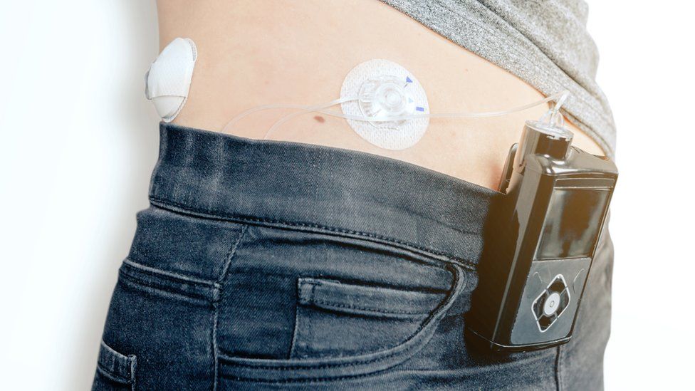
Published27 minutes agoShareclose panelShare pageCopy linkAbout sharingImage source, Getty ImagesBy Nick TriggleHealth correspondentTens of thousands of people with type 1 diabetes in England are to be offered a new technology, dubbed an artificial pancreas, to help manage the condition.The system uses a glucose sensor under the skin to automatically calculate how much insulin is delivered via a pump. Later this month, the NHS will start contacting adults and children who could benefit from the system. But NHS bosses warned it could take five years before everyone eligible had the opportunity to have one. This is because of challenges sourcing enough of the devices, plus the need to train more staff in how to use them.In trials, the technology – known as a hybrid closed loop system – improved quality of life and reduced the risk of long-term health complications.And at the end of last year, the National Institute of Health and Care Excellence (Nice) said the NHS should start using it.Type 1 diabetes: ‘People don’t know how hard it is’Artificial pancreas to revolutionise diabetes careType 1 diabetes trial to identify at-risk children Nearly 300,000 people in the UK have type 1 diabetes, including about 29,000 children. It means their pancreas fails to produce insulin, an important hormone that helps turn food into energy.They have to closely monitor levels of sugar, or glucose, in the blood and give themselves insulin every day of their lives, via either injections or a pump. And this new technology does that automatically, virtually mimicking the function of a pancreas – although it still requires information on food intake to be inputted at mealtimes to work accurately. The new technology is intended to help prevent people with type 1 diabetes experiencing life-threatening low or high blood glucose levels, which can lead to unconsciousness and can even be fatal. And it also helps to improve overall blood sugar control, which means the chance of complications – like heart disease, eyesight problems and kidney disease – decreases.Scotland is also offering the technology, and Wales and Northern Ireland could soon follow suit.Gemma Lavery, 38, from Plymouth, who is using the device after being part of an NHS pilot scheme, says it has transformed her life. “I no longer have to worry about work-related stress affecting my blood-glucose levels, as the closed loop helps to sort this out before it becomes a problem,” she says. “I can have a full night’s sleep without worrying about regular low glucose levels hindering my morning routine and I have found that my diabetes is more stable.” ‘Incredibly exciting’Prof Partha Kar, NHS national speciality advisor for diabetes, said the move was “great news for everyone with type 1 diabetes”. “This futuristic technology not only improves medical care but also enhances the quality of life for those affected,” he added.NHS England diabetes clinical director Dr Clare Hambling said the technology “holds the power to redefine the lives” of people with type 1 diabetes.She added: “Type 1 diabetes is an easily missed diagnosis, so if you are concerned about symptoms – the four ‘T’s – going to the toilet, passing urine more frequently, with thirst, feeling tired and getting thinner, please come forward for support.”Diabetes UK chief executive Colette Marshall said: “It’s incredibly exciting to see this technology being rolled out. “This really is a landmark moment.” Nice approved the NHS roll-out of the system last December, with the NHS later setting out a five-year plan for how to provide it for those eligible.Nice recommends its use for those with type 1 who are in certain categories, including children and under-18s, pregnant women, and those with a HbA1c reading – a way of recording long-term blood sugar levels – of 58 mmol/mol, or 7.5%, or higher.More on this storyArtificial pancreas to revolutionise diabetes carePublished1 April 2022Type 1 diabetes: ‘People don’t know how hard it is’Published9 May 2022Game-changing type 1 diabetes drug approved in USPublished18 November 2022Type 1 diabetes trial to identify at-risk childrenPublished14 November 2022Related Internet LinksType 1 diabetes – NHSJDRF UK – Type 1 diabetes researchDiabetes UKDiabetes Community, Support, EducationThe BBC is not responsible for the content of external sites.

Read more →

Reducing late-night alcohol sales curbed all violent crimes by 23% annually in a Baltimore neighborhood

A new study found that reducing alcohol hours of sale for bars and taverns in a Baltimore, Md. neighborhood also reduced homicides by 51 percent within the first month and by 40 percent annually, pointing to possible opportunities for other cities to address excessive drinking and crime.
Simply reducing the hours during which alcohol may be purchased can significantly reduce violent crime, according to a new study led by Boston University School of Public Health (BUSPH) and the Alcohol Research Group of Emeryville, Calif. The findings were published in JAMA Internal Medicine on Monday, April 1.
Substantial research has linked alcohol sales at liquor stores and other establishments to increased neighborhood crime, but this is the first study to look at the impact of changing the hours of sale in a low-income neighborhood on crime in that neighborhood.
The study found that shortening overnight operations by seven hours at bars and taverns in a Baltimore, Md. neighborhood resulted in a 51 percent immediate drop in homicides within the first month, followed by a 23 percent decline in all violent crimes annually in the surrounding area, compared to similar neighborhoods with no change in hours of sale. Homicide rates decreased by 40 percent in each subsequent year.
“We were able to take advantage of this natural experiment, and apply rigorous analytic methods to assess the effect of the change,” says study lead author Dr. Erika Rosen of the Alcohol Research Group. “While we expected to see some change, the size of the drop in crime was even more significant than we expected.”
The research team evaluated the impact of the Maryland Senate Bill 571 (SB571) passed by the state legislature in 2020. The bill reduced the hours of sale for alcohol in 2020, from 20 hours per day to 13 hours per day (from 6 a.m. to 2 am, to 9 a.m. to 10 p.m.).Their analysis also estimated that this reduction in crime saved the City of Baltimore an estimated $18.2 million in annual costs.
These findings suggest that reducing late-night hours of sale may be an effective way for cities to curb excessive drinking — a persistent problem that worsened nationwide during the COVID-19 pandemic — as well as homicides, assaults, and other crimes.

The study team utilized publicly available data to measure total violent crime incidents within 800 feet of bars and taverns in a Baltimore neighborhood from May 2018 to December 2022 — before and after the new legislation on hours of operation was implemented in September 2020. The team focused on total late-night crime occurrences between 8 p.m. and 4 a.m. around 26 bars and taverns, because these are the times at which crime is most likely to be associated with alcohol use. Then they compared these crimes to crimes happening near 41 other bars and taverns with unchanged hours of operation in demographically similar Baltimore neighborhoods.
The study assessed both violent crime — defined as homicide, robbery, aggravated assault, and forcible rape — and common assault, adjusting for neighborhood factors such as population size, percentage of Black and White residents, alcohol outlet totals per square mile, neighborhood disadvantage, and number of convenience stores.
Notably, the researchers also conducted additional sensitivity analyses that confirmed the decline of late-night crimes, suggesting that crimes did not shift to earlier hours of the day or to adjacent neighborhoods.
“Changing the hours of service and sale of alcohol is a relatively simple intervention,” said study co-author Dr. David Jernigan, professor of health law, policy & management at BUSPH. “Yet our findings suggest that, even in a period like the COVID-19 pandemic when alcohol consumption was rising, this policy has great promise for other cities and neighborhoods seeking to prevent and reduce crime.”
Additional research is needed to test this policy in other cities and for longer periods of time, but the researchers hope this evaluation serves as a potential model for other cities to consider implementing to decrease crime in their neighborhoods and support residents’ overall health and safety.
“Our study provides new and compelling evidence that supports the World Health Organization’s three “best buys” to reduce alcohol attributable morbidity and mortality through reducing availability of alcoholic beverages, along with increasing prices via taxation and banning alcohol marketing,” says study senior author Dr. Ziming Xuan, professor of community health sciences “These findings highlight the critical importance of population-based alcohol policies in violence prevention.”
Funding for the study was provided by a pilot research grant from BUSPH, the National Institute on Alcohol Abuse and Alcoholism, and the Centers for Disease Control and Prevention.

Read more →

Golfers’ risk from pesticides used on turfgrass is likely low

For many, spring heralds fresh air and exercise on the golf course. But do players risk exposure to unsafe levels of pesticides used to beautify and maintain a golf course’s green grass? To find out, researchers asked volunteers to play 18 holes on a simulated course sprayed with common pesticides. They report the results in ACS Agricultural Science & Technology, saying there is likely limited cause for concern over toxic exposure from pesticide-treated turf.
There are plenty of studies on pesticide exposure among people who tend and harvest crops grown in treated environments. But John M. Clark and colleagues couldn’t find much comparable information about individuals who play sports, including soccer and golf, in the great outdoors. So, his team designed a study to investigate golfers’ potential risks from four pesticides, which have low volatilities and relatively low toxicities for humans, and are commonly used on golf course turfgrass: cyfluthrin (insecticide), chlorothalonil (fungicide), MCPP-p (herbicide) and 2,4-D (herbicide).
For the study, the researchers created what they deemed a “worst-case-scenario” 18-hole course: All areas of a simulated golf course were treated with the manufacturers’ suggested maximum amount of all four pesticides. Then they recruited eight volunteer golfers to play a full round on the treated turf one hour after pesticide application and to remain on the course for four hours. To measure pesticide exposure, four of the volunteers wore cotton full-body suits with veils, socks and gloves that would pick up contact residues and personal air samplers that would capture airborne residues. The other four volunteers wore cut-off cotton suits over their own golfing clothes and submitted urine samples after the round.
After the volunteers finished golfing, Clark’s team measured pesticide residues on the dosimetry suits and air samplers and found that the hand and lower leg segments picked up the most residue while airborne residues contributed little to exposure. The researchers also measured the volunteers’ exposure risk from the levels of pesticides found on the suits and in the urine samples by calculating the hazard quotient (HQ). The team found that the HQ values from the golfers’ exposure indicated little risk to the four pesticides used in this study.
Finally, Clark and colleagues compared the insecticide cyfluthrin results to their 2008 Journal of Agricultural and Food Chemistry study with older, neurotoxic insecticides at the same simulated golf course site — and using the same protocols. Both studies’ HQ values were well below 1.0, the level that indicates potentially unsafe exposure. However, in the prior work, the volunteers’ urinalysis HQ values of 0.0318 and 0.054 for chlorpyrifos and carbaryl, respectively, were an order of magnitude greater than the volunteers’ urinalysis HQ of 0.0043 from this 2024 study with the insecticide cyfluthrin. The researchers say this comparison shows the potential benefit of using modern, lower volatility and less toxic pesticides, which could further reduce golfers’ risk of adverse effects from exposure.
The authors acknowledge funding from the United States Golf Association and the New England Regional Turf Foundation.

Read more →