Chat room helps Destiny-Rae, 5, find a new kidney

Published24 minutes agoShareclose panelShare pageCopy linkAbout sharingImage source, BBC NewsBy Michelle RobertsDigital health editorDestiny-Rae, who is five, is a record breaker. Her kidneys stopped working properly when she was 10 months old, making her the youngest patient to go on dialysis at Great Ormond Street Hospital.  She’s had to visit there three times a week for five-hour sessions of the treatment.  That adds up to nearly six months of her life rigged up to the machine – another hospital record as the longest time yet for someone so young.  Despite being on the transplant waiting list for years, the odds were stacked against her getting a new kidney.People from the same background are more likely to be a good blood and tissue match.But none of her family can donate and there is a national shortage of organs, particularly from donors belonging to ethnic minorities. In desperation, her mum, Maria sought friendship and support in a chat room.Image source, BBC NewsAnd by sharing her story, she met “guardian angel” Liffy, who was willing to donate a kidney to a special organ-swap scheme. Liffy is not a match for Destiny either – but her generosity resulted in Destiny receiving a kidney from another kind stranger who is. Become a living donorMaria, Destiny and Liffy invited BBC News to film as they found out if the UK Living Kidney Sharing Scheme, run by NHS Blood and Transplant (NHSBT), had managed to match them up with someone else – perhaps another parent who cannot donate a kidney to their own child who needs one. This was their third attempt, or matching round.Maria told me: “You can never force someone to willingly give you a kidney, because it’s literally an operation they don’t need. So I’m very understanding. “It’s a lot. It’s not an easy choice.”‘My inspiration’Without a transplant, Destiny’s future would be uncertain, other than lots more dialysis. “It’s sad because that’s her life,” Maria says.”Whereas other kids, maybe they get to go to do ballet or gymnastics… she doesn’t.”But she’s strong. She’s fierce. She’s intelligent. She’s my inspiration.”Massive decisionWhen Liffy heard about Destiny, she immediately wanted to help.”What really stuck out to me was that Destiny was actually the same age as my own daughter,” she says.”And I couldn’t imagine being in the same situation as Maria and Destiny and someone not wanting to help.”A healthy donor, like Liffy, can lead a normal life on one remaining kidney – but it is a massive decision.”It’s quite hard for people to get their heads round why I would donate my kidney to someone I’m not related to, to someone that I’ve not known for a huge amount of time and also someone that I’ve met online,” she says.”If I was ever in the same situation, I’m 100% sure someone out there would do what I’m doing.”You know, it’s not even something for me. I think it would mean the most to be being able to see Destiny grow up a healthier, happier little girl. “That’s enough for me.”Meanwhile, Maria says about Liffy: “She’s literally sent from heaven. “She’s my guardian angel.”Matching runsThe UK Living Kidney Sharing Scheme uses algorithms to match the 300 recipients registered at any one time. And four times a year, NHSBT carries out matching runs to find the best combinations of recipients and patients. The scheme is anonymous and confidential – people do not know who their donor or recipient is.And swaps go ahead only if every pair – up to three in total – can be matched. No patient misses out.Image source, BBC NewsIn the UK, about 5,700 people need a kidney transplant – 121 are childrenBlack patients wait an average of 10 months longer for a kidney than white, NHSBT figures show The number of registered organ donors belonging to ethnic minorities has risen in the UK since the introduction of the optout system, in recent yearsUnder that system, all adults are considered to have agreed to donate when they die, unless they have said otherwise – although, the bereaved family is still consulted About a third of all kidney transplants in the UK are from living donors – some 1,000 each year.They offer the best chance of success, because the kidney comes from a healthy person.Doctors never remove a kidney unless satisfied the risks are as low as possible.The donor may stay in hospital for two to four days, depending on their recovery, and then need up to three months off work to recuperate, depending on their occupation. They may be paid for expenses and loss of earnings but not for the kidney – it is a gift. While I was there, Liffy told Maria she had received a phone call from the hospital, that morning, with the best news possible. They had found a match.Image source, BBC NewsAnd, a few weeks on, I can report (without breaking donor anonymity guidelines) that the operations have successfully gone ahead and Destiny is now recovering at home.She cannot wait to start swimming lessons and wants to go to Disneyland – the first real holiday she will have had, because the dialysis routine has been so restrictive.Liffy may never meet the person who received her kidney – but, to her, it does not matter.”It would be lovely to see the face and see the person you have helped – but that aside, I know I’m going to see the face and see the person that I helped, because I see Destiny all the time,” she told me.”She calls me Auntie Liffy and I love it. “I’ve met like all of Maria and Destiny’s family. “They’ve also met mine. And my two children adore Destiny. “They know that what Mummy is doing is going to help Destiny. They’re super-happy with it.”About 60,400 people in the UK today are alive thanks to an organ transplant.Additional reporting by Nicki Stiastny and Philip ColdreyMore on this storyGirl receives UK’s first rejection-free kidneyPublished22 September 2023’We need parents to think the unthinkable’Published2 October 2022Related Internet LinksUK Living Kidney Sharing SchemeThe BBC is not responsible for the content of external sites.

Read more →
Bella moths use poison to attract mates: Scientists are closer to finding out how

Pyrrolizidine alkaloids are as bitter and toxic as they are hard to pronounce. They’re produced by several different types of plants and are among the leading causes of accidental death in cattle.
Plants that contain these alkaloids have made it very clear they don’t want to be consumed, but that hasn’t deterred bella moths (Utetheisa ornatrix). These day-flying moths exclusively eat the alkaloid-laden leaves and seeds of rattlebox plants. They then use the toxin to guard their eggs and deter predators in later life stages. They even use it to make pheromones that attract mates.
Exactly how bella moths and related species evolved the ability to safely consume pyrrolizidine alkaloids remains unknown.
In a new study published in the journal PNAS, researchers sequence the bella moth genome, which they used to pinpoint specific genes that may confer immunity to these toxins. They also sequenced genomes from 150 museum specimens — some more than a century old — to determine where bella moths and their close relatives originated. Finally, they combed through genetic data looking for clues that could help explain how the intricate wing patterns of bella moths evolved over time — the first study on moths or butterflies that has done so using dry museum collection specimens.
“We’ve managed to show that you can use museum specimens to answer genetic questions that normally require complicated laboratory techniques,” said study co-author Andrei Sourakov, collections coordinator at the Florida Museum of Natural History’s McGuire Center for Lepidoptera and Biodiversity. “This opens a window for future research of this kind.”
Sourakov has been studying bella moths for 15 years and said sequencing the genome for this species was the natural next step in the research he’s conducted so far. Many of the insights he gained during that time came from his work with undergraduate and high school students, whom he helped conduct short experiments, analyze data for science fairs and interpret the results in peer-reviewed papers.
In one such project, a student set out to determine the average lifespan of bella moth adults and inadvertently stumbled across the Methuselah of the moth world. “To our great surprise, they can live for up to 50 days, which is four to five times longer than the average moth,” Sourakov said.

Longevity is not a critically important trait in most moth species. Many breed once, then die shortly after, either from senescence or predation. But bella moths aren’t limited by the latter, making it more likely that genes conferring increased longevity will be beneficial and passed down to the next generation.
“It makes sense for something that’s chemically defended to live longer, because even if they’re caught, the predator most often lets go, and the moth can continue flying around.”
Bella moths live throughout much of eastern North America, Central America and the Caribbean and are often active during the day. Rather than using darkness as a shroud to avoid predators, bella moths make a point of being seen. Their wings are garbed with radiant pink, pearl, onyx and sulfur yellow scales, which birds and carnivorous insects can easily spot from a distance. Any predator unlucky enough to catch a bella moth quickly corrects its mistake.
“Banana spiders will cut them out of their webs,” Sourakov said, adding that wolf spiders and birds will go out of their way to avoid them. “When caught, they produce foaming liquid that tastes bad made almost entirely out of alkaloids.”
When ready to mate, females release a plume of aerosolized alkaloids derived from the plants they ate as caterpillars. Males are attracted to this scent, which they follow to its source. There, they perform a short but elaborate ritual in which they gently touch the female’s head with two fluffy and retractable structures that bear a strong resemblance to dandelions. Each filament in these structures is laced with pyrrolizidine alkaloids.
If the female decides the male has a sufficient quantity and quality of alkaloids stored up, the pair will mate. When finished, the male leaves behind a parting gift called a spermataphore, which contains sperm and, yes, more alkaloids. The female will use this and alkaloids from her own reserve to infuse the resulting eggs with toxins. This sort of biparental egg protection in insects is rare. In fact, when it was first observed in 1989 among bella moth adults, it was the only known example of a male moth or butterfly that invested any chemical resources in its progeny.

Bella moths are able to avoid the ill effects of pyrrolizidine alkaloids by using a special enzyme that oxidizes the molecule, rendering it harmless. If a predator eats a moth, however, the process is reversed, and the alkaloid regains its potency.
Pyrrolizidine alkaloids likely first evolved as a defense mechanism in plants, which then became a commodity for moths. Sourakov and his colleagues wanted to know how bella moths acquired this detoxifying enzyme and how they maintained it through an arms race millions of years long between plant and moth.
The authors discovered bella moths have not one but two copies of the gene that codes for their unique detoxifying enzyme. They may have acquired the second through a process of gene duplication, whereby other species, including many plants, have evolved new traits.
They also found two copies of a gene partially involved in antioxidant production and defense. Sourakov suspects these genes may be related both to the ability of bella moths to detoxify alkaloids and to their remarkable longevity.
“Certain types of stress on biological systems result in a longer lifespan. It could be that the interaction bella moths have with alkaloids is not only the reason why it makes sense for them to live long lives, but also one of the mechanisms behind it.”

Read more →

Joel Breman, Who Helped Stop an Ebola Outbreak in Africa, Dies at 87

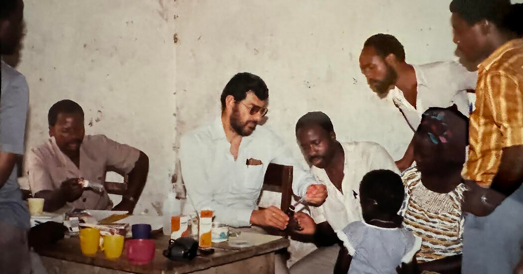
Part of a team flown in to fight the deadly virus in 1976, Dr. Breman also worked to stamp out tropical diseases like smallpox, malaria and Guinea worm.Dr. Joel Breman, a specialist in infectious diseases who was a member of the original team that helped combat the Ebola virus in 1976, died on April 6 at his home in Chevy Chase, Md. He was 87.His death was confirmed by his son, Matthew, who did not specify a cause.“We were scared out of our wits,” Dr. Breman, recollecting his pioneer mission, told a National Institutes of Health newsletter in 2014, as a new and even deadlier Ebola outbreak raged that year.Nearly 40 years earlier, his team of five had just landed in the interior of what is now the Democratic Republic of Congo, at a remote Roman Catholic mission hospital. They were up against a viral infection that had no name, whose origin was unknown, and that was accompanied by high fever and bleeding that led to a painful and quick death.Dr. Breman, dispatched by the Centers for Disease Control and Prevention, had only what he described to the N.I.H. as “the most basic protective equipment” against the disease, in contrast to the full-body spacesuit-like gear that was standard in the later outbreak. He and others on the team, laboring in intense heat and bitten by sand flies, “developed rashes and didn’t know if we would catch the virus too,” he said.But he calmly began deploying the techniques he had honed on earlier missions to Africa, on anti-smallpox initiatives in Guinea and Burkina Faso. He interviewed patients and witnesses, traveling from village to village and going from house to house. He and his colleagues, he recalled, soon determined that the infection was “spread by close contact with infected body fluids,” and that it had been propagated at a rural hospital that was using unsterilized needles.Over a long career, much of it spent at the Centers for Disease Control, the World Health Organization and the National Institutes for Health, Dr. Breman worked to stamp out deadly tropical diseases like smallpox, malaria and Guinea worm. But that initial Ebola outbreak, he told an interviewer in 2009, “was the scariest epidemic of my entire medical career and possibly of the last century.”We are having trouble retrieving the article content.Please enable JavaScript in your browser settings.Thank you for your patience while we verify access. If you are in Reader mode please exit and log into your Times account, or subscribe for all of The Times.Thank you for your patience while we verify access.Already a subscriber? Log in.Want all of The Times? Subscribe.

Read more →

Scientists discover the cellular functions of a family of proteins integral to inflammatory diseases

In a scientific breakthrough, Mount Sinai researchers have revealed the biological mechanisms by which a family of proteins known as histone deacetylases (HDACs) activate immune system cells linked to inflammatory bowel disease (IBD) and other inflammatory diseases.
This discovery, reported in Proceedings of the National Academy of Sciences (PNAS), could potentially lead to the development of selective HDAC inhibitors designed to treat types of IBD such as ulcerative colitis and Crohn’s disease.
“Our understanding of the specific function of class II HDACs in different cell types has been limited, impeding development of therapies targeting this promising drug target family,” says senior author Ming-Ming Zhou, PhD, Dr. Harold and Golden Lamport Professor in Physiology and Biophysics and Chair of the Department of Pharmacological Sciences at the Icahn School of Medicine at Mount Sinai. “Through our proof-of-concept study, we’re unraveling the mechanisms of class II HDACs, providing essential knowledge to explore their therapeutic potential for safer and more effective disease treatments.”
The Mount Sinai team focused specifically on class IIa HDACs, which exhibit more tissue-specific functions than class I HDACs, which act more broadly. Among the 18 histone deacetylases discovered to date in mammals, HDAC4 and HDAC7 — both class IIa HDACs — stand out for their roles in regulating the development and differentiation of Th17 cells. These cells are known for producing interleukin-17 (IL-17), a highly inflammatory cytokine associated with a spectrum of disorders, including IBD, multiple sclerosis, and rheumatoid arthritis. Given the strong correlation between excessive Th17 cell activity and human disease, scientists have focused on pharmacological or genetic interventions targeting HDAC4/7 to mitigate Th17 cell-mediated inflammation.
In their groundbreaking study, the Mount Sinai researchers delineated a previously unrecognized mechanism by which HDAC4 and HDAC7 operate independently yet cooperatively to govern Th17 cell differentiation and transcription. Transcription is the initial step of gene expression involving copying of the DNA sequence to generate RNA molecules; it is crucial for most biological processes.
“The role of class IIa HDACs in Th17 cells and inflammatory disease has been largely unexplored until now,” notes lead author Ka Lung Cheung, PhD, Assistant Professor of Pharmacological Sciences at Icahn Mount Sinai. “Mechanistically, we’ve discovered that class IIa HDACs orchestrate both gene transcriptional activation and repression to steer the process of Th17 cell differentiation. This significant revelation deepens our comprehension of the previously ambiguous role of class IIa HDACs in biology and human disease.”
As a critical aspect of their investigation, the research team found that a potent class IIa HDAC inhibitor, TMP269, influenced the differentiation of Th17 cells in a mouse model of ulcerative colitis. This pivotal discovery underscores the potential of pharmacological inhibition of class IIa HDACs as a promising therapeutic approach for addressing Th17-related inflammatory and autoimmune diseases, the study reported.
Expanding on this foundation of knowledge, researchers in the Zhou Lab and the Cheung Lab at Mount Sinai plan to concentrate on refining class IIa HDAC inhibitors with better efficacies for treating various types of Th17-mediated diseases.
“While our study primarily examined inflammatory bowel disease, specifically colitis,” says Dr. Zhou, “we believe our findings pave the way for extensive research into advanced therapies targeting severe inflammation in various other pathologies within the human body.”

Read more →

Lori and George Schappell, Long-Surviving Conjoined Twins, Die at 62

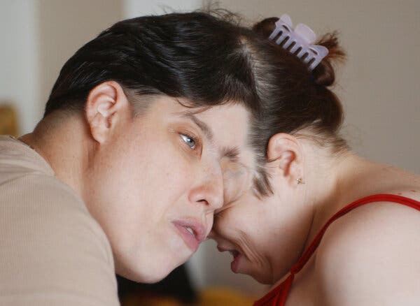
They were distinct people who pursued different lives. “Get past this already, everybody,” Lori said, “get past it and learn to know the individual person.”Lori and George Schappell, conjoined twins whose skulls were partly fused but who managed to lead independent lives, died on April 7 in Philadelphia. They were 62.Their death, at a hospital, was announced by a funeral home, which did not cite a cause.Dr. Christopher Moir, a professor of surgery at the Mayo Clinic, who has been on teams that separated six sets of conjoined twins — although none of them were joined at the head — said that when one of the Schappells died, the other would have almost certainly followed quickly.“Conjoined twins share circulation, he said, “so unless you somehow emergently divide their connection, it’s absolutely a fatal, nonviable process.”The Schappells lived much longer than had been expected when they were born as craniopagus twins, joined at the head, which is rare. They were cited as the second-oldest conjoined twins ever by Guinness World Records.They were connected at the sides of their foreheads and looked in opposite directions. Lori was able-bodied and pushed George, who had spina bifida, on a stool that had wheels. George was born female and changed her name in the 1990s to Reba, for the country singer Reba McEntire, but later identified as a trans male.The conjoined twins Lori, left, and George Schappell in 2003.John A. Secoges/Reading Eagle, via Associated PressWe are having trouble retrieving the article content.Please enable JavaScript in your browser settings.Thank you for your patience while we verify access. If you are in Reader mode please exit and log into your Times account, or subscribe for all of The Times.Thank you for your patience while we verify access.Already a subscriber? Log in.Want all of The Times? Subscribe.

Read more →

3 in 5 parents play short order cook for young children who don’t like family meal

While most parents of preschool and elementary aged children strive to give their children a balanced, nutritional diet, some of their strategies to promote healthy eating may backfire, a national poll suggests.
One in eight parents require children to eat everything on their plate, according to the University of Michigan Health C.S. Mott Children’s Hospital National Poll on Children’s Health. And while just one in three believe the standard American diet is healthy for kids, few have tried alternative, potentially more nutritional menus at home.
“Feeding young children can be difficult due to general pickiness, hesitancy to try unfamiliar foods and constantly evolving food preferences,” said Mott Poll co-director and Mott pediatrician Susan Woolford, M.D.
“The preschool and elementary age is an important time to establish healthy eating patterns. Yet parents’ concern about whether their child is eating enough or if they’re getting the nutrients they need may lead them to adopt practices that actually sabotage their efforts to get kids to have healthy eating habits in the short and long term.”
The nationally representative report is based on 1,083 responses of parents of children ages 3-10 surveyed in February.
More on poll findings:
Parents’ beliefs on nutritional diets vary
Just a third of parents think the standard American diet is healthy compared to half who seem to rank the Mediterranean higher in nutritional value. Still, few have tried alternative diets for their child.

“Parents may recognize the standard diet in the U.S. includes high amounts of saturated fats, added sugars, sodium, and refined carbohydrates, which can generate an excess intake of calories beyond nutritional needs and contribute to health problems,” Woolford said.
“However, despite this recognition and evidence suggesting that other diet options may help avoid many illnesses, only about 9% have tried the Mediterranean diet for their children and fewer have tried giving their children a vegetarian diet.”
Parents should ensure children are still getting adequate nutrition if they do try diets that eliminate certain food categories, she adds. Diets that limit animal products, for example, will require alternative protein sources such as meat substitutes, tofu, or legumes for children.
And while ketogenic diets have become popular among adults, they are generally not appropriate for children.
Family dining rules may promote or hinder a child’s healthy diet
Fifteen percent of parents say their family rule is that kids finish what’s on their plate, while more than half say children must try some of everything and a little less than a third say no to dessert if meals go unfinished.

But parents who try to force kids to eat may encourage portions that go beyond feeling full, Woolford cautions.
“Requiring children to eat everything on their plate, or withholding dessert unless all other foods are eaten, can lead to overconsumption, especially if portion sizes are too large for the child’s age,” she said.
She agrees with the recommendation that “parents provide, and the child decides.” This makes parents responsible for providing healthy options while allowing children to select which foods they will eat and the amount they want to consume.
Parents often play personal chef
Sixty percent of parents will make something separate if their child doesn’t like the food that’s on the dinner table — and this often leads to a less healthy alternative, Woolford says.
“Rather than allowing the child to choose an alternate menu, parents should provide a balanced meal with at least one option that their child is typically willing to eat,” she said.
“Then if their child chooses not to eat, parents should not worry as this will not cause healthy children any harm and they will be more likely to eat the options presented at the next meal.”
She points out that children learn through watching and imitating, so it’s beneficial for parents to model healthy eating through a well-balanced diet while their child’s eating habits and taste preferences mature.
Avoiding snacks between meals may also help children have a better appetite and increase willingness to eat offered foods.
Picky eating and protesting veggies among biggest battles
Parents describe their biggest challenges with making sure their child gets a healthy diet as the child being a picky eater, the higher cost of healthy food and food waste. Fewer say they don’t have time to prepare healthy food.
Nearly all parents polled report trying at least one strategy to get their child to eat vegetables as part of a healthy diet, such as serving vegetables every day, fixing vegetables how their child prefers, trying vegetables their child hasn’t had before and letting children pick out vegetables at the grocery store.
Others involve children with preparing the vegetables, hide vegetables in other foods or offer a reward for finishing vegetables.
“Unsurprisingly, parents said pickiness and getting kids to eat veggies were among major challenges during mealtimes,” Woolford said.
“Parents should try to include children in meal decisions, avoid pressuring food consumption and provide a variety of healthy options at each meal so kids feel more control.”
Right sizing food may be difficult
Portion size is key to mitigating the risk of childhood obesity, but it can be hard for parents to “right-size” a child portion.
In determining portion size for their child, nearly 70% of parents polled give their child slightly less than adults in the family while fewer let their child choose how much to take, use predetermined portions from the package or give their child the same portions as adults.
Woolford recommends parents seek sources to help. The U.S. Department of Agriculture, for example, provides a visual called “MyPlate” that can help parents estimate the recommended balance of the major food groups and offers guidance on estimating portion size.
Healthy eating starts at the grocery store
When grocery shopping or planning meals, parents polled say they try to limit the amount of certain foods to help their child to maintain a healthy diet, with more than half limiting foods with added sugars and processed foods.
But it may be difficult to identify unhealthy food. Added sugars or processing may be present in foods marketed or packaged as healthy, Woolford says.
Parents should read labels, avoiding the marketing on the front of packages and focusing instead on the details on the back. They should pay particular attention to nutrition information and ingredient lists — especially if they’re long with unrecognizable items — as well as sodium, added sugars, and fat.
Woolford also encourages involving children in grocery trips, spending time in the produce section and asking them what they may like to try.
“Have them help in the process of choosing the healthiest options, not ones that necessarily directly advertise to children, but foods that they are willing to try that are lower in sugar, fat and salt,” she said.
“Spend most of the time in the produce section and try to make it fun by maybe selecting new options from different parts of the world that they haven’t tried before.”

Read more →

Study explores possible future for early Alzheimer’s diagnostics

Digital memory test and a blood sample — this combination will be tested for its potential to identify early Alzheimer’s disease in a new research study. Over a hundred healthcare centers are part of the study that is now inviting participants to sign up. At least 3,000, preferably many more participants are needed for the study to be successful.
The REAL AD study is the first of its kind in terms of focus and scope. Principal investigators are the University of Gothenburg and the Västra Götaland Region, VGR, which represents a model region for Swedish healthcare. All hundred-plus care centers within VGR Närhälsan, one of the largest primary care providers in Sweden, are included in the study, together with some additional sampling sites.
REAL AD addresses all people aged 50-80 who do not have a diagnosis of dementia and who can go to a care center within VGR Närhälsan. Anyone who meets the criteria can participate regardless of which health center they are listed at.
Tests of memory and thinking ability at home
Starting point is a digital study portal, available in Swedish, English, Finnish and Arabic, where participants receive all information about the study and clear instructions about the next steps. First, cognition, i.e. memory and thinking ability, is tested at home using digital tools for three months. Participants are then invited to provide a blood sample at any of the 111 sampling points around VGR. Participants can complete the digital cognition tests in three additional rounds, after 18, 27 and 36 months, and provide a second blood sample after 18 months. The tests are relatively quick and can be done in all four languages.
The study is led by Michael Schöll, professor at Gothenburg University and research group leader in close collaboration with the co-investigators and a team of project leaders.
– REAL AD is a seriously ambitious project, and it has been an enormous challenge to democratize the study design. It must reflect both urban and rural areas, be accessible to as many participants as possible and involve the entire VGR Närhälsan, which means that even the most remote healthcare centers must be able to participate in terms of sample handling and transport, he says.

Signs of Alzheimer’s in a simple blood test
Central hub of the study is a laboratory environment in neurochemistry, located at the Sahlgrenska Academy at the University of Gothenburg and the Sahlgrenska University Hospital Mölndal, with professors Kaj Blennow and Henrik Zetterberg at the helm. The researchers will study so-called Alzheimer’s biomarkers in the participants’ blood samples, which have been shown to be early signs of the disease.
In addition, a separate study will then be conducted enrolling a smaller number of randomly selected participants who are also thoroughly examined clinically at Sahlgrenska University Hospital to confirm the results of the digital cognition tests and blood analyses. The clinical part of the study is carried out at the university hospital memory clinic in collaboration with professor and senior physician Silke Kern.
Knowledge base for healthcare and research
The research focuses on the potential for early diagnosis based on digital cognitive tests and blood markers. If the combination of the tools works to detect early signs of disease in the general population, the hope is that they will be used in primary care in the future to follow individuals over time and identify Alzheimer’s with greater certainty and much earlier than is often the case today.
“The need for earlier diagnosis is widely accepted, also in view of the new treatments that are around the corner. Many if not most diagnoses are made in primary care in Sweden, so diagnostics must be strengthened there, and knowledge is needed about whether it will be feasible to screen for Alzheimer’s in the general population. In the short term, society does not have the resources to establish a lot of new specialized memory clinics,” says Michael Schöll.

“The study is important, and the timing is perfect. We are closer than ever before to a treatment option for Alzheimer’s, but we are not sufficiently prepared,” he says.
The study needs at least 3,000 participants but has capacity to enroll up to 10,000 volunteers. It is accompanied by information via Närhälsan and several other marketing efforts.
Research ethics do not allow researchers to share individual information with participants since experimental tools are used. Individuals do thus not receive a cognitive rating or diagnosis, and their participation is unpaid.
“What we are offering is participation in a community where we will actively inform about progresses in Alzheimer’s research, also via information meetings, which we know many people are asking for. By participating in the study, you also help our healthcare to prepare for an enormous challenge,” concludes Michael Schöll.

Read more →

New approach to tackle muscle loss in aging

Sarcopenia, a prevalent condition among the elderly, is characterized by a progressive decline in muscle mass and function, which can significantly diminish their quality of life and increase the risk of falls, injuries, and dependency. A greater understanding of this phenomenon is crucial for devising effective strategies that enable the ageing population to remain healthy and independent.
A study co-led by Dr. Antonio Zorzano, from IRB Barcelona, and Dr. David Sebastián, now a Professor at the University of Barcelona (UB), reveals that a protein named TP53INP2 could be pivotal in combatting sarcopenia. Conducted in partnership with Parc Sanitari Sant Joan de Déu, the study indicates that increased levels of this protein in muscle correlate with greater muscular strength and healthier ageing in humans.
Loss of muscle mass typically starts around the age of 55, and it has a detrimental effect on people’s ability to perform daily tasks and on their health. Sarcopenia leads to increased frailty physical disability and the need for long-term care.
By conducting experiments on mouse models and analysing human muscle tissue samples, the researchers observed a decrease in TP53INP2 levels with age. However, artificially boosting the presence of this protein in muscles — whether continuously in young mice or temporarily in older mice through genetic engineering techniques — led to a significant improvement in both muscle mass and function.
These findings suggest that promoting the activity of TP53INP2 and, consequently, autophagy in the muscle, could be an effective strategy to tackle sarcopenia, thereby contributing to a more active and healthier ageing process. In studies involving humans, high levels of TP53INP2 were associated with greater strength and enhanced physical performance, thereby highlighting the potential of this protein as an indicator of healthy ageing.
“This study not only underscores the importance of keeping autophagy active in muscles to prevent muscle mass loss but also gives us hope regarding potential treatments that could improve the condition or at least mitigate the effects of ageing on our muscles,” explains Dr. Zorzano, who is also a Professor at the Faculty of Biology at the UB and a member of CIBERDEM. “Furthermore, the activation of autophagy through TP53INP2 improved the quality of mitochondria, essential organelles in energy generation — a process that we had previously shown to be disrupted during ageing,” highlights Dr. Sebastián, Professor in the Department of Biochemistry and Physiology at the Faculty of Pharmacy and Food Sciences of the UB.
The partnership with Parc Sanitari Sant Joan de Déu has been pivotal, providing access to biologic samples from well-characterized patients. This allowed the researchers to establish the link between TP53INP2 muscle expression and healthy ageing in humans. This advance not only paves the way for further research in ageing but also points to potential interventions to greatly improve the quality of life of seniors, helping them to maintain their independence and vitality.
Moving forward, the researchers will continue to explore whether TP53INP2 levels in each person are influenced by genetic factors and physical activity, or, whether other habits, like nutrition, play a significant role.
This work has been possible thanks to funding from the Ministry of Science, Innovation, and Universities, the Generalitat de Catalunya, and the Carlos III Health Institute.

Read more →

Predicting cardiac arrhythmia 30 minutes before it happens

Atrial fibrillation is the most common cardiac arrhythmia worldwide with around 59 million people concerned in 2019. This irregular heartbeat is associated with increased risks of heart failure, dementia and stroke. It constitutes a significant burden to healthcare systems, making its early detection and treatment a major goal. Researchers from the Luxembourg Centre for Systems Biomedicine (LCSB) of the University of Luxembourg have recently developed a deep-learning model capable of predicting the transition from a normal cardiac rhythm to atrial fibrillation. It gives early warnings on average 30 minutes before onset, with an accuracy of around 80%. These results, published in the scientific journal Patterns, pave the way for integration into wearable technologies, allowing early interventions and better patient outcomes.
During atrial fibrillation, the heart’s upper chambers beat irregularly and are out of sync with the ventricles. Reverting to a regular rhythm can require intensive interventions, from shocking the heart back to normal sinus rhythm to the removal of a specific area responsible for faulty signals. Being able to predict an episode of atrial fibrillation early enough would allow patients to take preventive measures to keep their cardiac rhythm stable. However, current methods based on the analysis of heart rate and electrocardiogram (ECG) data are only able to detect atrial fibrillation right before its onset and do not provide an early warning.
“In contrast, our work departs from this approach to a more prospective prediction model,” explains Prof. Jorge Goncalves, head of the Systems Control group at the LCSB. “We used heart rate data to train a deep learning model that can recognise different phases — sinus rhythm, pre-atrial fibrillation and atrial fibrillation — and calculate a “probability of danger” that the patient will have an imminent episode.” When approaching atrial fibrillation, the probability increases until it crosses a specific threshold, providing an early warning.
This artificial intelligence model, called WARN (Warning of Atrial fibRillatioN), was trained and tested on 24h-recordings collected from 350 patients at Tongji Hospital (Wuhan, China) and gave early warnings, on average 30 minutes before the start of atrial fibrillation, with great accuracy. Compared to previous work on arrhythmia prediction, WARN is the first method to provide a warning far from onset.
“Another interesting aspect is that our model has a high performance using only R-to-R intervals, basically just heart rate data, that can be acquired from easy-to-wear and affordable pulse signal recorders such as smartwatches,” highlights Dr Marino Gavidia, first author of the publication, who worked on this project during his PhD within the Systems Control group and the Doctoral Training Unit CriTiCS (see box below). “These devices can be used by patients on a daily basis, so our results open possibilities for the development of real-time monitoring and early warnings from comfortable wearable devices,” adds Dr Arthur Montanari, a LCSB researcher involved in the project.
Additionally, the deep-learning model developed by the researchers could be implemented in smartphones to process the data from a smartwatch. This low computational cost makes it ideal for integration into wearable technologies. The long-term objective is for patients to be able to continuously monitor their cardiac rhythm and receive early warnings that can provide sufficient time to take antiarrhythmic medication or use some targeted treatments to prevent the onset of atrial fibrillation. This in turn would reduce emergency interventions and improve patient outcomes.
“Moving forward, we will focus on developing personalised models. The daily use of a simple smartwatch constantly provides new information on personal heart dynamics, enabling us to continuously refine and retrain our model for that patient to achieve enhanced performance with even earlier warnings,” concludes Prof. Goncalves. “Eventually, this approach could even lead to new clinical trials and innovative therapeutic interventions.”

Read more →

Protein network dynamics during cell division

An international team led by researchers at the University of Toronto has mapped the movement of proteins encoded by the yeast genome throughout its cell cycle. This is the first time that all the proteins of an organism have been tracked across the cell cycle, which required a combination of deep learning and high-throughput microscopy.
The team applied two convolutional neural networks, or algorithms, called DeepLoc and CycleNet, to analyze images of millions of live yeast cells. The result was a comprehensive map identifying where proteins are located and how they move and change in abundance within the cell during each phase of the cell cycle.
“We found proteins that regularly increase and decrease in concentration within the cell tend to be involved in regulating the cell cycle, while proteins with predictable movement through the cell tend to facilitate the cycle’s biophysical implementation” said Athanasios Litsios, first author on the study and postdoctoral fellow at U of T’s Donnelly Centre for Cellular and Biomolecular Research.
The study was recently published in the journal Cell.
The cell cycle is understood to be the stages through which a cell progresses to ultimately divide into separate cells. It is this process that underlies the proliferation of life — and is ongoing in all living things.
At the molecular level, the cell cycle depends on the coordination of many proteins to ferry the cell from growth and DNA replication all the way to cell division. Dysregulation of proteins can throw a wrench into the cell cycle, with its disruption potentially leading to diseases like cancer.
The researchers observed that around a quarter of mapped yeast proteins followed regular patterns of emergence and disappearance or movement to specific areas of the cell. Most proteins followed these patterns for either concentration or movement, but not both.

“We identified around 400 proteins with only periodic localization during the cell cycle and around 800 with only periodic concentration,” said Litsios. “This means that proteins are being regulated at multiple levels to ensure the cell cycle occurs as programmed.”
The research team used fluorescence microscopy to track around 4,000 proteins in images of yeast cells to classify the cell cycle phase as well as the location of proteins within 22 categorized areas of the cell, such as the nucleus, cytoplasm and mitochondria. Phase and protein location identification were automated through the use of convolutional neural networks, with a cell cycle phase prediction accuracy of more than 93 per cent.
“We analyzed images of more than 20 million live yeast cells, which we assigned to different cell cycle stages using machine learning,” said Brenda Andrews, principal investigator on the study and university professor of molecular genetics at the Donnelly Centre and the Temerty Faculty of Medicine. “We then developed and applied a second computational pipeline to survey how proteins change in localization and concentration during the cell cycle. This study produced a unique dataset that offers a genome-scale view of molecular changes that occur during cell division.”
“The yeast cell is a great model for eukaryotic biology,” said Litsios. “There are certain things we can do with yeast cells but not with other organisms that are either more simple or complex. We can use yeast cells to observe processes at a large-scale, which makes it the perfect organism for studying the cell cycle — in the hopes of better understanding the human cell cycle.”
This research was supported by the Canada Foundation for Innovation, the Canadian Institutes of Health Research, the National Institutes of Health and the Ontario Research Fund.

Read more →