Larry Young, Who Studied the Chemistry of Love, Dies at 56

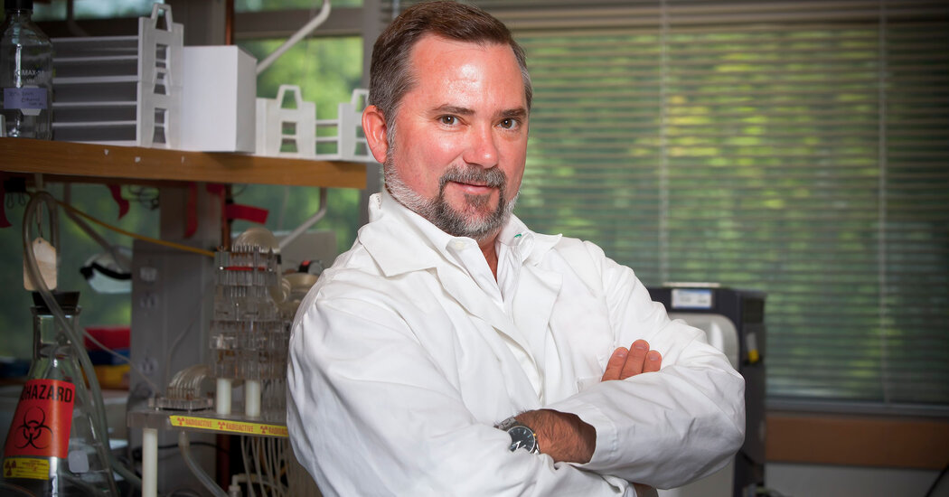
Professor Young’s experiments with prairie voles revealed what poets never could: how the brain processes that fluttering feeling in the heart.Prairie voles are stocky rodents and Olympian tunnellers that surface in grassy areas to feast on grass, roots and seeds with their chisel-shaped teeth, sprouting migraines in farmers and gardeners.But to Larry Young, they were the secret to understanding romance and love.Professor Young, a neuroscientist at Emory University in Atlanta, used prairie voles in a series of experiments that revealed the chemical process for the pirouette of heart-fluttering emotions that poets have tried to put into words for centuries.He died on March 21 in Tsukuba, Japan, where he was helping to organize a scientific conference. He was 56. The cause was a heart attack, his wife, Anne Murphy, said.With their beady eyes, thick tails and sharp claws, prairie voles are not exactly cuddly. But among rodents, they are uniquely domestic: They are monogamous, and the males and females form a family unit to raise their offspring together.“Prairie voles, if you take away their partner, they show behavior similar to depression,” Professor Young told The Atlanta-Journal Constitution in 2009. “It’s almost as if there’s withdrawal from their partner.”That made them ideal for laboratory studies examining the chemistry of love.Males and female prairie voles are known to form a family unit to raise their offspring together.Todd Ahern/Emory University, via Associated PressIn a study published in 1999, Professor Young and his colleagues exploited the gene in prairie voles associated with the signaling of vasopressin, a hormone that modulates social behavior. They boosted vasopressin signaling in meadow voles, which are highly promiscuous.“With their vasopressin receptor levels boosted in this brain region,” Scientific American reported, “these normally solitary and promiscuous voles gained a new propensity to cuddle with a mate.”Headline writers were amused. “Gene Swap Turns Lecherous Mice Into Devoted Mates,” The Ottawa Citizen declared. The Fort Worth Star-Telegram: “Genetic Science Makes Mice More Romantic.” The Independent in London: “‘Perfect Husband’ Gene Discovered.”Professor Young followed up with other prairie vole studies that focused on oxytocin, a hormone that stimulates contractions during childbirth and is involved in the bonding between mothers and newborns.“Because we knew that oxytocin was involved in mother-infant bonding, we explored whether oxytocin might be involved in this partner bonding,” he said in an interview with the Australian Broadcasting Corporation in 2019.It was.“If you take two prairie voles, a male and a female, put them together, and this time you don’t let them mate and you just give them a little bit of oxytocin, they will bond,” Professor Young said. “So that was our first set of experiments to show that oxytocin was involved in things other than maternal bonding.”He also injected female prairie voles with a drug that blocks oxytocin, which made them temporarily polygamous.“Love doesn’t really fly in and out,” Professor Young wrote in “The Chemistry Between Us: Love, Sex and the Science of Attraction” (2012, with Brian Alexander). “The complex behaviors surrounding these emotions are driven by a few molecules in our brains. It’s these molecules, acting on defined neural circuits, that so powerfully influence some of the biggest, most life-changing decisions we’ll ever make.”Professor Young always cautioned that prairie voles weren’t humans (obviously). But in the same way that mouse studies have led to medical breakthroughs, he thought his research with prairie voles had intriguing implications.“Perhaps genetic tests for the suitability of potential partners will one day become available, the results of which could accompany, and even override, our gut instincts in selecting the perfect partner,” Professor Young wrote in Nature. He added, “Drugs that manipulate brain systems at whim to enhance or diminish our love for another may not be far away.”In recent years, Professor Young was exploring whether increasing oxytocin in certain conditions would help children with autism who struggle in social interactions.Professor Young in 2021. He became interested in genetics after dissecting a fruit fly in a biochemistry class.Center for Translational Social NeuroscienceLarry James Young was born on June 16, 1967, in Sylvester, a rural town in southwest Georgia. His father, James Young, and his mother, Margaret (Giddens) Young, were peanut farmers.As a child, he had a cow named Bessie.“It was a really rural lifestyle,” Ms. Murphy said. “His aspiration was to go work at the gas station down the street and become a manager.”He attended the University of Georgia on a Pell Grant with plans to become a veterinarian. One day, in biochemistry class, he dissected a fruit fly.“And that’s when he fell in love with genetics and just wanted to figure out the genetic basis of behavior,” Ms. Murphy said. “That’s what drove him the rest of his life.”After graduating in 1989 with a degree in biochemistry, he received a Ph.D. in zoology from the University of Texas at Austin in 1994, and then took a postdoctoral position at Emory. He never left the university, eventually becoming division chief of behavioral neuroscience and psychiatric disorders at the Emory National Primate Research Center.Professor Young married Michelle Willingham in 1985; they later divorced. He married Ms. Murphy in 2002. She is a neuroscientist at Georgia State University in Atlanta.In addition to his wife, he is survived by three daughters from his first marriage, Leigh Anna, Olivia and Savannah Young; two stepsons, Jack and Sam Murphy; a brother, Terry Young; and two sisters, Marcia Young-Whitacre and Robyn Hicks.Professor Young in 2010. He predicted that one day there might be a drug that would increase the urge to fall in love.Emory UniversityAround Emory’s campus, Professor Young was also known as the Love Doctor. He was popular on Valentine’s Day — not just with Ms. Murphy. Reporters around the world would ask him to explain the chemistry of romance.One day, he said, there might even be a drug that would increase the urge to fall in love.“It would be completely unethical to give the drug to someone else,” he told The New York Times, “but if you’re in a marriage and want to maintain that relationship, you might take a little booster shot yourself every now and then.”

Read more →

Robert Oxnam, China Scholar Beset by Multiple Personalities, Dies at 81

Through psychotherapy, recounted in a memoir, he learned that he had 11 personalities, or fractured parts of his identity. One of them told of childhood abuse.Robert B. Oxnam, an eminent China scholar who learned through psychotherapy that his years of erratic behavior could be explained by the torment of having multiple personalities, died on April 18 at his home in Greenport, N.Y., on the North Fork of Long Island. He was 81.His wife, Vishakha Desai, said the cause was complications of Alzheimer’s disease.In the 1ate 1980s, Dr. Oxnam was president of the Asia Society, a television commentator and an accomplished sailor. But his psyche was exceedingly frail. He had myriad problems, including intermittent rages, bulimia, memory blackouts and depression, but it was for excessive drinking that he first sought treatment, from Dr. Jeffery Smith, a psychiatrist.The first personality to emerge in that therapy was Tommy, an angry boy, followed by others, like Bobby, an impish teenager, and Baby, who revealed what appeared to have been abuse when Dr. Oxnam was very young.In his 2005 book, “A Fractured Mind: My Life With Multiple Personality Disorder,” Dr. Oxnam recalled the session when Tommy first spoke to Dr. Smith. All that Dr. Oxnam could remember from the 50-minute session, he wrote, was telling the psychiatrist that he didn’t think the therapy was working for him. But Dr. Smith told him that he had been speaking to Tommy all that time.“He’s full of anger,” Dr. Smith told him. “And he’s inside you.”“You’re kidding?” Dr. Oxnam replied.His 11 personalities took up residence inside Dr. Oxnam’s brain and acted out in real life, and nearly all appeared during therapy with Dr. Smith. Wanda had a Buddhist-like presence who was once submerged in the cruel personality known as the Witch. Bobby, who loved Rollerblading with bottles balanced on his head, had an affair with a young woman, a revelation that startled Dr. Oxnam and his wife.In his 2005 memoir, Dr. Oxnam recounted therapy sessions in which his psychiatrist would find himself speaking to one or another of Dr. Oxnam’s multiple personalities. Hachette Books“It can get really noisy in there, a din,” Dr. Oxnam told The New York Times in a profile about him in 2005.Dr. Smith said in an interview, “There was a lot going on in his head, like if one personality was about to do something destructive, another was liable to say, ‘That’s not OK.’”In the book, Dr. Oxnam described how the personalities inhabited a vivid internal world — a castle with rooms, dungeons, walkways and a library behind iron-locked doors. Tommy described the castle to Dr. Smith, telling him it was “Middle Ages-style, standing on a large hill,” and was made of “gray stones and topped with long walkways and towers at the corners.”Dr. Oxnam did not reveal in the book who had abused him. But through Dr. Smith’s conversations with Baby, he wrote, Baby was “crystal clear” that the severe traumas that Robert had experienced as a boy were not inflicted by his parents.“Our vow to hide the abusers’ identity was easier said than done,” Dr. Oxnam wrote. “To be honest, when rage reigns in the Castle, it has been hard to keep quiet. But over time, I have found that withholding the abusers’ names, and refusing to stay in an angry state, actually helps the healing process.”Therapy eventually helped merge the 11 personalities into a more manageable three, he said.Dr. Oxnam with Dr. Jeffery Smith, a psychiatrist who helped identify his multiple personalities, in 2005.Hiroko Masuike/The New York TimesMultiple personality disorder — now called dissociative identity disorder — affects about one percent of the population and usually emerges after severe trauma early in life, said Dr. David Spiegel, a professor of psychiatry at the Stanford University School of Medicine. He coined the name change, which appeared in the fifth edition of “Diagnostic and Statistical Manual of Mental Disorders” (2013).Dr. Spiegel said that the personalities that Dr. Oxnam experienced are better known as fragmentations of his identity.“You’re a different guy talking to me than you are at a party, but there’s a smooth continuity between the two,” he said in an interview. “In people with D.I.D., they experience themselves as different components that get filed into different identities.”The disorder was the basis for the best-selling 1973 book “Sybil,” by Flora Rheta Schreiber, about a woman who was said to have had 16 personalities. It was adapted for a 1976 made-for-television movie starring Sally Field and Joanne Woodward.Robert Bromley Oxnam was born on Dec. 14, 1942, in Los Angeles. His father, also named Robert, was president of Drew University in New Jersey and, before that, the Pratt Institute in Brooklyn. His mother, Dalys (Houts) Oxnam, ran the household.He graduated from Williams College in Massachusetts in 1964 with a bachelor’s degree in history. His father urged him to consider graduate work in international studies, and Robert surmised that China would be playing a greater role on the world stage. At Yale University, he earned a master’s degree in East Asian studies in 1966 and a Ph.D. in 1969, with a dissertation on China’s 17th-century Oboi Regency.“For two years, I ferreted through court documents, biographies and local histories, all in classical Chinese, trying to find the patches of historical forest in the midst of dense linguistic trees” on the Oboi Regency, he wrote in 2014 in Perspectives on History, the American Historical Association’s newsmagazine.Dr. Oxnam at his home on the Upper West Side of Manhattan in 2005. The artwork was part of a collection of 18th- and 19th-century Japanese wood block prints. He was president of the Asia Society for 11 years. Hiroko Masuike/The New York TimesIn 1969, Dr. Oxnam began a six-year run as an associate professor of Chinese and Japanese history at Trinity College in Connecticut before being recruited to the Asia Society, a cultural, educational and research organization in Manhattan. He was the founder of its China Council, which issued papers and briefs about China as it began reopening to the West after President Richard M. Nixon’s visit there in 1972.As director of the society’s Washington center from 1979 to 1981, Dr. Oxnam started the organization’s first contemporary affairs department, to focus on government policy. He was named the society’s president in 1981. Over the next 11 years, he expanded its corporate, contemporary affairs and cultural programming to include 30 Asian countries and helped guide the opening of the Asia Society Hong Kong Center in 1990.Marshall Bouton, a former Asia Society executive, said Dr. Oxnam had helped transform the organization “from a gathering spot for Upper East Siders who were interested in Asia to a more professional organization that dealt with Asia’s most pressing challenges.”Mr. Bouton said that he had not been aware of the full extent of Dr. Oxnam’s alcoholism and that he had had inklings about his behavioral problems. He said that it was remarkable that Dr. Oxnam had been able to work through them.But in 1992, Dr. Oxnam told the society’s board that he was going to resign.“The Bob part of me was touched that they pressured me to reconsider,” he wrote in his book. But he left.In addition to his wife, whom he married in 1993 and who was president of the Asia Society from 2004 to 2012, his survivors include his daughter, Deborah Betsch, and his son, Geoff Oxnam, both from his marriage to Barbara Foehl, which ended in divorce in 1993, and four grandchildren.After leaving the Asia Society, Dr. Oxnam hosted and wrote a series about China for “The MacNeil/Lehrer NewsHour” on PBS in 1993; taught a graduate seminar on U.S.-Asia relations at Beijing University from 2003 to 2004 (where his Bobby personality lectured in Chinese), and advised the Bessemer Trust, a wealth management firm.He also wrote “Ming: A Novel of Seventeenth-Century China” (1995) and turned to art, crafting found wood into sculptures inspired by Chinese philosophy and taking photographs of glacial rocks.“In Chinese tradition, the term ‘qi’ has many meanings, but for me, it means an invisible but palpable source of creative energy,” Dr. Oxnam told Hamptons Art Hub, an online publication, in 2018. He added, “I have suffered from dissociation all my life, but somehow the linkage between ‘qi’ and art has given me focus and hope.”

Read more →

Scientists track ‘doubling’ in origin of cancer cells

Working with human breast and lung cells, Johns Hopkins Medicine scientists say they have charted a molecular pathway that can lure cells down a hazardous path of duplicating their genome too many times, a hallmark of cancer cells.
The findings, published May 3 in Science, reveal what goes wrong when a group of molecules and enzymes trigger and regulate what’s known as the “cell cycle,” the repetitive process of making new cells out of the cells’ genetic material.
The findings could be used to develop therapies that interrupt snags in the cell cycle, and have the potential to stop the growth of cancers, the researchers suggest.
To replicate, cells follow an orderly routine that begins with making a copy of their entire genome, followed by separating the genome copies, and finally, dividing the replicated DNA evenly into two “daughter” cells.
Human cells have 23 pairs of each chromosome — half from the mother and half from the father, including the sex chromosomes X and Y — or 46 total, but cancer cells are known to go through an intermediate state that has double that number — 92 chromosomes. How this happens was a mystery.
“An enduring question among scientists in the cancer field is: How do cancer cell genomes get so bad?” says Sergi Regot, Ph.D., associate professor of molecular biology and genetics at the Johns Hopkins University School of Medicine. “Our study challenges the fundamental knowledge of the cell cycle and makes us reevaluate our ideas about how the cycle is regulated.”
Regot says cells that are stressed after copying the genome can enter a dormant, or senescent stage, and mistakenly run the risk of copying their genome again.

Generally and eventually, these dormant cells are swept away by the immune system after they are “recognized” as faulty. However, there are times, especially as humans age, when the immune system can’t clear the cells. Left alone to meander in the body, the abnormal cells can replicate their genome again, shuffle the chromosomes at the next division, and a growing cancer begins.
In an effort to pin down details of the molecular pathway that goes awry in the cell cycle, Regot and graduate research assistant Connor McKenney, who led the Johns Hopkins team, focused on human cells that line breast ducts and lung tissue. The reason: These cells generally divide at a more rapid pace than other cells in the body, increasing the opportunities to visualize the cell cycle.
Regot’s lab specializes in imaging individual cells, making it especially suited to spot the very small percentage of cells that don’t enter the dormant stage and continue replicating their genome.
For this new study, the team scrutinized thousands of images of single cells as they went through cell division. The researchers developed glowing biosensors to tag cellular enzymes called cyclin dependent kinases (CDKs), known for their role in regulating the cell cycle.
They saw that a variety of CDKs activated at different times during the cell cycle. After the cells were exposed to an environmental stressor, such as a drug that disrupts protein production, UV radiation or so-called osmotic stress (a sudden change in water pressure around cells), the researchers saw that CDK 4 and CDK 6 activity decreased.
Then, five to six hours later, when the cells started preparations to divide, CDK 2 was also inhibited. At that point, a protein complex called the anaphase promoting complex (APC) was activated during the phase just before the cell pulls apart and divides, a step called mitosis.

“In the stressed environment in the study, APC activation occurred before mitosis, when it’s usually been known to activate only during mitosis,” says Regot.
About 90% of breast and lung cells leave the cell cycle and enter a quiet state when exposed to any environmental stressors.
In their experimental cells, not all of the cells went quiet.
The research team watched as about 5% to 10% of the breast and lung cells returned to the cell cycle, dividing their chromosomes again.
Through another series of experiments, the team linked an increase in activity of so-called stress activated protein kinases to the small percentage of cells that skirt the quiet stage and continue to double their genome.
Regot says there are ongoing clinical trials testing DNA-damaging agents with drugs that block CDK. “It’s possible that the combination of drugs may spur some cancer cells to duplicate their genome twice and generate the heterogeneity that ultimately confers drug resistance,” says Regot.
“There may be drugs that can block APC from activating before mitosis to prevent cancer cells from replicating their genome twice and prevent tumor stage progression,” says Regot.
Other researchers who contributed to the study include Yovel Lendner, Adler Guerrero-Zuniga, Niladri Sinha, Benjamin Veresko and Timothy Aikin from Johns Hopkins.
Funding for the study was provided by the National Institutes of Health National Institute of General Medical Sciences (T32-GM007445, 1R35GM133499) and National Cancer Institute (1R01CA279546), the National Science Foundation and the American Cancer Society.

Read more →

Cancer patients gain important benefits from genome-matched treatments

In 2016, The Jackson Laboratory (JAX), a National Cancer Institute-designated Cancer Center and at the forefront of cancer research, launched the Maine Cancer Genomics Initiative (MCGI) to bring the latest progress in cancer care to rural Maine patients. Now, after successfully expanding access to genome tumor testing and targeted cancer treatments throughout Maine, the MCGI team provides compelling evidence that genome-matched treatments can provide significant patient benefit.
The MCGI report, published recently in npj Precision Oncology, presents data showing that only 17% of patients received genome-matched treatment through MCGI, signaling a large gap between testing and delivery of treatment based on genomic information. However, those who did receive genome-matched treatment were 31% less likely to die within one year compared to those who did not receive matched treatment. While this is an observational study, the findings clearly point to the potential for a significant one-year survival benefit from genomic tumor testing and matched treatments.
In this observational study, there were many reasons why cancer patients didn’t receive genome-matched treatments after the program sequenced the tumor’s DNA. A certain percentage of patients did not have an actionable tumor variant detected, so they received standard of care.
“For the rest, it was a matter of care delivery,” said Jens Rueter, M.D., chief medical officer of JAX and medical director of MCGI. “Patients may have had an actionable tumor variant but only through participation in a clinical trial that isn’t available in rural Maine, or a patient’s community hospital may not have been able to deliver a treatment that’s already on the market.”
Spearheaded by Rueter, who is also associate director for translational education at JAX Cancer Center and Edison Liu, M.D., former JAX President and CEO., the impetus for launching MCGI in 2016 was the lack of local access to recently developed genomic testing and targeted therapy strategies for cancer patients in Maine. In addition, most patients lacked the time and means to travel to Boston or New York for care. Therefore, JAX created MCGI to bring the latest technology in precision oncology and treatment to patients.
In only four years, through 2020, MCGI had partnered with every oncology practice in Maine (there are 13 of them) and enrolled more than 1,600 of their patients. Leah Graham, program director of MCGI, described that early work focused on providing genomic education to oncologists and other healthcare professionals, free access to genomic tumor testing for their patients, and detailed consultation about test results with precision oncology experts through a genomic tumor board.
Follow up with the MCGI patients revealed that of the 1,052 who did not receive genome-matched treatments, 399 (37.9%) died within 365 days of consent. This compares with 30.6% (63 of 206) in the genome-matched group. After adjusting for baseline characteristics, the analysis showed that the genome-matched group was 31% less likely to die within the first year than those who received standard care, even though only 9% were able to participate in a clinical trial, a smaller share than previously reported by other studies and might be explained by the rurality of Maine.
The percentage of tested patients who received genome-matched treatments in the MCGI study — 17% — exactly matched the figure found in a larger 2019 Veteran Affairs study, suggesting that delivery of cancer care is not just limited to Maine. Moving forward, the MCGI program will focus more on enabling effective precision oncology care delivery, whether it’s through its Genomic Tumor Board program, providing access to more biomarker-driven clinical trials within Maine or using mobile outreach to bring treatments directly to patients who might not be able to access it otherwise.
The study carries with it several limitations. Patients were primarily white and non-Hispanic, reflecting the population characteristics in Maine; the genomic tumor testing was provided free of charge, potentially expanding its use; the study population also had variable cancer sites and stages.
“Nonetheless, we’ve been offering this program for seven years now, and we can see some really positive impacts on patient outcomes,” said Rueter. “And in the future, we want to do on a population level what we’re now doing in Maine with MCGI, meaning that every patient receives genomic tumor testing and a thorough biomarker analysis. How we deliver care and how we expand access to clinical trials through MCGI can be a blueprint for other states across the country, especially those with significant rural areas.”

Read more →

Small molecule shows early-stage promise for repairing myelin sheath damage

When treated with a novel protein function inhibitor called ESI1, mice that mimic the symptoms of multiple sclerosis (MS) and lab-prepared human brain cells both demonstrated the ability to regenerate vital myelin coatings that protect healthy axon function.
This breakthrough, published May 2, 2024, in Cell, appears to overcome difficulties that have long frustrated previous attempts to reverse a form of nerve damage that robs people with MS of motor control and gradually blunts cognitive functions for many people as they age.
“Currently, there are no effective therapies to reverse myelin damage in devastating demyelinating diseases such as MS,” says corresponding author Q. Richard Lu, PhD, a top brain research expert at Cincinnati Children’s. “These findings are significant as they offer new pathways for treatment that potentially shift the therapeutic focus from just managing symptoms to actively promoting repair and regeneration of myelin.”
Promoting healing by clearing a roadblock
A critical insight driving the new findings was observing that brain regions damaged by MS still possessed a type of cell needed to repair myelin damage, but the disease activates other cell types and signals that combine forces to silence the repair function.
These useful cells in the brain, called oligodendrocytes, are responsible for producing myelin sheaths that wrap around cable-like parts of nerve cells called axons, much like the plastic insulation around a wire. When the protective myelin gets damaged, be it by disease or the wear and tear of age, nerve signaling gets disrupted. Depending on where the damaged nerves lead, the disruptions can affect movement, vision, thinking and so on.
Essentially, the research team found a way to unsilence the silenced repair process, setting the oligodendrocytes (OLs) free to do their jobs.

Pinning down the genetic changes and signals involved in the repair silencing process and finding a small molecule compound that can reverse the silencing was a complex undertaking. The project, which spanned over five years, involved four co-first authors and 29 contributing co-authors from Cincinnati Children’s, the University of Cincinnati, and 14 other institutions including universities in Australia, China, Germany, India, Singapore, and the United Kingdom.
Among the team’s key findings:
Identifying the mechanism preventing myelin production in MS
Analysis of stored autopsy tissues revealed that OLs within MS lesions lacked an activating histone mark called H3K27ac, while expressing high levels of two other repressive histone marks H3K27me3 and H3K9me3 associated with silencing gene activity.
Finding a compound that can reverse the silencing
The research team scoured a library of hundreds of small molecules known to target enzymes that could modify gene expression and influence the silenced OLs. The team determined that the compound ESI1 (epigenetic-silencing-inhibitor-1) was nearly five times more powerful than any other compounds they considered.

The compound tripled the levels of the desired H3K27ac histone mark in OLs while sharply reducing levels of the two repressive histone marks. Additionally, the research reveals a new way in which ESI1 promotes the creation of special membrane-less regulatory hubs known as “biomolecular condensates” within the cell nucleus that control fat and cholesterol levels. These hubs act as central points to boost the production of essential fats and cholesterol needed to make myelin, a crucial component of nerve fibers.
Demonstrating benefits in mice and lab-grown human tissue
In both aging mice and mice mimicking MS, the ESI1 treatment prompted myelin sheath production and improved lost neurological function. Testing included tracking gene activation, measuring the microscopic new myelin sheaths surrounding axons, and observing that treated mice were quicker at navigating a water maze.
Then the team tested the treatment on lab-grown human brain cells. The team used a type of brain organoid, myelin organoids, that is far more simplified than a full brain but still produces complex myelinating cells. When the organoids were exposed to ESI1, the treatment extended the myelin sheath of myelinating cells, the study reports.
Implications and next steps
MS is the most common and best known of several major neurodegenerative diseases. The new findings may spark a new approach to stopping the degenerative effects of these conditions, Lu says.
Myelin regeneration treatment also could be helpful for people recovering from brain and spinal cord injuries.
But the most far-reaching implication of the study is the possibility of using ESI1, or similar compounds, to help slow or even reverse cognitive losses that often occur during aging. Many studies have shown that myelin loss plays a role in age-related loss of cognitive function, Lu says.
However, more research is needed to determine whether human clinical trials can be launched to evaluate ESI1 as a potential treatment. For example, the effects of ESI1 may need to be modulated by adjusting the dose, treatment duration, or using “pulsed therapy” during specific time windows. More study also is needed to determine whether even more effective compounds than ESI1 might be designed from scratch.
“This study is a beginning,” Lu says. “Prior to finding ESI1, most scientists believed that remyelination failure in MS was due to the stalled development of precursors. Now we show a proof of concept that reversing the silencing activity in OLs present in the damaged brain can enable myelin regeneration.”

Read more →

Low intensity light to fight the effects of chronic stress

Some neurological disorders can be improved through photobiomodulation, a non-invasive technique based on the application of low-intensity light to stimulate altered functions in specific regions of the body. Now, a study published in the Journal of Affective Disorders reveals how photobiomodulation applied to the brain-gut axis is effective in recovering some cognitive alterations and sequelae caused by chronic stress. The study opens up new perspectives for applying the technique in future therapies for the treatment of neurological diseases in patients.
The article, based on the study of laboratory animal models, is led by Professor Albert Giralt, from the Faculty of Medicine and Health Sciences and the Institute of Neurosciences (UBneuro) of the University of Barcelona. Teams from the UB’s Centre for the Production and Validation of Advanced Therapies (CREATIO) and the University of Girona, as well as from the University of Montpellier and the company REGEnLIFE (France) are also participating.
Low-intensity light for activating the gut-brain axis
In clinical practice, photobiomodulation applies light from lasers or other low-intensity sources to stimulate the activity of an organ with an altered physiology. Now, the new study applies, for the first time in the field of depression, the use of combined photobiomodulation to stimulate different organs, specifically the brain and the gut.
“This is one of the most innovative scientific contributions of the study: to co-stimulate in a coordinated way the brain and the gut at the same time, i.e. the gut-brain axis. Today, the area of research into the gut-brain axis is generating great scientific interest and is a very promising field for the possible treatment of diseases of the nervous system,” says Professor Albert Giralt, member of the August Pi i Sunyer Biomedical Research Institute (IDIBAPS) and the Network Centre for Biomedical Research on Neurodegenerative Diseases (CIBERNED).
“The new therapeutic approach focuses on this now rediscovered scenario of intervention and manipulation of the gut-brain axis to address neurological and psychiatric disorders,” says Giralt. “Photobiomodulation is a non-invasive technology that is very well tolerated by patients and lacks the side effects of pharmacological treatments. In addition, this advance could also be useful in the treatment of pathologies without clear or incomplete medical coverage, such as the treatment-resistant subtype of depression,” adds the expert.
The devices for the application of photobiomodulation, developed by the company REGEnLIFE, have been adapted from previous studies related to Alzheimer’s patients. They combine multiple stimulation sources (laser, LED, etc.) associated with a magnetic ring to stabilise the emission of light in a pulsed — and not continuous — manner to avoid overheating the tissues, and are adapted for clinical application in patients.

Psychiatric disorders: beyond the brain
Another scientific objective of the study is to prove that psychiatric disorders are not only centred in the brain, “but that other tissues and organs also play a decisive role in their pathophysiology. If new therapies take all these factors into account, it is very likely that we will be able to obtain very satisfactory results in the future,” says the researcher.
But do both photobiomodulation and photobiomodulation act on the cervical-intestinal axis? To date, there have only been descriptive studies of the modifications induced by photobiomodulation. Now, the study delves into the molecular mechanisms and reveals how photobiomodulation is able to reverse the cognitive effects of chronic stress by restoring the sirta1 pathway, “related to senescence and neuronal death, the modulation of negative pyramiding and normalisation of diversity in the intestinal microbiota,” notes researcher Anna Sancho-Balcells (UB-UBneuro-CIBERNED), first author of the article.
“From other studies — she continues — it was known that the SIRT1 pathway is altered in preclinical models of stress and depression. However, the mechanisms by which photobiomodulation exerts its beneficial effects remained a mystery. In our study, we found that the SIRT1 pathway is the most altered physiological pathway in certain brain regions under chronic stress, and photobiomodulation has the capacity to restore it.”
In the digestive system, photobiomodulation would activate changes in the intestinal microbiota, effects that are superior in the case of dual brain-gut stimulation compared to treatment of the gut alone. As Professor Xavier Xifró, from the TargetsLab research group at the Faculty of Medicine of the University of Girona, explains, “the associated cellular mechanisms seem to be linked to the improvement of neuroinflammatory processes. Thus, the changes observed in the microbiota are strongly associated with some changes in neuroinflammation (for example, microgliosis and astrogliosis, which occur through the inflammation of specific cells of the nervous system).”
Combined photobiomodulation in patients with depression
Photobiomodulation could become a potential adjunctive treatment to be administered in coordination with pharmacological therapy in cases of major depressive disorders. In future research, the team would like to promote the design of clinical trials to test the efficacy of combined photobiomodulation in patients with depression.
“Photobiomodulation is likely to be particularly suitable for specific forms of depression, such as treatment-resistant depression. We also want to explore the relationship with neuroinflammatory processes: this is one of the best-rescued parameters after photobiomodulation and treatment-resistant depression is strongly associated with neuroinflammation,” concludes the research team.

Read more →

Dietary changes may treat pulmonary hypertension

Blood vessels in the lungs aren’t like the others in the body. This difference becomes clear in pulmonary hypertension, in which only the lungs’ blood vessels stiffen progressively, leading to chronic lung disease, heart failure and death. The underlying reasons for this organ-specific vessel stiffening remained a mystery until University of Pittsburgh researcher Stephen Chan and colleagues made a surprising discovery about these blood vessel cells in patients with pulmonary hypertension — they’re hungry.
Chan, Vitalant Chair in Vascular Medicine and Professor of Medicine in the Division of Cardiology at the University of Pittsburgh, and his team collaborated with the team of Thomas Bertero at the Université Côte d’Azur in France. They found that hypertensive pulmonary blood vessel cells have a voracious appetite for two amino acids, glutamine and serine, and — as happens with any unbalanced diet — there are consequences. This metabolism of glutamine and serine is a key driver of pulmonary hypertension disease progression.
Amino acids are the building blocks of proteins, which help build cellular structures, carry out biological functions, and regulate tissue and organ function. As hypertensive pulmonary blood vessels metabolize glutamine and serine, they create two new amino acids, called proline and glycine. Proline and glycine are the primary building blocks of collagen protein, which makes up 30% of our body’s total protein and provides a structural framework for our skin, muscles, bones and connective tissues. The appetite for glutamine and serine and the resulting elevated levels of proline and glycine in hypertensive pulmonary blood vessel cells drive the overproduction of collagen, which leads to vessel stiffening and impaired function — the hallmark feature of pulmonary hypertension.
Using rodent models for the disease, the researchers saw that drugs that limit cellular uptake of glutamine and serine deprived hypertensive pulmonary blood vessels of their craving. In turn, the lack of cellular glutamine and serine metabolism halted the excess production of collagen building blocks and collagen production. Knowing amino acids are most often absorbed through our diets, the team also discovered that reducing the dietary intake of glutamine- and serine-rich foods helped reduce collagen overproduction.
“For the first time, we have a dietary maneuver that may serve as an effective therapy for the disease,” says Chan, who also directs the Vascular Medicine Institute and Center for Pulmonary Vascular Biology and Medicine at the University of Pittsburgh School of Medicine and UPMC.
For patients with pulmonary hypertension, avoiding foods rich in serine and glutamine, or eating foods with these amino acids depleted, might bolster the effectiveness of current medications. “It opens up a new way that we could treat this disease, because now — instead of just relying on medications and transplantation — there are possibly effective lifestyle interventions,” says Chan.
Chan’s team also harnessed the characteristic appetite of these cells to create a new diagnostic test for pulmonary hypertension using positron emission tomography (PET) scan technology and a glutamine imaging tracer. The imaging tracer acts like a GPS monitor to track where glutamine goes in the body. As a result, cells hungry for the amino acid light up on the PET scan, and the intensity of that light shows how ravenous cells are for glutamine and where those cells are in the body. This screening will enable earlier disease diagnosis and implementation of lifestyle and pharmacological interventions and allow doctors to check the efficacy of medications in slowing disease progression.

Read more →

‘Baby asteroid’ just a toddler in space years

An asteroid discovered last November is in fact a solar system toddler — just 2-3 million years old, a Cornell University-led research team estimates using novel statistical calculations.
The team derived the age of Selam, a “moonlet” circling the small asteroid Dinkinesh in the main asteroid belt between Mars and Jupiter, based only on dynamics, or how the pair moves in space. Their calculation agrees with one by NASA’s Lucy mission based on an analysis of surface craters, the more traditional method for dating asteroids.
The new method complements that work and has some advantages: It doesn’t require an expensive spacecraft to capture close-up images; could be more accurate in cases where asteroid surfaces have undergone recent changes; and can be applied to the secondary bodies in dozens of other known binary systems, which account for 15% of near-Earth asteroids, the researchers said.
“Finding the ages of asteroids is important to understanding them, and this one is remarkably young when compared to the age of the solar system, meaning it formed somewhat recently,” said Colby Merrill, a doctoral student in the field of aerospace engineering. “Obtaining the age of this one body can help us to understand the population as a whole.”
Merrill is the first author of “Age of (152830) Dinkinesh-Selam Constrained by Secular Tidal-BYORP Theory,” published in Astronomy & Astrophysics.
Merrill, a dynamics expert who was part of NASA’s Double Asteroid Redirection Test (DART) mission, was watching closely when the Lucy spacecraft flew by Dinkinesh on Nov. 1, 2023, and unexpectedly found Selam. The latter turned out to be “an extraordinarily unique and complex body,” Merrill said — a so-called “contact binary” consisting of two lobes that are essentially rubble piles stuck together, and the first of its kind seen orbiting another asteroid.
Binary asteroids are dynamically complex and fascinating objects that are engaged in a sort of tug of war, the researchers said. Gravity acting on the objects causes them to physically bulge and results in tides, which slowly reduce the system’s energy. Meanwhile, the sun’s radiation also alters the binary system’s energy with an effect termed the Binary Yarkovsky-O’Keefe-Radzievskii-Paddack (BYORP) effect. Eventually, the system will reach an equilibrium where tides and BYORP are equally strong — a stalemate in the tug of war.
Assuming those forces were in equilibrium and plugging in asteroid data shared publicly by the Lucy mission, the researchers calculated how long it would have taken for Selam to reach its current state after forming from surface material ejected by a rapidly spinning Dinkinesh. Along the way, the team said it improved upon preexisting equations that assumed both bodies were equally dense and ignored the secondary body’s mass. Running roughly 1 million calculations with varying parameters, the results produced a median age for Selam of 3 million years old, with 2 million being the most likely result.
Researchers hope to apply their new aging method to other binary systems where dynamics have been well characterized, even without close flybys.
“Used in tandem with crater counting, this method could help better constrain a system’s age,” said Alexia Kubas, a doctoral student in the field of astronomy and space sciences and paper co-author. “If we use two methods and they agree with each other, we can be more confident that we’re getting a meaningful age that describes the current state of the system.”

Read more →

Stay active — or get active — to boost quality of life while aging, study suggests to middle-aged women

Consistent adherence to physical activity guidelines throughout middle-age is associated with a higher health-related quality of life in women, according to a new study publishing May 2 in the open-access journal PLOS Medicine by Binh Nguyen of University of Sydney, Australia, and colleagues.
The evidence for an association between physical activity and health-related quality of life has been based primarily on cross-sectional studies and short-term randomized controlled trials. Few longitudinal studies have measured physical activity at more than one time point and examined the long-term causal effects of exercise.
In the new study, researchers used data collected at three-year intervals beginning in 1996 from 11,336 participants in the Australian Longitudinal Study on Women’s Health. Women were born in 1946 through 1951, making them 47 to 52 years old at the study outset. Participants were classified as either meeting WHO physical activity guidelines — of 150 minutes of activity a week — consistently throughout the fifteen-year exposure period, not initially meeting the guidelines but starting to meet them at age 55, 60 or 65, or never meeting the guidelines. Health-related quality of life was assessed using the physical health composite score (PCS) and mental health composite score (MCS) from the Short Form 36 Health Survey, which includes 36 questions about functional health and well-being.
On average, people who consistently met physical activity guidelines and those who first started to meet guidelines at age 55 had a three-point higher PCS (46.93 [95% CI 46.32 to 47.54] and 46.96 [95% CI 45.53 to 48.40], respectively), compared to those that did not meet physical activity guidelines (43.90 [95% CI 42.79 to 45.01]). The effect of physical activity on the PSC was significant even after controlling for socioeconomic factors and pre-existing health diagnoses. However, there was no significant association between physical activity and MCS.
“Combined with existing evidence, this study contributes to growing evidence of the benefits of maintaining or adopting an active lifestyle in mid-age,” the authors say. “An important public health message is that being active for as many years as possible, even if women start to meet physical activity guidelines in their mid-50s, could have important health benefits in terms of physical health, especially in physical functioning.”
The authors add, “Our study shows that it’s important for women to be active throughout mid-age to gain the most benefits for physical health in later life. Ideally, women should increase their activity levels to meet the guidelines by age 55.”

Read more →

Deeper understanding of malaria parasite development unlocks opportunities to block disease spread

For the first time, the developmental stages of the deadliest human malaria parasite have been mapped in high resolution, allowing researchers to understand this ever-adapting adversary in more detail than previously possible.
The study, published today (2 May) in Science, details the critical developmental stages of the malaria parasite, Plasmodium falciparum, using single-cell RNA sequencing. This gives detailed information on the life stages of this parasite as it matures, changing from an asexual state to a sexual state, which is necessary before the parasite can be transmitted to mosquitoes.
The research from the Wellcome Sanger Institute, the Malaria Research and Training Center (MRTC) in Mali, and other collaborators, adds to the freely available Malaria Cell Atlas1. The Atlas provides information for researchers worldwide to investigate and generate tools to track the disease.
The novel insights accessible through the Malaria Cell Atlas can also help identify new ways to block the parasite’s development, including through new drugs or vaccines that can prevent transmission.
Malaria is a life-threatening disease with an estimated 249 million cases and 608,000 deaths globally in 20222. It is caused by the Plasmodium parasite, with P. falciparum being the deadliest type of this parasite and the most prevalent on the African continent2.
P. falciparum is a single-celled parasite that evolves quickly, making it difficult to develop long-lasting and effective diagnostics, drugs and vaccines to protect against it. Malaria parasites have a huge amount of genetic diversity and people are frequently infected with multiple different parasite strains. In Mali, around 80 per cent of people infected with malaria carry multiple genetically distinct parasite strains3.
Malaria parasites are found in either an asexual or sexually developed form in the human host. Asexual replication in humans is what causes the symptoms of malaria, but to transmit, parasites have to develop and become either a male or female reproductive cell, known as a gametocyte.

Sexual commitment and development are controlled by transcription factors, which are proteins that regulate gene activity. The mature sexual forms of the parasite circulate in the bloodstream until they are taken up by mosquitoes.
In the latest research, from the Wellcome Sanger Institute and the MRTC in Mali researchers used both long-read and short-read single-cell RNA sequencing to map the sexual development stages of P. falciparum in the laboratory. This allowed them to track the gene expression levels and highlight which genes are involved in each stage of the process.
The team then applied this approach to parasites from blood samples collected from four people naturally infected with malaria in Mali. This is the first time that these technologies have been applied to real-time infection strains at such a high resolution.
By comparing the laboratory data with the natural infection data, the researchers found parasite cell types not previously seen in laboratory strains, highlighting the importance of real-world data.
The team compared different natural P. falciparum strains within each donor to identify genes of interest.
Some of the genes that were overexpressed in particular strains in the sexual development stages are involved in the survival of the parasite in the
ito, including one that plays a role in dampening mosquito immunity. The next step will be to assess the impact these genes have on transmission.

Jesse Rop, co-first author from the Wellcome Sanger Institute, said: “This is the first time that we have been able to map the sexual development stages of malaria parasites in both laboratory and natural strains, allowing us to gain deeper insight into the similarities and differences. Our research uncovered new biology present in the naturally occurring strains that are not seen in laboratory strains, improving our understanding of how malaria develops and spreads.”
Dr Sunil Dogga, co-first author from the Wellcome Sanger Institute, said: “Our research adds to the growing Malaria Cell Atlas, giving a high-quality, open-access genomic resource for researchers worldwide. This high-resolution atlas can be used by scientists to gain a clear understanding of the genes they are investigating, combine research efforts, and help us more effectively prevent, control, and treat malaria. Working together as a scientific community is the only way we are going to successfully control and treat malaria.”
Professor Abdoulaye Djimdé, co-author from the Malaria Research and Training Centre, University of Bamako, Mali, and Honorary Faculty at the Wellcome Sanger Institute, said: “Malaria has a huge global impact, affecting millions of people each year, and attempts to control and treat the disease are quickly overcome by the parasite. Understanding more about the parasite’s life cycle, the genes involved, and the factors that control these, can be vital to ongoing malaria research. Our research highlights key points in the sexual development of the parasite, which if targeted in future drug development could break the cycle of transmission and help minimise the spread.”
Dr Mara Lawniczak, senior author from the Wellcome Sanger Institute, said: “This new focus of the Malaria Cell Atlas project on natural infections coincides with malaria vaccines being used for the first time and a continued rise of drug resistance. Single-cell RNA sequencing gives us a window into parasite gene usage that is not possible with any other approach, while also providing a much clearer understanding of just how genetically diverse parasites are, even within the same person. The Malaria Cell Atlas is a resource we hope will be increasingly useful on the path to malaria elimination.”

Read more →