We need to normalise death and dying, says carer

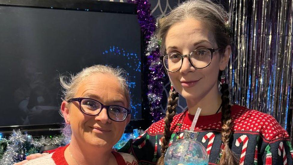
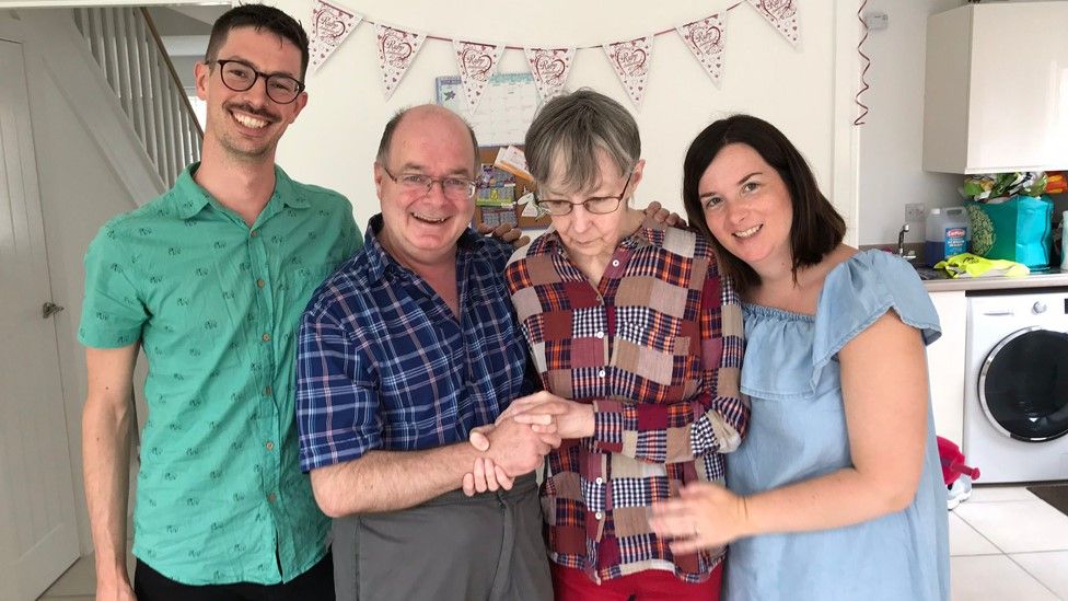
Published58 minutes agoShareclose panelShare pageCopy linkAbout sharingImage source, Abi JenkinsBy Antonia MatthewsBBC News More open conversations about dying should become the norm to better support people at the end of their lives, health experts and carers say.It comes as charity Hospice UK launches a week-long campaign focusing on the language we use around death.Abi Jenkins, who cared for a friend with terminal cancer, said “normalising” death and dying might help people.”We have this really weird taboo around death,” she said.Ms Jenkins was an end-of-life carer for Erin Corner, who died of ampullary cancer in January, aged 31.Ms Corner, who had also lost her youngest son Zac in a caravan fire in 2020, had written birthday and wedding cards for her two surviving sons before her death. Abi said she offered practical help, but also helped Ms Corner navigate her end-of-life care.Image source, Abi JenkinsThe 40-year-old, from Holyhead, Anglesey, said she and Erin, from Pontrhydfendigaid, Ceredigion, grew close following Zac’s death.”She knew how she wanted to die. We talked about what she wanted that to look like,” Ms Jenkins said.She said Ms Corner might have made some different decisions had she been better informed about things like sedation and terminal agitation, which can occur in the final days before death and leads to patients becoming restless and anxious.”Lots of the stuff that happened, we had no idea that that was going to happen,” she said.”We need to talk about it, whether we like it or not.”End-of-life: How home nurses helped ease my husband’s last daysExtreme financial pressure on hospices, says bossFear holiday park by kids hospice will taunt families”There should be far more discussion,” Ms Jenkins said. “People don’t know what to say. They draw back.”Conversations she had with nurses helped Ms Corner with the care she was receiving, Ms Jenkins said, but also made her realise how ill-informed many people are when it comes to palliative care.Laura Hugman, clinical team manager at Tŷ Paul Sartori, which offers a hospice at home service in Pembrokeshire, agreed there was a taboo around death that was sometimes unhelpful.”It’s a challenge. I don’t think we’re very good at talking about dying at all,” she said.Image source, Paul Sartori FoundationHow much people want to know about dying itself is “a real mix and a personal choice”, she said.She said it was key for people to know what services were available, particularly at a time when accessing a GP is not always easy.’Clearer message’Mark Taubert, a consultant in palliative medicine at Velindre University NHS Trust in Cardiff, said it was important to let patients know gently what might happen to them, but also how good planning could provide them with better care.In his experience, 95% of patients want more detail about what will happen to them towards the end of their lives.”Improving the language and getting the message across in a much clearer way is important,” he said. But the approach needs to be individual for every patient and every family, he added, with some patients less willing to hear certain details than others.Image source, Dr Mark Taubert”If they feel in control of the conversation about end-of-life care and the future… that can be very, very helpful,” said Dr Taubert.”You nearly need an end-of-life palliative care ‘personal assistant’ who is ‘bespoke’ to you.” ‘Battle language’Dr Taubert said there was a tendency among many patients and their loved ones towards constant positivity “battle” language, praising the patient for “fighting” and “being strong”.But such language can be problematic as people near the end of their lives, in his view.”When you’re tired, all your hair has fallen out, you’re feeling nauseous and you’re in pain, then the battle language I think lets you down a bit,” he said.”You need a trusted person to be able to confide in and to talk to and let out these things,” he said.Signalling to patients – even when they are not yet dying – that a conversation about end-of-life would be useful to have in the near future can help them prepare for it and get family members involved, he said.Image source, Paul Sartori FoundationMs Hugman said Tŷ Paul Sartori nurses provide information to carers about nutrition and mobility in a patient’s final days. The charity, which helps 40 to 50 patients a week by offering respite visits, also makes sure discussions around managing pain relief take place, ensuring patients and carers understand the extent to which that will affect the patient’s cognition.Family members were often very protective of each other, Ms Hugman said, sometimes making it hard to have open conversations.”People don’t want to come near death and dying… people are never quite ready to think about it,” she said.Ultimately, there’s no right or wrong way to look after a bereaved family, said Ms Hugman.”It always comes as a shock when someone passes away, even if the family is completely prepared.”More on this storyMum with cancer dies after losing son in firePublished9 January’What terminal cancer has taught me about life’Published27 JanuaryMums who met at children’s hospice climb mountainsPublished30 April 2023

Read more →

‘I have a 50:50 chance of developing Alzheimer’s in my 50s’

Published11 minutes agoShareclose panelShare pageCopy linkAbout sharingImage source, Jennings familyBy Lucy WallisBBC NewsWhen John Jennings’ mother started showing signs of Alzheimer’s Disease at the age of 50, he and his sister Emily knew they too would also have a 50:50 chance of developing the condition that causes dementia. But a letter their mother wrote almost 40 years ago could hold the key to changing the course of the future for their family and thousands of others.”You know you tend to get older people and they’re buying a pair of shoes and they’re like, ‘Those will see me out’. I’ve basically always had that attitude,” says 39-year-old John Jennings. “I’m like, ‘Shall I buy a new laptop?’ – because I’ve had Macs that have lasted 10 years in the past.”John, who lives in Edinburgh with his husband Matt, is not bothered by material possessions because he doesn’t know if he has inherited a faulty gene from his mother that could trigger early-onset Alzheimer’s Disease.”I’ve just found that the thing that makes me happiest is being around people who love me,” he says. “So we just spend a lot of time building relationships and that seems to be the most fulfilling part of life.”But these social connections are the things John is most scared of losing if he does have the gene mutation and goes on to develop the condition in his 50s, like his mother Carol did.”I’m trying to learn loads of languages and I’m obsessively exercising, I know for most people that would dramatically reduce my risk of getting Alzheimer’s,” says John, “but the fact of the matter is, for me, it doesn’t make any difference.” For the majority of people, advancing age is the biggest risk factor in Alzheimer’s. In every 100 people with the condition, less than 1% will have the inherited form.Image source, Jennings familyIn the 1980s, it was widely thought Alzheimer’s Disease had no familial link, but when John’s grandfather – Carol’s dad – and his four siblings were all diagnosed with the condition in their 50s, Carol knew this couldn’t be a coincidence.A new documentary for BBC Two tells the story of how Carol, a teacher from Coventry, helped change the face of Alzheimer’s Disease research with one handwritten letter. She had always tried to find solutions to problems, says John, it was “her way of getting some control of the situation”. In 1986, Carol wrote to a team at University College London (UCL) who were studying the disease. By analysing the genetics of her family, the team identified a gene in 1991 that all her affected family members shared. A mutation to the amyloid precursor protein (APP) gene meant too much amyloid protein built up in the brain, which clumps together to form plaques and causes brain cells to die.Carol passed down a 50% chance to John and his older sister Emily, 42, of inheriting the gene mutation that triggers early-onset Alzheimer’s Disease.”If someone has the gene, they will develop disease at around the same time that their family members did,” says consultant neurologist, Dr Cath Mummery, head of clinical trials at the Dementia Research Centre, at UCL. “So they are aware of the ticking time bomb, especially as they near that age.”Image source, Jennings family”It’s tempting to think if I discovered that I had it, that Emily didn’t, and vice versa,” says John. “But it could be that both of us do. It could be that neither of us do.”For those at risk of genetic Alzheimer’s Disease, a blood test can be taken – after specialist consultation and counselling – to show if the genetic abnormality that causes the disease is present. John’s mother Carol chose not to take the test, as she believed there was no point in worrying, says John, but while he respects his mother’s choice, it’s not the route he wants to go down. “We could have planned better if we knew that she had,” says John. Image source, Jennings familyHe was 21 when his mother first started showing symptoms in the mid-2000s. Her condition slowly deteriorated until she became bedbound and couldn’t speak. Every stage of his mother’s decline was “like a new tonne of bricks that you have to deal with”, he says.Carol passed away in March this year. She asked for her brain to be donated for scientific research.John says he will definitely take the blood test at some point – it’s just when – and he will decide on the timing with his sister. Now, is not the time. “I think if one of us had the test then the other one would probably follow pretty quickly afterwards,” says John. “So it almost feels like it’s a decision we need to make together.”Image source, Jennings familyThe one thing that might bring forward John’s decision to take the test would be if he needs to know his gene status to start any medical treatment.He is optimistic about new Alzheimer’s drugs that could be licensed this year. They have been engineered to help the immune system clear amyloid from the brain and slow the progress of the disease. There are potentially serious side effects and their effectiveness depends on early diagnosis – so it is still early days in terms of knowing what impact they will have.While genetic Alzheimer’s Disease is rare, it is similar in many ways to more typical Alzheimer’s Disease that develops in other people later in life, says Dr Mummery, and can be used as a model to find new therapies. “If we can find a treatment that works in this genetic form, then we might be able to extrapolate from that to a treatment for the more common, non-genetic Alzheimer’s Disease,” she says.All this has stemmed from the work Carol started with the team at UCL, says Dr Mummery.While work continues on the development of treatments, John’s coping mechanism is to remain resilient and have a positive outlook. He also recommends sharing experiences with others at support groups. He is continuing his mother’s legacy, helping to change the course of the future, by taking part in Alzheimer’s Disease research and having regular brain scans.John says he feels we are “on the cusp” of seeing treatments that can help stop the disease in its tracks.”I’d really like to live long enough to see that – and I think I might.”The Jennings v Alzheimer’s will be broadcast on Monday 13 May at 21:00 BST on BBC Two. Or catch up afterwards on BBC iPlayer.If you are affected by any of the issues raised in this story, support and advice is available via the BBC Action LineMore on this storyWhat is Alzheimer’s and how common is it?Published4 AprilNHS exploring Alzheimer’s disease blood testsPublished4 AprilNHS ‘not ready’ for new Alzheimer’s drugsPublished12 FebruaryNew drugs for Alzheimer’s hailed as turning pointPublished17 July 2023

Read more →

How the NHS has run out of jobs for new doctors

Published20 minutes agoShareclose panelShare pageCopy linkAbout sharingImage source, EmmaBy Nick TriggleHealth correspondentThe NHS needs more doctors, and this year saw a record number of applications from medical students to start junior doctor training. But problems behind the scenes have meant many have not yet been found jobs. What has gone wrong?When the email of congratulations arrived in her inbox last month, Emma could not have been any more excited. Inside was meant to be the details of her first job as a junior doctor.On the brink of graduation from Warwick Medical School, the 29-year-old was due to find out about where she would working for the next two years.But despite the email being titled congratulations, next to each of her placements for her two-year foundation course, the first rung on the doctor training ladder, was the abbreviation TBC.She had been given what is called a placeholder job. “I’ve been told I will have a job, but that they don’t know where yet and it could take until three weeks before I am due to start in August for me to find out. All I know is it will be somewhere in the West Midlands South region.”It is incredibly unsettling. It makes you feel unvalued and an inconvenience. The NHS is short of doctors and yet they cannot find me a job.”I am renting at the moment, but depending on where I am placed I may have to give notice to my landlord and move.”Emma was one of more than 1,000 medical students in this position when the emails initially went out. That was nearly one in nine of those who have applied to start junior doctor training this summer.Since then the numbers have dropped to 800 as NHS trusts have created new roles and some students have already decided not to take up their place.Part of the problem is that there has been an increase in medical students this summer to start junior doctor training, with just over 9,700 accepted by the UK Foundation Programme, up from 8,655 in 2023.Some of these will have come from abroad, and under immigration rules the NHS cannot prioritise UK-trained students.The increase is welcome given the need to train more doctors, with the government in England aiming to double the number of medical student places by 2031.But it has meant NHS trusts have struggled to keep up and ensure they have enough training posts for new graduates. ‘A lottery’The British Medical Association has this week written to the health secretary to warn it is “exceedingly concerned” about the placeholder situation, after being inundated with complaints from worried medical students.Its letter says many are reporting they may only be told where they will be three weeks before their August start date, when work schedules are meant to be confirmed at least eight weeks in advance.All they know is what part of the UK they are going to be working in after being allocated to one of 18 foundation school areas in March. Some cover very large areas. There is one each for Wales, Scotland and Northern Ireland, while in England, one covers the whole of Yorkshire and Humber. Medical students have also had to cope with a change in the way their regions are allocated. As part of the process, they have to list each foundation school in order of preference.Previously people were allocated according to merit – with each student ranked according to how they had performed during their studies and in an application test.But this year that has changed and has been done randomly. The logic behind it was that the previous system was stressful for students and was particularly unfair on those from deprived backgrounds and ethnic minorities. They tended to perform less well, and therefore were more likely to be posted to regions they did not favour, according to the UK Foundation Programme.However, overall it has resulted in more students not ending up with one of their top five choices – more than 730 compared to just over 430 last year.Image source, Getty ImagesBMA medical students committee deputy chairman Rob Tucker says while students understood the logic behind changing the system, he feels “something has gone wrong” given what has happened. “Students feel like they are in a lottery. It all depends what number the computer algorithm spits out.”And he is worried it will lead to more people dropping out and not progressing on to the next stage of training.”Having more students with their bottom choices, and so many with placeholder jobs, increases the chances of more medical students walking away.”It’s very hard if you end up in a part of the country you don’t want to work [in] and then if you don’t know where you are going to work in that particular region until as little as a few weeks before you start, that’s not fair.”It costs a lot of money to train a doctor. It’s good value if they work for the NHS for 30 or 40 years, but not if you lose them straight away.”‘Bottlenecks’There is sympathy for their plight among those running the system.Speaking at the Royal College of Physicians annual conference last month, North London Foundation School deputy director Dr Celia Bielawski said the deaneries were working hard with NHS trusts to create the new junior doctor posts needed.”We have an increasing number of international medical graduates applying for foundation training every year, and the government is very keen that we should take these doctors as we need to train up additional doctors to go up through the ranks. “At the moment we don’t have places for all of them, but we are all actively working, creating additional posts and I’m confident that they will all be placed. “We have to get trusts to agree to take them, that’s one of the fundamental issues and bottlenecks with all of this. But we’ll get there, I am sure.” Prof Sheona Macleod, director of education and training at NHS England, is confident the situation will be resolved too, saying: “We will get there because we’ve committed to finding them a post.”But she also pointed out that under the previous system, medical students could still find themselves without a job initially as some would be placed on a national reserve list after the first round of job offers.”We have made sure people know where they are going, as in what region, which is different to the past.” But she added: “The numbers have meant that the anxiety for the students looking at finding places hasn’t gone away and we’re very aware of it.”Additional reporting by Catherine Snowdon.

Read more →

Widening Racial Disparities Underlie Rise in Child Deaths in the U.S.

New research finds that the death rate among Black youths soared by 37 percent, and among Native American youths by 22 percent, between 2014 and 2020, compared with less than 5 percent for white youths.The NewsThanks to advancements in medicine and insurance, mortality rates for children in the United States had been shrinking for decades. But last year, researchers uncovered a worrisome reversal: The child death rate was rising.Now, they have taken their analysis a step further. A new study, published Saturday in The Journal of the American Medical Association, revealed growing disparities in child death rates across racial and ethnic groups. Black and Native American youths ages 1 to 19 died at significantly higher rates than white youths — predominantly from injuries such as car accidents, homicides and suicides.Dr. Coleen Cunningham, chair of pediatrics at the University of California, Irvine, and the pediatrician in chief at Children’s Hospital of Orange County, who was not involved in the study, said the detailed analysis of the disparities documented “a sad and growing American tragedy.”“Almost all are preventable,” she said, “if we make it a priority.”Flowers for Karon Blake, 13, who was shot and killed in Washington, D.C., in January 2023. Gun-related deaths were two to four times higher among Black and Native American youth than among white youth.Carolyn Kaster/Associated PressSome Context: A frightening trend examined more closely.Researchers at Virginia Commonwealth University and Children’s Hospital of Richmond had previously revealed that mortality rates among children and adolescents had risen by 18 percent between 2019 and 2021. Deaths related to injuries had grown so dramatically that they eclipsed all public health gains.The group, seeking to drill deeper into the worrying trend, obtained death certificate data from the Centers for Disease Control and Prevention’s public WONDER database and stratified it by race, ethnicity and cause for children ages 1 to 19. They found that Black and American Indian/Alaska Native children were not only dying at significantly higher rates than white children but that the disparities — which had been improving until 2013 — were widening.The data also revealed that while the mortality rates for children overall took a turn for the worse around 2020, the rates for Black, Native American and Hispanic children had begun increasing much earlier, around 2014.Between 2014 and 2020, the death rates for Black children and teenagers rose by about 37 percent, and for Native American youths by about by about 22 percent — compared with less than 5 percent for white youths.“We knew we would find disparities, but certainly not this large,” said Dr. Steven Woolf, a professor of family medicine at the V.C.U. School of Medicine, who worked on the research. “We were shocked.”We are having trouble retrieving the article content.Please enable JavaScript in your browser settings.Thank you for your patience while we verify access. If you are in Reader mode please exit and log into your Times account, or subscribe for all of The Times.Thank you for your patience while we verify access.Already a subscriber? Log in.Want all of The Times? Subscribe.

Read more →

Aligned peptide ‘noodles’ could enable lab-grown biological tissues

A team of chemists and bioengineers at Rice University and the University of Houston have achieved a significant milestone in their work to create a biomaterial that can be used to grow biological tissues outside the human body. The development of a novel fabrication process to create aligned nanofiber hydrogels could offer new possibilities for tissue regeneration after injury and provide a way to test therapeutic drug candidates without the use of animals.
The research team, led by Jeffrey Hartgerink, professor of chemistry and bioengineering, has developed peptide-based hydrogels that mimic the aligned structure of muscle and nerve tissues. Alignment is critical for the tissues’ functionality, but it is a challenging feature to reproduce in the lab, as it entails lining up individual cells.
For over ten years, the team has been designing multidomain peptides (MDPs) that self-assemble into nanofibers. These resemble the fibrous proteins found naturally in the body, much like a spiderweb at nanoscale.
In their latest study, published online and featured on the cover of the journal ACS Nano, the researchers discovered a new method to create aligned MDP nanofiber “noodles.” By first dissolving the peptides in water and then extruding them into a salty solution, they were able to create aligned peptide nanofibers — like twisted strands of rope smaller than a cell. By increasing the concentration of ions, or salt, in the solution and repeating the process, they achieved even greater alignment of the nanofibers.
“Our findings demonstrate that our method can produce aligned peptide nanofibers that effectively guide cell growth in a desired direction,” explained lead author Adam Farsheed, who recently received his Ph.D. in bioengineering from Rice. “This is a crucial step toward creating functional biological tissues for regenerative medicine applications.”
One of the key findings of the study was an unexpected discovery: When the alignment of the peptide nanofibers was too strong, the cells no longer aligned. Further investigation revealed that the cells needed to be able to “pull” on the peptide nanofibers to recognize the alignment. When the nanofibers were too rigid, the cells were unable to exert this force and failed to arrange themselves in the desired configuration.
“This insight into cell behavior could have broader implications for tissue engineering and biomaterial design,” said Hartgerink. “Understanding how cells interact with these materials at the nanoscale could lead to more effective strategies for building tissues.”
Additional study co-authors from Rice include chemistry department Ph.D. graduates Tracy Yu and Carson Cole, graduate student Joseph Swain, and undergraduate researcher Adam Thomas. Bioengineering undergraduate researcher Jonathan Makhoul, graduate student Eric Garcia Huitron, and Professor K. Jane Grande-Allen were also co-authors on the study. The team of researchers from the University of Houston includes Ph.D. student Christian Zevallos-Delgado, research assistant Sajede Saeidifard, research assistant professor Manmohan Singh and engineering professor Kirill Larin.
This work was supported in part by grants from the National Institutes of Health (R01DE021798, R01EY022362, R01HD095520, R01EY030063), the National Science Foundation (2129122), the National Science Foundation Graduate Research Fellowship Program, and the Welch Foundation (C-2141). The content in this news release is solely the responsibility of the authors and does not necessarily represent the official views of the funding organizations.

Read more →

Quantifying U.S. health impacts from gas stoves

Households with gas or propane stoves regularly breathe unhealthy levels of nitrogen dioxide, a study of air pollution in U.S. homes found.
“I didn’t expect to see pollutant concentrations breach health benchmarks in bedrooms within an hour of gas stove use, and stay there for hours after the stove is turned off,” said Stanford Doerr School of Sustainability Professor Rob Jackson, senior author of the May 3 study in Science Advances. Pollution from gas and propane stoves isn’t just an issue for cooks or people in the kitchen, he said. “It’s the whole family’s problem.”
Among other negative health effects, breathing high levels of nitrogen dioxide, or NO2, over time can intensify asthma attacks and has been linked to decreased lung development in children and early deaths.
Although most exposure to NO2 is caused by cars and trucks burning fossil fuels, the researchers estimate that the mix of pollutants coming from gas and propane stoves overall may be responsible for as many as 200,000 current childhood asthma cases. One quarter of these can be attributed to nitrogen dioxide alone, according to the paper’s authors, who include scientists from Central California Asthma Collaborative, PSE Healthy Energy, and the Harvard T.H. Chan School of Public Health.
“We found that just how much gas you burn in your stove is by far the biggest factor affecting how much you’re exposed. And then, after that, do you have an effective range hood — and do you use it?” said lead study author Yannai Kashtan, a PhD student in Earth system science.
Little room for additional exposure
Beyond asthma cases, the long-term exposure to NO2 in American households with gas stoves is high enough to cause thousands of deaths each year — possibly as many as 19,000 or 40% of the number of deaths linked annually to secondhand smoke. This estimate is based on the researchers’ new measurements and calculations of how much nitrogen dioxide people breathe at home because of gas stoves and the best available data on deaths from long-term exposure to outdoor NO2, which is regulated by the U.S. Environmental Protection Agency.

The death toll estimate is approximate in part because it does not factor in the harmful effects of repeated exposure to extremely high levels of nitrogen dioxide in short bursts, as occurs in homes with gas stoves. It also relies on past studies of health impacts from nitrogen dioxide encountered outdoors, where additional pollutants from vehicles and power plants are present.
The researchers used sensors to measure concentrations of NO2 throughout more than 100 homes of various sizes, layouts, and ventilation methods, before, during, and after stove use. They incorporated these measurements and other data into a model powered by National Institutes for Standards and Technology (NIST) software known as CONTAM for simulating airflow, contaminant transport, and room-by-room occupant exposure in buildings. This allowed them to estimate nationwide averages and short-term exposures under a range of realistic conditions and behaviors, and cross-check model outputs against their home measurements.
The results show that nationwide, typical use of a gas or propane stove increases exposure to nitrogen dioxide by an estimated 4 parts per billion, averaged over a year. That’s three quarters of the way to the nitrogen dioxide exposure level that the World Health Organization recognizes as unsafe in outdoor air. “That’s excluding all outdoor sources combined, so it makes it much more likely you’re going to exceed the limit,” said Kashtan.
Understanding how gas stoves affect health
The study is the latest in a series from Jackson’s group at Stanford looking at indoor air pollution from gas stoves. Earlier studies documented the rate at which gas stoves emit other pollutants, including the greenhouse gas methane and the carcinogen benzene. But to understand the implications of stove emissions for human health, the researchers needed to find out how much pollutants spread through a home, build up, and eventually dissipate. “We’re moving from measuring how much pollution comes from stoves to how much pollution people actually breathe,” said Jackson, who is the Michelle and Kevin Douglas Provostial Professor in Earth System Science.
With any fuel source, particle pollution can rise from food cooking in a hot pan. The new research confirms that food emits little or no nitrogen dioxide as it cooks, however, and electric stoves produce no NO2. “It’s the fuel, not the food,” said Jackson. “Electric stoves emit no nitrogen dioxide or benzene. If you own a gas or propane stove, you need to reduce pollutant exposures using ventilation.”
Home size matters

Even in larger homes, concentrations of nitrogen dioxide routinely spiked to unhealthy levels during and after cooking even if a range hood was on and venting air outdoors. But people who live in homes smaller than 800 square feet — about the size of a small two-bedroom apartment — are exposed to twice as much nitrogen dioxide over the course of a year compared to the national average, and four times more compared to those living in the largest homes, upwards of 3,000 square feet.
Because home size makes such a difference, there are also differences in exposure across racial, ethnic, and income groups. Compared to the national average, the researchers found long-term NO2 exposure is 60% higher among American Indian and Alaska Native households, and 20% higher among Black and Hispanic or Latino households. This exposure to indoor air pollution from gas stoves compounds the fact that exposure to outdoor sources of nitrogen dioxide pollution, such as vehicle exhaust, is also typically higher among people in poorer, often minority, communities.
“People in poorer communities can’t always afford to change their appliances, or perhaps they rent and can’t replace appliances because they don’t own them,” Jackson said. “People in smaller homes are also breathing more pollution for the same stove use.”
This research was supported by HT, LLC.

Read more →

Newly discovered mechanism of T-cell control can interfere with cancer immunotherapies

Activated T cells that carry a certain marker protein on their surface are controlled by natural killer (NK) cells, another cell type of the immune system. In this way, the body presumably curbs destructive immune reactions. Researchers from the German Cancer Research Center (DKFZ) and the University Medical Center Mannheim (UMM) now discovered that NK cells can impair the effect of cancer therapies with immune checkpoint inhibitors (ICI) in this way. They could also be responsible for the rapid decline of therapeutic CAR-T cells. Interventions in this mechanism could potentially improve the efficacy of these cellular cancer immunotherapies.
The T cells of the immune system are the main players in the defense against viral infections and tumor cells. On the other hand, they attack the body’s own healthy tissue in autoimmune reactions, which can even be fatal. The body must therefore keep a tight control on T cell activity.
A large number of molecules and messenger are involved in the highly complex regulation of T cell activity. Only recently have researchers discovered that another group of immune cells contibutes to the control of T cell activity. Natural killer cells (NK cells) are part of the innate immunity, i.e. the rapid response force that quickly detects and eliminates infected or malignant cells.
“Studies have shown that NK cells can also kill activated T cells and thus limit their proliferation,” says Michael Platten, Head of Department at the DKFZ and Director of the Neurological University Clinic Mannheim. “However, until now we did not know which feature characterizes T cells as a target for the NK cell.”
When screening activated T cells from healthy donors, Platten’s team identified the protein B7H6 as a recognition molecule for NK cell attacks in a new study. Activated T cells from the blood of patients with autoimmune diseases, cancer or viral infections expose large amounts of B7H6 on their surface. Co-culture experiments in the culture dish showed that NK cells recognize the activated T cells by their B7H6 expression. In contrast, T cells whose B7H6 gene was destroyed with the CRISPR-Cas were protected from the lethal attack of the NK cells.
“The elimination of T cells by NK cells is triggered by an intrinsic mechanism of the T cells. The activated T cells temporarily identify themselves as targets for NK-induced cell lysis,” explains Michael Kilian, first author of the publication, and adds: “This may limit excessive activation and expansion of T cells as a control mechanism to curb destructive immune responses.”
Immune checkpoint inhibitor therapies are neutralized by NK cells
“We now know a number of so-called checkpoint molecules that reduce or enhance the activation of T cells and thus modulate the course of immune reactions. B7H6 can now be classified as a further inhibitory immune checkpoint on T cells,” explains study leader Platten.

Some widespread cancer therapies with drugs from the checkpoint inhibitor (ICI) group are targeting certain inhibitory checkpoint molecules. They activate the immune system against the tumor by releasing the immune brakes. Could the B7H6-mediated elimination of tumor-reactive T cells possibly counteract the effect of ICI cancer immunotherapy? The researchers tested this using tissue samples from patients with esophageal cancer who had received ICI therapy. Those patients who had not responded to ICI had a higher number of NK cells in the tumor tissue and actually had a shorter progression-free survival time.
Cellular immunotherapy more effective in the absence of NK cells
Cellular immunotherapies are becoming increasingly important in cancer medicine. For example, some forms of blood cancer are now often treated with so-called CAR-T cells, which are equipped with customized receptors against the cancer. However, the success of therapy is often limited as the number of therapeutic cells in the patient’s body declines rapidly.
The therapeutic CAR-T cells also carry B7H6 on the cell surface. Could NK cells be responsible for the rapid decline in their numbers after the start of therapy? Experiments with a humanized mouse model suggest this: if NK cells were added during CAR-T cell treatment of leukaemia, the number of therapeutic cells decreased, while the tumour load increased.
“NK control of T cells has the potential to interfere with various forms of cancer immunotherapy. By specifically intervening in this process, it may be possible to modulate T cell immune responses in the future,” explains Michael Platten, head of the current study. With the help of the CRISPR-Cas gene scissors, the researchers now want to protect CAR-T cells from elimination by NK cells in a clinical trial together with the Department of Haematology and Oncology at Heidelberg University Hospital and thus improve the effectiveness of cellular immunotherapy.

Read more →

New Mutations Identified in Bird Flu Virus

A genetic analysis sheds light on when the outbreak began, how the virus spread and where it may be going.The bird flu virus sweeping across dairy farms in multiple states has acquired dozens of new mutations, including some that may make it more adept at spreading between species and less susceptible to antiviral drugs, according to a new study.None of the mutations is a cause for alarm on its own. But they underscore the possibility that as the outbreak continues, the virus may evolve in ways that would allow it to spread easily between people, experts said.“Flu mutates all the time — it’s what, sort of, flu does,” said Richard Webby, an influenza expert at St. Jude Children’s Research Hospital, who was not involved in the work.“The real key would be if we start to see some of these mutations getting more prevalent,” Dr. Webby said. “That would raise the risk level.”The virus, called H5N1, has infected cows in at least 36 herds in nine states, raising fears that milk could be infectious — concerns now largely put to rest — and highlighting the risk that many viruses might jump across species on crowded farms.The study was posted online on Wednesday and has not been peer reviewed. It is among the first to provide details of a Department of Agriculture investigation that has been mostly opaque until now, frustrating experts outside the government.We are having trouble retrieving the article content.Please enable JavaScript in your browser settings.Thank you for your patience while we verify access. If you are in Reader mode please exit and log into your Times account, or subscribe for all of The Times.Thank you for your patience while we verify access.Already a subscriber? Log in.Want all of The Times? Subscribe.

Read more →

Pan-cancer analysis uncovers a new class of promising CAR T–cell immunotherapy targets

Targeting anti-cancer therapy to affect cancer cells but not healthy cells is challenging. For chimeric antigen receptor (CAR) T-cell immunotherapy, where a patient’s own immune cells are re-engineered to attack cancer cells, many solid and brain cancers lack an effective target. St. Jude Children’s Research Hospital scientists have identified 156 potential targets through a comprehensive analysis paired with an experimental validation in vivo. The findings were published today in Nature Communications.
“We discovered targets for cancer immunotherapy, which hopefully can be translated in the future into curative approaches,” said co-corresponding author Stephen Gottschalk, MD, St. Jude Department of Bone Marrow Transplantation and Cellular Therapy chair. “We’ve given the field a large set of potential targets and validated at least one, COL11A1.”
While COL11A1 was one of the 156 targets identified that the researchers validated in mouse models, others showed promise in cell lines such as anti-fibronectin CAR T cells. Most targets have yet to be tested but are publicly available for other researchers to pursue.
“In addition to validating the selected targets, we have built a data resource for the community,” said co-corresponding author Jinghui Zhang, PhD, St. Jude Department of Computational Biology. “The final list is available in a web portal. Others can evaluate the evidence and pursue any of these targets.”
The portal, named SCE-Miner, is freely available on the St. Jude Cloud platform. External researchers can access the data and use the internal analysis tools to fuel their own research.
“Our portal empowers the users to explore these targets and the underlying data,” Gottschalk said. “Other scientists can say, ‘Is this target present in a particular pediatric cancer subtype?’ They can access that information in the Cloud and do their own analysis.”
Fishing for CAR T-cell targets
The researchers found COL11A1 and the other targets by comprehensively analyzing the subparts of genes called exons. When a gene is transcribed to RNA, only exonic regions are retained to form a mature product, which will be used as a template for protein translation. The researchers looked at whole transcriptome sequencing data of 1,532 pediatric tumor samples with 7,460 normal tissue samples, finding which exons were more highly selective or uniquely expressed in cancer cells than normal cells. Such cancer-specific exons can potentially be targeted by CAR T cells without causing harm to healthy cells. The final target list also includes several targets identified in previous analyses and are being pursued clinically, increasing confidence in the group’s analytical approaches.

The St. Jude method differs in several ways compared to previous CAR target searches. The sample size was much larger, and the study involved all major cancer types. Therefore, the candidate pool was expanded. To ensure the inclusion of all relevant candidates to CAR T targets, the researchers focused on proteins existing inside the membrane, a traditional approach, and those sitting on top of the membrane in a place called the extracellular matrix, which was unusual.
“Normally, groups looking for new CAR targets looked only at membrane-associated proteins,” Gottschalk said. “But one of our targets told us that we needed to broaden our criteria to include proteins of the extracellular matrix. Most of those proteins are not anchored to the cell, but it turns out they stick to the cell surface. When the CAR comes, it can still recognize that protein and kill the cancer cell.”
The researchers also found that some of these genes have different exons expressed inside them, resulting in a different version of a protein called an isoform. Think of a gene as a movie; an isoform would be like a director’s cut, with different scenes cut, extended or inserted while maintaining major similarities to the original. Cancer cells are prone to alternative splicing of exons, which creates isoforms, which the St. Jude analysis detected in a way that had been difficult in earlier screens.
“We used a more straightforward and robust analysis than prior approaches,” Zhang said. “We could see if a subset or all set of exons within a gene were differentiated between cancer and normal cells, which allowed us to evaluate cancer-specificity at the isoform versus gene level.”

Read more →

New immunosuppressive mechanism found in brain cancer

The Wistar Institute assistant professor Filippo Veglia, Ph.D., and team, have discovered a key mechanism of how glioblastoma — a serious and often fatal brain cancer — suppresses the immune system so that the tumor can grow unimpeded by the body’s defenses. The lab’s discovery was published in the paper, “Glucose-driven histone lactylation promotes the immunosuppressive activity of monocyte-derived macrophages in glioblastoma,” in the journal Immunity.
“Our study shows that the cellular mechanisms of cancer’s self-preservation, when sufficiently understood, can be used against the disease very effectively,” said Dr. Veglia. “I look forward to future research on metabolism-driven mechanisms of immunosuppression in glioblastoma, and I’m hopeful for all that we will continue to learn about how to best understand and fight this cancer.”
Until now, it has been poorly understood how monocyte-derived macrophages and microglia create an immunosuppressive tumor microenvironment in glioblastoma. The Veglia lab investigated the cellular “how” of glioblastoma immunosuppression and identified that, as glioblastoma progressed, monocyte-derived macrophages came to outnumber microglia — which indicated that monocyte-derived macrophages’ eventuality to becoming the majority in the tumor microenvironment was advantageous to the cancer’s goal of evading immune response. Indeed, monocyte-derived macrophages, but not microglia, blocked the activity of T cells (immune cells that destroy tumor cells), in preclinical models and patients. The team confirmed this finding when they assessed preclinical models of glioblastoma with artificially reduced numbers of monocyte-derived macrophages. And as the group expected, the models with fewer malicious macrophages in the tumor microenvironment showed improved outcomes relative to the standard glioblastoma models.
Glioblastoma accounts for slightly more than half of all malignancies that originate in the brain, and the prognosis for those diagnosed with the cancer is quite poor: only 25% of patients who receive a glioblastoma diagnosis will survive beyond a year. Glioblastoma is inherently dangerous due to its location in the brain and its immunosuppressive tumor microenvironment, which renders glioblastoma resistant to promising immunotherapies. By programming certain immune cells like macrophages, (such as monocyte-derived macrophages and microglia), to work for — rather than against — the tumor, glioblastoma fosters a tumor microenvironment for itself that enables the cancer to grow aggressively while evading anticancer immune responses.
Having confirmed the role of monocyte-derived macrophages, the Veglia lab then sought to understand just how the cancer-allied immune cells were working against the immune system. They sequenced the macrophages in question to see whether the cells had any aberrant gene expression patterns that could point to which gene(s) could be playing a role in immunosuppression, and they also investigated the metabolic patterns of macrophages to see whether the macrophages’ nonstandard gene expression could be tied to metabolism.
The team’s twin gene expression & metabolic analysis led them to glucose metabolism. Through a series of tests, the Veglia lab was able to determine that monocyte-derived macrophages with enhanced glucose metabolism and expressing GLUT1, a major transporter for glucose (a key metabolic compound), blocked T cells’ function by releasing interleukin-10 (IL-10). The team demonstrated that glioblastoma-perturbed glucose metabolism in these macrophages induced their immunosuppressive activity.
The team discovered the key to macrophages’ glucose-metabolism-driven immunosuppressive potency lies in a process called “histone lactylation.” Histones are structural proteins in the genome that play a key role in which genes — like IL-10 — are expressed in which contexts. As rapidly glucose-metabolizing cells, monocyte-derived macrophages produce lactate, a by-product of glucose metabolism. And histones can become “lactylated” (which is when lactate becomes incorporated into histones) in such a way that the histones’ organization further promotes the expression of IL-10 — which is effectively produced by monocyte-derived macrophages to help cancer cells to grow.
But how can the glucose-driven immunosuppressive activity of monocyte-derived macrophages be stopped? Dr. Veglia and his research team identified a possible solution: PERK, an enzyme they had identified as regulating glucose metabolism and GLUT1 expression in macrophages. In preclinical models of glioblastoma, targeting PERK impaired histone lactylation and immunosuppressive activity of macrophages, and in combination with immunotherapy blocked glioblastoma progression and induced long-lasting immunity that protected the brain from tumor re-growth — a sign that targeting PERK-histone lactylation axis may be a viable strategy for fighting this deadly brain cancer.
Note: The work detailed in this publication was initiated at The H. Lee Moffitt Cancer Center during Dr. Veglia’s time there and continued at Wistar.

Read more →