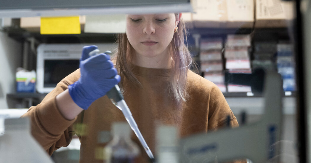
How Does Pregnancy Change the Brain? Clues Are Emerging.

As hormones surge, some brain areas shrink in what scientists say may be a fine-tuning that helps mothers bond with and care for their babies.Research is revealing intriguing clues about how pregnancy changes the brain. Studies scanning women’s brains before and after pregnancy have found that certain brain networks, especially those involved in social and emotional processing, shrink during pregnancy, possibly undergoing a fine-tuning process in preparation for parenting. Such changes correspond with surges in pregnancy hormones, especially estrogen, and some last at least two years after childbirth, researchers have found.A new study, published Monday in the journal Nature Neuroscience, adds to the picture by documenting with M.R.I.s brain changes throughout one woman’s pregnancy. It confirms previous results and adds detail, including that white matter fibers showed greater ability to efficiently transmit signals between brain cells, a change that evaporated once the baby was born.“What’s very interesting about this current study is that it provides such a detailed mapping,” said Elseline Hoekzema, a neuroscientist who heads the Pregnancy and the Brain Lab at Amsterdam University Medical Center and has helped lead studies analyzing brain scans of more than 100 women before and after pregnancy.Dr. Hoekzema, who was not involved in the new study, said it showed that along with previously documented “longer-lasting changes in brain structure and function, more subtle, transient changes also occur.”Dr. Ronald Dahl, director of the Institute of Human Development at the University of California, Berkeley, who was not involved in the new study, said the emerging research reflected the key role of hormones in transitions like puberty and pregnancy, guiding neurological shifts in priorities and motivations.“There is that sense that it’s affecting so many of these systems,” he said. The study participant, Elizabeth Chrastil, is a neuroscientist at the University of California, Irvine. She became pregnant in 2019, at 38, after in vitro fertilization. That allowed precise tracking of her pregnancy from the start.We are having trouble retrieving the article content.Please enable JavaScript in your browser settings.Thank you for your patience while we verify access. If you are in Reader mode please exit and log into your Times account, or subscribe for all of The Times.Thank you for your patience while we verify access.Already a subscriber? Log in.Want all of The Times? Subscribe.

Read more →

First Day of a ‘New Life’ for a Boy With Sickle Cell

There was supposed to be a special party for Kendric Cromer, 12, last Wednesday, but it had to be postponed because he was too groggy to celebrate.It was meant to mark the first day of his new life — the day he became one of the first children ever to be treated with a newly approved gene therapy that will free him from the sickle cell disease that has stolen his childhood.On Sept. 11, despite the excitement of the moment, Kendric was unable to keep his eyes open as he lay in his hospital bed at Children’s National Hospital in Washington because of the drugs he had been given in preparation for his treatment.His life with the disease has been punctuated by episodes of excruciating pain, requiring days in the hospital as doctors tried to control it. Sickle cell eroded his hip bones. It prevented him from riding a bike or playing soccer or even going outside when the temperature was below 55 degrees Fahrenheit because cold often brought on intense pain.Now he could see a future — in a month or so — without pain from sickle cell.“I can’t wait to start my new life,” he told his mother, Deborah Cromer.His disease is caused by an inherited genetic mutation that leads to blood cells that form crescent shapes — sickles — instead of discs. Trapped in blood vessels and organs, the cells cause damage and pain. Gene therapy fixes that problem by giving the patient a new, normal hemoglobin gene.We are having trouble retrieving the article content.Please enable JavaScript in your browser settings.Thank you for your patience while we verify access. If you are in Reader mode please exit and log into your Times account, or subscribe for all of The Times.Thank you for your patience while we verify access.Already a subscriber? Log in.Want all of The Times? Subscribe.

Read more →

Dr. George Berci, Whose Innovations Changed Surgery, Dies at 103

He conceived many of the techniques and tools that have revolutionized minimally invasive operations and procedures.Dr. George Berci, a Hungarian-born Holocaust survivor who changed the way modern surgery is conducted by developing techniques and instruments that help doctors better see and treat the inside of the body while minimizing cutting, died on Aug. 30 in Thousand Oaks, Calif. He was 103.His death, in a hospital, was confirmed by his daughter Katherine DeFevere.Dr. Berci brought a precise eye and an inventor’s zeal to innovations that enabled doctors to better visualize the bladder, colon, esophagus, prostate, common bile duct and other body parts. Until earlier this summer, he was the senior director of minimally invasive surgery research at Cedars-Sinai Medical Center in Los Angeles, where he had worked since 1969.His innovations were critical to the revolution in minimally invasive endoscopies and laparoscopies, which dramatically reduced the need for surgeons to make large incisions.In endoscopies, doctors use a flexible tube with a light and a camera to examine the upper and lower digestive system. Dr. Berci focused mainly on the area around the throat and vocal cords.In laparoscopies, surgeons place a thin rod with a video camera attached at the end through a small abdominal incision. Carbon dioxide is then used to inflate the space to give doctors enough room to use small instruments to, among other things, remove gallbladders, cysts, tumors, appendixes and spleens; diagnose endometriosis; and repair hernias.Working at a hospital in Melbourne, Australia, Dr. Berci adapted an existing miniaturized television camera, which he attached to an endoscope. It allowed procedures to be shown on monitors so the medical team could work from the screen. One of his early broadcasts was the bronchoscopy of a dog.We are having trouble retrieving the article content.Please enable JavaScript in your browser settings.Thank you for your patience while we verify access. If you are in Reader mode please exit and log into your Times account, or subscribe for all of The Times.Thank you for your patience while we verify access.Already a subscriber? Log in.Want all of The Times? Subscribe.

Read more →