Making the role of AI in medicine explainable

Researchers at Charité — Universitätsmedizin Berlin and TU Berlin as well as the University of Oslo have developed a new tissue-section analysis system for diagnosing breast cancer based on artificial intelligence (AI). Two further developments make this system unique: For the first time, morphological, molecular and histological data are integrated in a single analysis. Secondly, the system provides a clarification of the AI decision process in the form of heatmaps. Pixel by pixel, these heatmaps show which visual information influenced the AI decision process and to what extent, thus enabling doctors to understand and assess the plausibility of the results of the AI analysis. This represents a decisive and essential step forward for the future regular use of AI systems in hospitals. The results of this research have now been published in Nature Machine Intelligence.
Cancer treatment is increasingly concerned with the molecular characterization of tumor tissue samples. Studies are conducted to determine whether and/or how the DNA has changed in the tumor tissue as well as the gene and protein expression in the tissue sample. At the same time, researchers are becoming increasingly aware that cancer progression is closely related to intercellular cross-talk and the interaction of neoplastic cells with the surrounding tissue — including the immune system.
Although microscopic techniques enable biological processes to be studied with high spatial detail, they only permit a limited measurement of molecular markers. These are rather determined using proteins or DNA taken from tissue. As a result, spatial detail is not possible and the relationship between these markers and the microscopic structures is typically unclear. “We know that in the case of breast cancer, the number of immigrated immune cells, known as lymphocytes, in tumor tissue has an influence on the patient’s prognosis. There are also discussions as to whether this number has a predictive value — in other words if it enables us to say how effective a particular therapy is,” says Prof. Dr. Frederick Klauschen of Charité’s Institute of Pathology.
“The problem we have is the following: We have good and reliable molecular data and we have good histological data with high spatial detail. What we don’t have as yet is the decisive link between imaging data and high-dimensional molecular data,” adds Prof. Dr. Klaus-Robert Müller, professor of machine learning at TU Berlin. Both researchers have been working together for a number of years now at the national AI center of excellence the Berlin Institute for the Foundations of Learning and Data (BIFOLD) located at TU Berlin.
It is precisely this symbiosis which the newly published approach makes possible. “Our system facilitates the detection of pathological alterations in microscopic images. Parallel to this, we are able to provide precise heatmap visualizations showing which pixel in the microscopic image contributed to the diagnostic algorithm and to what extent,” explains Prof. Müller. The research team has also succeeded in significantly further developing this process: “Our analysis system has been trained using machine learning processes so that it can also predict various molecular characteristics, including the condition of the DNA, the gene expression as well as the protein expression in specific areas of the tissue, on the basis of the histological images.
Next on the agenda are certification and further clinical validations — including tests in tumor routine diagnostics. However, Prof. Klauschen is already convinced of the value of the research: “The methods we have developed will make it possible in the future to make histopathological tumor diagnostics more precise, more standardized and qualitatively better.”

Story Source:
Materials provided by Charité – Universitätsmedizin Berlin. Note: Content may be edited for style and length.

Read more →

Lung Cancer Scans Are Recommended for People 50 and Older With Shorter Smoking Histories

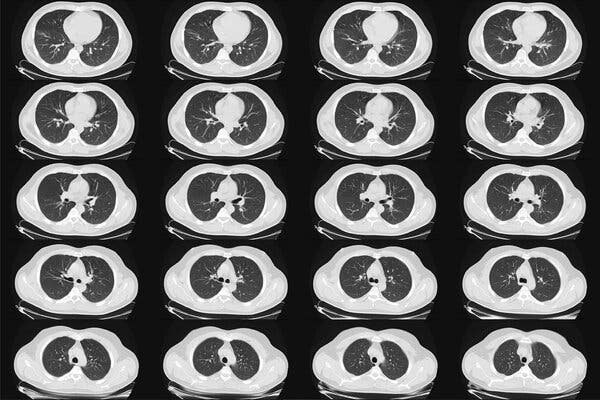
AdvertisementContinue reading the main storySupported byContinue reading the main storyYearly Lung Cancer Scans Are Advised for People 50 and Over With Shorter Smoking HistoriesNew advice from an influential panel will make more women and African-Americans eligible for CT scans, but some who need them most may not be able to afford them.A computed tomography scan of the lungs of a healthy adult man. The panel recommended low-dose CT scans, so-called because they involve a relatively small amount of radiation, and cost about $300.Credit…Alamy Stock PhotoMarch 9, 2021Updated 11:21 a.m. ETNew guidelines from medical experts will nearly double the number of people in the United States who are advised to have yearly CT scans to screen for lung cancer, and will include many more African-Americans and women than in the past.The disease is the leading cause of U.S. cancer deaths, and the goal of the expanded screening is to find it early enough to cure it in more people at high risk because of smoking. In those individuals, annual CT scans can reduce the risk of death from the cancer by 20 to 25 percent, large studies have found.The new recommendations, by the U.S. Preventive Services Task Force, include people ages 50 to 80 who have smoked at least a pack a day for 20 years or more, and who still smoke or have quit within the past 15 years.The advice, published on Tuesday in the medical journal JAMA, differs in two major ways from the task force’s previous guidelines, issued in 2013: It lowers the age when screening should start, to 50 from 55, and it reduces the smoking history to 20 years, from 30.Those changes will add more women and African-Americans to the pool eligible for screening, because they tend to smoke less heavily than the white male study participants on whom earlier guidelines were based. Women and Black Americans also tend to develop lung cancer earlier and from less tobacco exposure than do white men, experts said.Why the risk appears to differ by race and gender is not known.“Some studies have alluded to some hormonal influences in women,” Dr. Mara Antonoff, a lung surgeon at the M.D. Anderson Cancer Center in Houston, said in an interview. “In terms of racial differences, we don’t have an answer. We have population-based data to show they have a tendency to develop lung cancer younger and with less exposure to tobacco, but we don’t have a mechanism.”Under the new criteria, 14.5 million people in the United States will qualify for the screening, an increase of 6.4 million.The task force includes 16 physicians, scientists and public health experts who periodically evaluate screening tests and preventive treatments. Members are appointed by the director of the federal Agency for Healthcare Research and Quality, but the group is independent and its recommendations often help shape U.S. medical practice.The use of chest X-rays to detect lung cancer was largely abandoned decades ago because they could not find the disease early enough to be useful.The CT scans, called low-dose CT — because they involve a relatively small amount of radiation — cost about $300. Patients are advised to stop the screening once they have not smoked for 15 years, or if they develop health problems that would substantially shorten their life expectancy or make them unable to have lung surgery if needed.Patients have not flocked to clinics for this screening. Researchers estimate that only 6 to 18 percent of those who qualify and could be helped by the screening have taken advantage of it. Some cannot afford it.“Part of the low uptake is simply lack of access to care,” said Dr. Robert Smith, a screening expert at the American Cancer Society. “Smoking in general is increasingly concentrated in lower-income populations.”The Affordable Care Act does require that insurers cover any screening broadly recommended by the task force, with no out-of-pocket costs.Bettye Givens, right, of south Kansas City, reviewing lung scans with her doctor in 2017. She received a low-dose CT scan that detected her lung cancer at its earliest stage and was successfully treated.Credit…Andy Marso/Kansas City Star/TNS/Alamy Live NewsBut researchers have found that half the population eligible for lung-cancer screening had either no insurance, or Medicaid, Dr. Smith said. Not all Medicaid plans have covered the screening, according to an editorial in JAMA.“There could be a 15-year period when you might quality for screening and not have any insurance,” Dr. Smith said.He and other researchers also said that patients may be missing out on lung-cancer screening because they just don’t know about it. It has not received as much attention as other cancer screenings, like mammograms, colonoscopies and Pap tests. Some doctors may not encourage it as strongly, and especially with former smokers, may not take the time to calculate a patient’s smoking history to see if it matches the guidelines.The changes in the criteria for smoking history and screening age were based on new data from multiple studies, Dr. Alex H. Krist, the task force chairman and a professor of family medicine and population health at Virginia Commonwealth University, said in an interview.“Lung cancer is the No. 1 cancer killer in America,” Dr. Krist said, adding that with the new data, “we have even more confidence that screening does save lives.”Like other kinds of screenings affected by the pandemic, those for lung cancer remain below 2019 levels, according to an analysis of Medicare data by Avalere Health, a consulting firm, conducted for Community Oncology Alliance, which represents independent cancer specialists.While the number of screenings had started to rebound in the summer, the fresh spike in Covid cases later in the year caused them to fall again. In November, screenings were down by 30 percent, compared to 2019, and the number of lung biopsies had also dropped, indicating cases were not being diagnosed.Using its own grading system, the task force gave its recommendation a B, saying there was “moderate certainty” that annual screening was of “moderate net benefit.”That may not sound like a ringing endorsement, given that a grade of A means “high certainty that the net benefit is substantial.” But anything with an A or B grade should be offered to patients, according to task force rules.“There is building evidence that a pretty simple, five-minute, low-dose, low radiation scan can really save a lot of people’s lives,” said Dr. Bernard J. Park, a lung surgeon and the clinical director of the lung-screening service at Memorial Sloan Kettering Cancer Center in New York. About 75 to 85 percent of the cancers found with this screening are Stage 1, and curable with just surgery or radiation, he estimated.Dr. Park said that many people who signed up for the screening had quit smoking or were trying to stop, but that a few regarded clear scans as a sign that they could keep smoking.Dr. Smith said that the American Cancer Society was due to revise its own guidelines for lung-cancer screening, and that its advice would probably be similar to that of the task force.In 2013, the American Academy of Family Physicians declined to recommend for or against CT screening for lung cancer, saying there was insufficient evidence. But the president, Dr. Ada Stewart, said in an emailed statement on Monday that the academy would review the new task force evidence and decide whether to update its own recommendation to its members.Globally, there were 2.09 million new cases of lung cancer in 2018, and the disease is also the leading cause of cancer deaths, killing 1.76 million people that year, according to the World Health Organization.There were 228,820 new cases of lung cancer in the United States in 2020, and 135,720 people died from it, according to the National Cancer Institute. About 90 percent of cases occur in people who smoke, and current smokers’ risk of developing the disease is about 20 times that of nonsmokers.Only about 20.5 percent of patients survive five years after the diagnosis. Most cases are diagnosed late, after the cancer has begun to spread. But if it can be found and treated early, cure is possible, doctors say.CT screening has risks, and doctors say those must be explained to patients, who may decide to decline the testing. The scans detect tiny nodules in the lungs that may be early cancers — or maybe not. A suspicious-looking spot could be just a minor infection, inflammation or a benign growth, Dr. Park said.Often, the nodules can just be monitored with repeat scans, but it can be nerve-racking for patients to spend months waiting for the next test, knowing there is something in their lung that might be malignant.False positive rates, when something harmless is mistaken for cancer, have ranged from 3.9 to 25 percent and higher in studies, but tend to decrease over time, as the patient has more annual scans.A major concern about false positives is that they can lead to invasive procedures like lung biopsies. One large study found that invasive procedures were performed needlessly in 1.7 percent of the patients who were screened. The task force report said that standards created by radiology societies for evaluating the scans could help to prevent some unnecessary procedures spurred by false positives.Another possible risk from screening is the chance that the cumulative radiation exposure could cause cancer. But the dose is low, and the risk is thought to be small, especially when compared with the risk of lung cancer caused by smoking.In theory, screening could also lead to unneeded invasive tests and treatment for a cancer that would not have progressed or harmed the patient. How often that might occur is not known, but it is considered rare.Reed Abelson contributed to this article.AdvertisementContinue reading the main story

Read more →

Study reveals process to explain how maternal stress triggers idiopathic preterm birth

Preterm birth is a leading cause of infant deaths and illness in the U.S. — yet its underlying molecular causes remain largely unclear. About 40 to 50% of preterm births, defined as births before 37 weeks of pregnancy, are estimated to be “idiopathic,” meaning they arise from unexplained or spontaneous labor. And, maternal stress linked to depression and post-traumatic stress disorders as well as fetal stress have been strongly implicated in preterm births with no known cause.
Now, for the first time, a University of South Florida Health (USF Health) preclinical study has uncovered a mechanism to help explain how psychological and/or physiological stress in pregnant women triggers idiopathic preterm birth. A research team at the USF Health Morsani College of Medicine Department of Obstetrics and Gynecology shows how cortisol — the “fight-or-flight” hormone critical for regulating the body’s response to stress — acts through stress-responsive protein FKBP51 binding to progesterone receptors to inhibit progesterone receptor function in the uterus. This reduced progesterone receptor activity stimulates labor.
The findings were reported online first March 8 in Proceedings of the National Academy of Sciences (PNAS).
“This new study fills in some longstanding mechanistic gaps in our understanding of how normal labor begins and how stress causes preterm birth,” said the paper’s senior author Charles J. Lockwood, MD, senior vice president of USF Health, dean of the USF Health Morsani College of Medicine, and a professor of obstetrics and gynecology specializing in maternal-fetal medicine.
Dr. Lockwood was a co-principal investigator for the study along with the paper’s lead author Ozlem Guzeloglu-Kayisli, PhD, a USF Health associate professor of obstetrics and gynecology. Nihan Semerci, MSc, a senior biological scientist, shares the lead authorship with Dr. Guzeloglu-Kayisli.
Progesterone reduces contractions of the uterus and sustained levels are essential to prevent a baby from being born too early. Reduced uterine progesterone receptor expression and signaling stimulates labor. In the brain, elevated FKBP51 expression has been strongly associated with increased risk for stress-related disorders.

advertisement

Previous work by the USF Health team showed that normal human labor starting at term (between 37 and 42 weeks of pregnancy) was associated with reduced expression of progesterone receptors and increased expression of FKBP51, specifically in maternal decidual cells (specialized cells lining the uterus).
For the current study focused on maternal stress-induced idiopathic preterm birth, the researchers combined experiments in human maternal decidual cells and a mouse model in which FKBP5, the gene that makes FKBP51, had been removed, or “knocked out.” Altogether, their results revealed a novel functional progesterone withdrawal mechanism, mediated by maternal stress-induced uterine FKBP51 overexpression and enhanced FKPB51-progesterone receptor binding, that decreased progestational effects and triggered preterm birth. The researchers found that Fkbp5 knockout mice (with depletion of the gene encoding for FKBP51) exhibit prolonged gestation and are completely resistant to maternal stress-induced preterm birth.
Among the USF Health team’s key findings:
– FKPB51 levels were greater and FKPB51 binding to progesterone receptors was significantly increased in the decidual cells of women with idiopathic preterm birth, compared to decidual cells of gestational age-matched controls.
– The study reports for the first time that Fkbp5-deficient (knockout) mice are completely resistant to maternal stress-induced preterm birth and exhibit prolonged pregnancies accompanied by slower decline in systemic progesterone levels. This indicates that FKBP51 plays a crucial role in the length of pregnancy and initiation of labor and delivery.
– In contrast, mice with the FKPB5 gene intact and normal levels of FKPB51 protein (wild type mice) delivered earlier when exposed to maternal stress than either non-stressed wild type mice or FKPB5 knockout mice under nonstressed or stressed conditions.

advertisement

“Collectively, these results suggest that FKBP51 plays a pivotal role both in term labor and stress-associated preterm parturition (birth) and that inhibition of FKBP51 may prove to be a novel therapy to prevent idiopathic preterm birth,” the study authors conclude.
Currently, injectable progesterone is the only drug approved to help prevent preterm birth in high-risk women who have had a previous preterm birth. However, its effectiveness was not confirmed by a recent large clinical trial, sparking debate in the health care community. The authors finding that progesterone receptor activity was reduced in idiopathic preterm birth may explain the apparent lack of effectiveness of supplemental progesterone.
Babies born before 37 weeks, particularly those born before 34 weeks, have more health problems and may face long-term health complications, including childhood lung or heart disease and neurodevelopmental delays, Dr. Guzeloglu-Kayisli said. The likelihood of poor outcomes decreases as gestational age (length of the pregnancy) increases.
“Prevention of idiopathic preterm birth by extending gestation even two or three weeks can benefit the newborn, because it provides critical time needed for the fetus’s lungs and brain to mature,” Dr. Guzeloglu-Kayisli said. “Our research indicates the importance of investigating the potential use of FKBP51 inhibitors as a targeted therapy to reduce the risk of stress-related preterm birth.”
The USF Health study was supported in part by The March of Dimes Prematurity Research Center Ohio Collaborative grant.

Read more →

New brain sensor offers Alzheimer's answers

Scientists at the University of Virginia School of Medicine have developed a tool to monitor communications within the brain in a way never before possible, and it has already offered an explanation for why Alzheimer’s drugs have limited effectiveness and why patients get much worse after going off of them.
The researchers expect their new method will have tremendous impact on our understanding of depression, sleep disorders, autism, neurological diseases and major psychiatric conditions. It will speed scientific research into the workings of the brain, they say, and facilitate the development of new treatments.
“We can now ‘see’ how brain cells communicate in sharp detail in both the healthy and diseased brains,” said lead researcher J. Julius Zhu, PhD, of UVA’s Department of Pharmacology.
Unexpected Transmissions
The new method developed by Zhu and his collaborators lets scientists examine transmissions inside the brain at both the microscopic level and the far, far smaller nanoscopic level. It combines a biological “sensor” with two different forms of cutting-edge imaging.
The approach can quantify “neuromodulatory” transmissions, which are associated with major brain disorders, including addiction, Alzheimer’s, depressive disorders and schizophrenia. They’re also linked to autism, epilepsy, eating disorders and sleep disorders.
Neuromodulatory transmissions are the “slower” transmissions in the brain. They’re typically thought to involve lots of neurons in large regions. That’s in contrast to the much faster transmissions that happen neuron-to-neuron.
But Zhu’s new tool has already shown it’s not that simple.
In Alzheimer’s disease, Zhu and his colleagues discovered a surprising degree of “fine control and precision” in the supposedly shotgun neuromodulatory transmissions. Widely used Alzheimer’s drugs known as acetylcholinesterase inhibitors may inhibit this precise communication, the scientists report. That may explain the limited effectiveness of the drugs, they say.
The researchers went on to identify potential changes in the brain that could be brought about by long-term use of the drugs, which could explain why patients often get much worse when they stop taking them. “The new method points out Alzheimer’s defects in the unprecedented spatial and temporal resolution, defining the precise targets for medicine,” Zhu said.
Alzheimer’s, the researchers say, is just the tip of the iceberg. The new system has “broad applicability” across the spectrum of neurological and psychiatric diseases and disorders, they report. In the years to come, the scientists predict, it will help doctors understand neurological illnesses and psychiatric problems, screen drugs for potential treatments, identify disease-causing genes and develop better, more personalized medicine tailored for specific patient needs.
“If we see problems,” Zhu said, “we will be ready to treat them.”

Story Source:
Materials provided by University of Virginia Health System. Note: Content may be edited for style and length.

Read more →

COVID-19 risk increases with airborne pollen, study finds

When airborne pollen levels are higher, increased SARS-CoV-2 infection rates can be observed. These results were determined by a large-scale study conducted by an international team headed by researchers at the Technical University of Munich (TUM) and the Helmholtz Zentrum München. Members of high-risk groups could protect themselves by watching pollen forecasts and wearing dust filter masks.
In the spring of 2020, the outbreak of the coronavirus pandemic appeared to coincide with the tree pollen season in the northern hemisphere. These observations prompted an international team of researchers to conduct an extensive investigation: The scientists wanted to know whether there is a demonstrable link between airborne pollen concentrations and SARS-CoV-2 infection rates.
Pollen is a significant environmental factor influencing infection rates
Under the leadership of first author Athanasios Damialis, the team at the Chair of Environmental Medicine at TUM collected data on airborne pollen concentrations, weather conditions and SARS-CoV-2 infections — taking into consideration the variation of infection rates from one day to another and the total number of positive tests. In their calculations, the team also included data on population density and the effects of lockdown measures. The 154 researchers analyzed pollen data from 130 stations in 31 countries on five continents.
The team showed that airborne pollen can account for, on average, 44 percent of the variation in infection rates, with humidity and air temperature also playing a role in some cases. During intervals without lockdown regulations, infection rates were on average 4 percent higher with every increase of 100 grains of airborne pollen per cubic meter. In some German cities, concentrations of up to 500 pollen grains per cubic meter per day were recorded during the study — which led to an overall increase in infection rates of more than 20 percent. In regions where lockdown rules were in effect, however, the infection numbers were on average only half as high at comparable pollen concentrations.
Airborne pollen weakens immune response
High pollen concentrations lead to a weaker immune response in airways to viruses that can cause coughs and colds. When a virus enters the body, infected cells usually send out messenger proteins. This is also the case with SARS-CoV-2. These proteins, known as antiviral interferons, signal nearby cells to escalate their antiviral defenses to keep the invaders at bay. Additionally, an appropriate inflammation response is activated to fight the viruses.
But if airborne pollen concentrations are high, and pollen grains are inhaled with the virus particles, fewer antiviral interferons are generated. The beneficial inflammatory response itself is also affected. Therefore, on days with a high concentration of pollen, it can lead to an increase in the number of respiratory illnesses. This also holds true for Covid-19. Whether individuals are allergic to the different pollen types is irrelevant.
“You cannot avoid exposure to airborne pollen,” says Stefanie Gilles who is also first author of the study. “People in high-risk groups should, therefore, be informed that high levels of airborne pollen concentrations lead to an increased susceptibility to viral respiratory tract infections.” Athanasios Damialis emphasizes: “When studying the spread of SARS-CoV-2, environmental factors such as pollen must be taken into account. Increased awareness of these effects are an important step in preventing and mitigating the impact of Covid-19.”
Particle filtering masks provide protection
What can vulnerable people do to protect themselves? Claudia Traidl-Hoffmann, last author and a professor of environmental medicine, advises people at high-risk to monitor pollen forecasts over the coming months. Claudia Traidl-Hoffmann states: “Wearing a particle filtering mask when pollen concentrations are high can keep both the virus and pollen out of the airways.”

Story Source:
Materials provided by Technical University of Munich (TUM). Note: Content may be edited for style and length.

Read more →

Israeli Study Offers First Real-World Glimpse of COVID-19 Vaccines in Action

Credit: Getty Images/Hispanolistic

There are many reasons to be excited about the three COVID-19 vaccines that are now getting into arms across the United States. At the top of the list is their extremely high level of safety and protection against SARS-CoV-2, the coronavirus that causes COVID-19. Of course, those data come from clinical trials that were rigorously conducted under optimal research conditions. One might wonder how well those impressive clinical trial results will translate to the real world.

A new study published in the New England Journal of Medicine [1] offers an early answer for the Pfizer/BioNTech vaccine. The Pfizer product is an mRNA vaccine that was found in a large clinical trial to be up to 95 percent effective in preventing COVID-19, leading to its Emergency Use Authorization last December.

The new data, which come from Israel, are really encouraging. Based on a detailed analysis of nearly 600,000 people vaccinated in that nation, a research team led by Ran Balicer, The Clalit Research Institute, Tel Aviv, found that the risk of symptomatic COVID-19 infection dropped by 94 percent a week after individuals had received both doses of the Pfizer vaccine. That’s essentially the same very high level of protection that was seen in the data gathered in the earlier U.S. clinical trial.

The study also found that just a single shot of the two-dose vaccine led to a 57 percent drop in the incidence of symptomatic COVID-19 infections and a 62 percent decline in the risk of severe illness after two to three weeks. Note, however, that the protection clearly got better after folks received the second dose. While it’s too soon to say how many lives were saved in Israel thanks to full vaccination, the early data not surprisingly suggest a substantial reduction in mortality.

Israel, which is about as large as New Jersey with a population of around 9 million, currently has the world’s highest COVID-19 vaccination rate. In addition to its relatively small size, Israel also has a national health system and one of the world’s largest integrated health record databases, making it a natural choice to see how well one of the new vaccines was working in the real world.

The study took place from December 20, 2020, the start of Israel’s first vaccination drive, through February 1, 2021. This also coincided with Israel’s third and largest wave of COVID-19 infections and illness. During this same period, the B.1.1.7 variant, which was first detected in the United Kingdom, gradually became Israel’s dominant strain. That’s notable because the U.K. variant spreads from person-to-person more readily and may be associated with an increased risk of death compared with other variants [2].

Balicer and his colleagues reviewed data on 596,618 fully vaccinated individuals, ages 16 and older. A little less than one third—about 170,000—of the people studied were over age 60. To see how well the vaccine worked, the researchers carefully matched each of the vaccinated individuals in the study to an unvaccinated person with similar demographics as well as risks of infection, severe illness, and other important health attributes.

The results showed that the vaccine works remarkably well. In fact, the researchers determined that the Pfizer/BioNTech vaccine is similarly effective—94 percent to 96 percent—across adults in different age groups. It also appears that the vaccine works about equally well for individuals age 70 and older as it does for younger people.

So far, more than 92 million total vaccine doses have been administered in the U.S. With the Janssen COVID-19 vaccine (also called the Johnson & Johnson vaccine) now coming online, that number will rise even faster. For those of you who haven’t had the opportunity just yet, these latest findings should come as added encouragement to roll up your sleeve for any one of the authorized vaccines as soon as your invitation arrives.

References:

[1] BNT162b2 mRNA Covid-19 Vaccine in a Nationwide Mass Vaccination Setting. Dagan N, Barda N, Kepten E, Miron O, Perchik S, Katz MA, Hernán MA, Lipsitch M, Reis B, Balicer RD. N Engl J Med. 2021 Feb 24.

[2] Emerging SARS-CoV-2 Variants. Centers for Disease Control and Prevention.

Links:COVID-19 Research (NIH)

Clalit Research Institute (Tel Aviv, Israel)

Ran Balicer (Clalit Research Institute)

Read more →

Majority of women can still give birth naturally if their water breaks early

About 11% of women who carry to term will experience prelabor rupture of membrane — a condition where the amniotic sac breaks open early, but labor doesn’t begin.
Typically, when a woman’s water breaks but labor doesn’t start, labor is induced. But a new University of Michigan study found that expectant management — waiting a period of time after the water breaks for labor to begin spontaneously — did not significantly increase risk to the fetus or the mother in healthy pregnancies.
Therefore, both induction and expectant management should be considered, and the decision should be made in the context of the mother’s wishes and health, said study co-author Ruth Zielinski, a nurse midwife and U-M clinical professor in nursing.
The American College of Obstetricians and Gynecologists recommends induction, but in healthy pregnancies carried to term the American College of Nurse Midwives recommends expectant management be offered as an option.
During pregnancy, the fetus is surrounded by a fluid-filled membrane called the amniotic sac. At some point at the beginning of or during labor, this sac ruptures and contractions typically begin soon after. The goal of the study was to examine rates of induction, maternal infection, neonatal outcomes and time to birth in women who carried to term, and were expectantly managed at home or in the hospital.
Zielinski and colleagues looked at 2,357 women cared for by a midwestern midwifery service between January 2016 and December 2018. The amniotic sac ruptured early in 281 women (12%). Among that group, 150 (53%) opted to wait for labor onset at home; 102 (36%) were expectantly managed in the hospital; 21 (7.5%) were admitted for immediate induction of labor; and 8 (3%) were admitted for immediate cesarean birth.

advertisement

Of the women who opted to wait, the majority (65%) went into labor on their own and did not need to be induced. Rates of maternal and infant infection were no different between the groups of women with prelabor rupture of membranes.
Labor is typically induced when the water breaks early because the prevailing wisdom is that as the time between the amniotic sac rupturing and the beginning of labor grows, so does the risk of infection.
“The risk of infection does increase with prolonged ruptured membranes, which is why with prelabor rupture of membranes, when the mother is a Group B strep carrier, the recommendation is a shorter duration of expectant management,” Zielinski said.
Group B strep is a common bacteria that does not cause maternal infections, and providing antibiotic prophylaxis during labor is recommended to decrease the risk of transmission to the newborn.
The majority of newborns will not get sick but if they do, they get quite sick, which is why antibiotics are recommended, Zielinski said.
“Twenty-six years ago when I graduated from midwifery school, I assumed everyone wanted to avoid induction, but this is definitely not the case,” she said. “Often, patients want to get things going and are fine with induction. However, with healthy, term pregnancies, waiting for a period of time for labor to start is reasonable and should be offered.”
It is important for women to discuss their options with their provider, she said.

Story Source:
Materials provided by University of Michigan. Note: Content may be edited for style and length.

Read more →

Membrane around tumors may be key to preventing metastasis

For cancer cells to metastasize, they must first break free of a tumor’s own defenses. Most tumors are sheathed in a protective “basement” membrane — a thin, pliable film that holds cancer cells in place as they grow and divide. Before spreading to other parts of the body, the cells must breach the basement membrane, a material that itself has been tricky for scientists to characterize.
Now MIT engineers have probed the basement membrane of breast cancer tumors and found that the seemingly delicate coating is as tough as plastic wrap, yet surprisingly elastic like a party balloon, able to inflate to twice its original size.
But while a balloon becomes much easier to blow up after some initial effort, the team found that a basement membrane becomes stiffer as it expands.
This stiff yet elastic quality may help basement membranes control how tumors grow. The fact that the membranes appear to stiffen as they expand suggests that they may restrain a tumor’s growth and potential to spread, or metastasize, at least to a certain extent.
The findings, published this week in the Proceedings of the National Academy of Sciences, may open a new route toward preventing tumor metastasis, which is the most common cause of cancer-related deaths.
“Now we can think of ways to add new materials or drugs to further enhance this stiffening effect, and increase the toughness of the membrane to prevent cancer cells from breaking through,” says Ming Guo, a lead author of the study and associate professor of mechanical engineering at MIT.

advertisement

Guo’s co-authors include first author Hui Li of Beijing Normal University, Yue Zheng and Shengqiang Cai of the University of California at Santa Diego, and MIT postdoc Yu Long Han.
Blowing up
The basement membrane envelopes not only cancerous growths but also healthy tissues and organs. The film — a fraction of the thickness of a human hair — serves as a physical support that holds tissues and organs in place and helps to shape their geometry, while also keeping them separate and distinct.
Guo’s group specializes in the study of cell mechanics, with a focus on the behavior of cancer cells and the processes that drive tumors to metastasize. The researchers had been investigating how these cells interact with their surroundings as they migrate through the body.
“A critical question we realized hasn’t gotten enough attention is, what about the membrane surrounding tumors?” Guo says. “To get out, cells have to break this layer. What is this layer in terms of material properties? Is it something cells have to work really hard to break? That’s what motivated us to look into the basement membrane.”
To measure the membrane’s properties, scientists have employed atomic force microscopy (AFM), using a tiny mechanical probe to gently push on the membrane’s surface. The force required to deform the surface can give researchers an idea of a material’s resistance or elasticity. But, as the basement membrane is exceedingly thin and tricky to separate from underlying tissue, Guo says it’s difficult to know from AFM measurements what the resistance of the membrane is, apart from the tissue underneath.

advertisement

Instead, the team used a simple technique, similar to blowing up a balloon, to isolate the membrane and measure its elasticity. They first cultured human breast cancer cells, which naturally secrete proteins to form a membrane around groups of cells known as tumor spheroids. They grew several spheroids of various sizes and inserted a glass microneedle into each tumor. They injected the tumors with fluid at a controlled pressure, causing the membranes to detach from the cells and inflate like a balloon.
The researchers applied various constant pressures to inflate the membranes until they reached a steady state, or could expand no more, then turned the pressure off.
“It’s a very simple experiment that can tell you a few things,” Guo says. “One is, when you inject pressure to swell this balloon, it gets much bigger than its original size. And as soon as you release the pressure, it gradually shrinks back, which is a classical behavior of an elastic material, similar to a rubber balloon.”
Elastic snap
As they inflated each spheroid, the researchers observed that, while a basement membrane’s ability to inflate and deflate showed that it was generally elastic like a balloon, the more specific details of this behavior were surprisingly different.
To blow up a latex balloon typically requires a good amount of effort and pressure to start up. Once it gets going and starts to inflate a bit, the balloon suddenly becomes much easier to blow up.
“Typically, once the radius of a balloon increases by about 38 percent, you don’t need to blow any harder — just maintain pressure and the balloon will expand dramatically,” Guo says.
This phenomenon, known as snap-through instability, is seen in balloons made of materials that are linearly elastic, meaning their inherent elasticity, or stiffness, does not change as they deform or inflate.
But based on their measurements, the researchers found that the basement membrane instead became stiffer, or more resistant as it inflated, indicating that the material is nonlinearly elastic, and able to change its stiffness as it deforms.
“If snap-through instability were to occur, a tumor would become a disaster — it would just explode,” Guo says. “In this case, it doesn’t. That indicates to me that the basement membrane provides a control on growth.”
The team plans to measure the membrane’s properties at different stages of cancer development, as well as its behavior around healthy tissues and organs. They are also exploring ways to modify the membrane’s elasticity to see whether making it stiffer will prevent cancer cells from breaking through.
“We are actively following up on how to modify the mechanics of these membranes, and apply perturbations on breast cancer models, to see if we can delay their invasion or metastasis,” Guo says. “This is an analogy to making a stiffer balloon, which we plan to try.”
This research was supported, in part, by the Alfred Sloan Foundation and the National Cancer Institute.

Read more →

Opinions and attitudes can last when they are based on emotion

Depending on the topic, people’s attitudes can change from moment to moment or last a lifetime. The factors that make one opinion long-lasting and another ephemeral, however, are not always clear.
Past studies have demonstrated that opinions based on hard facts and data can remain constant over time, but new research published in the journal Psychological Science reveals that attitudes based on feelings and emotions can also stand the test of time. This research has implications for both predicting whose attitudes are fixed versus fleeting and how to nudge people to form more long-lasting opinions.
“We have known that encouraging people to think carefully and rationally can produce attitudes that change less in the future,” said Matthew Rocklage, a researcher with the University of Massachusetts, Boston, and co-lead author on the paper. “Our research, however, shows that opinions based on people’s emotional reactions can be particularly long-lasting as well.”
As part of their study, the researchers asked more than 1,000 people to what extent they believed attitudes based on feelings or emotional reactions were more stable over time than those based on thinking and rational analysis. Only 15% expressed any belief that attitudes based on emotion would be more stable over time.
To test the role that emotion plays in forming long-lasting attitudes, the researchers conducted seven independent studies involving more than 20,000 participants in a variety of real-world situations.
The first survey, which was conducted the day after Christmas, measured feelings about recently received gifts. The timing of this survey allowed the researchers to measure real-world reactions to a relatively newly formed attitude.
The participants were given a list of adjectives to describe their attitudes toward their gifts. Adjectives like “worthwhile” were associated with a practical reaction to the gift, whereas words like “delightful” were more strongly associated with an emotional reaction.
One month later, the participants completed a follow-up survey to test the endurance of their opinions. The results showed that the stronger the initial positive emotional reaction, the more likely that opinion remained fixed one month later.
The researchers conducted similar tests using virtually the same procedure but involving other scenarios, such as how much the participants supported consumer brands over time and how favorable their online restaurant reviews were between visits.
In the final test, participants read one of two messages about a fictitious aquatic animal. One message contained encyclopedic facts about the animal (low-emotion condition). The other message was about a swimmer’s underwater interaction with the animal (high-emotion condition). The participants in the high-emotion condition showed significantly less change in their attitude across time.
“Emotionality is an unappreciated predictor of long-lasting attitudes,” said Andrew Luttrell, a researcher at Ball State University and the other lead author on the paper. “These findings are important for understanding why some opinions are so difficult to change as well as how to create opinions that stick.”

Story Source:
Materials provided by Association for Psychological Science. Note: Content may be edited for style and length.

Read more →

Steroid abuse by men leads to long-lasting impaired testicular function

Illegal use of anabolic steroids not only has dangerous side effects during use but also can harm of men’s testicular function years after they stop abusing steroids, according to a study published in the Endocrine Society’s Journal of Clinical Endocrinology & Metabolism.
Anabolic steroids are synthetic forms of testosterone, and their abuse is prevalent among athletes worldwide. Some people use these steroids without a prescription to improve athletic performance or get a more muscular look. Known side effects of these drugs in men include breast growth, hair loss, shrunken testicles and lower testosterone levels. Also called hypogonadism, low testosterone can cause decreased sex drive, poor erections and a low sperm count.
“It is still debated whether illicit use of anabolic steroids causes long-lasting testosterone deficiency,” said Jon J. Rasmussen, M.D., Ph.D., the study’s principal investigator and a scientist at Rigshospitalet in Copenhagen, Denmark.
Researchers at the hospital have identified a hormone made by Leydig cells — cells in the testicles that produce testosterone — as a promising biological marker of testicular function, Rasmussen said. Because blood levels of testosterone can vary greatly during the day and vary by body composition, Rasmussen and his co-workers are investigating a more stable marker than testosterone, called serum insulin-like factor 3 (INSL3).
For this study, supported by Anti Doping Denmark, the research team included 132 participants from another study: men who did recreational strength training. Their ages ranged from 18 to 50 and averaged 32. Three study groups consisted of 46 men currently using anabolic steroids, 42 former steroid users and 44 who had never used these steroids. On average, former users had reportedly not taken anabolic steroids for 32 months.
Among current steroid users, INSL3 was markedly suppressed compared with former users and never-users, Rasmussen said. Compared with never-users, the former steroid users had lower INSL3 concentrations: 0.39 versus 0.59 microgram micrograms per liter. Furthermore, the longer the duration that the men reportedly used steroids, the lower their INSL3 levels, the researchers found.
“Our results suggest a long-lasting impaired gonadal capacity in previous anabolic steroid users,” Rasmussen said.
Although the clinically relevant difference in INSL3 levels is not yet known, because INSL3 measurement is primarily for research, he said their findings indicate that prior steroid users may have an increased risk of hypogonadism later in life.
“The results,” Rasmussen said, “raise the question whether some previous anabolic steroid users should receive medical stimulation therapy to increase Leydig cell capacity in the testicles.”
This therapy would include drugs used to block estrogen production or its conversion to testosterone, such as aromatase inhibitors and selective estrogen receptor modulators, he noted.

Story Source:
Materials provided by The Endocrine Society. Note: Content may be edited for style and length.

Read more →