Damage to white matter is linked to worse cognitive outcomes after brain injury

A new University of Iowa study challenges the idea that gray matter (the neurons that form the cerebral cortex) is more important than white matter (the myelin covered axons that physically connect neuronal regions) when it comes to cognitive health and function. The findings may help neurologists better predict the long-term effects of strokes and other forms of traumatic brain injury.
“The most unexpected aspect of our findings was that damage to gray matter hubs of the brain that are really interconnected with other regions didn’t really tell us much about how poorly people would do on cognitive tests after brain damage. On the other hand, people with damage to the densest white matter connections did much worse on those tests,” explains Justin Reber, PhD, a UI postdoctoral research fellow in psychology and first author on the study. “This is important because both scientists and clinicians often focus almost exclusively on the role of gray matter. This study is a reminder that connections between brain regions might matter just as much as those regions themselves, if not more so.”
The new study, published in PNAS, analyzes brain scans and cognitive function tests from over 500 people with localized areas of brain damage caused by strokes or other forms of brain injury. Looking at the location of the brain damage, also known as lesions, the UI team led by Reber and Aaron Boes, MD, PhD, correlated the level of connectedness of the damaged areas with the level of cognitive disability the patient experienced. The findings suggest that damage to highly connected regions of white matter is more predictive of cognitive impairment than damage to highly connected gray matter hubs.
Network hubs and brain function
Research on cognition often focuses on networks within the brain, and how different network configurations contribute to different aspects of cognition. Various mathematical models have been developed to measure the connectedness of networks and to identify hubs, or highly connected brain regions, that appear to be important in coordinating processing in brain networks.
The UI team used these well accepted mathematical models to identify the location of hubs within both gray and white matter from brain imaging of normal healthy individuals. The researchers then used brain scans from patients with brain lesions to find cases where areas of damage coincided with hubs. Using data from multiple cognitive tests for those patients, they were also able to measure the effect hub damage had on cognitive outcomes. Surprisingly, damage to highly connected gray matter hubs did not have a strong association with poor cognitive outcomes. In contrast, damage to dense white matter hubs was strongly linked to impaired cognition.
“The brain isn’t a blank canvas where all regions are equivalent; a small lesion in one region of the brain may have very minimal impact on cognition, whereas another one may have a huge impact. These findings might help us better predict, based on the location of the damage, which patients are at risk for cognitive impairment after stroke or other brain injury,” says Boes, UI professor of pediatrics, neurology, and psychiatry, and a member of the Iowa Neuroscience Institute. “It’s better to know those things in advance as it gives patients and family members a more realistic prognosis and helps target rehabilitation more effectively.”
UI registry is a unique resource for neuroscientists
Importantly, the new findings were based on data from over 500 individual patients, which is a large number compared to previous studies and suggests the findings are robust. The data came from two registries; one from Washington University in St. Louis, which provided data from 102 patients, and the Iowa Neurological Registry based at the UI, which provided data from 402 patients. The Iowa registry is over 40 years old and is one of the best characterized patient registries in the world, with close to 1000 subjects with well characterized cognition derived from hours of paper and pencil neuropsychological tests, and detailed brain imaging to map brain lesions. The registry is directed by Daniel Tranel, PhD, UI professor of neurology, and one of the study authors.
Reber notes that the study also illustrates the value of working with clinical patients as well as healthy individuals in terms of understanding relationships between brain structure and function.
“There is a lot of really excellent research using functional brain imaging with healthy participants or computer simulations that tell us that these gray matter hubs are critical to how the brain works, and that you can use them to predict how well healthy people will perform on cognitive tests. But when we look at how strokes and other brain damage actually affect people, it turns out that you can predict much more from damage to white matter,” he says. “Research with people who have survived strokes or other brain damage is messy, complicated, and absolutely essential, because it builds a bridge between basic scientific theory and clinical practice, and it can improve both.
I cannot stress enough how grateful we are that these patients have volunteered their time to help us; without them, a lot of important research would be impossible,” he adds.
Story Source:
Materials provided by University of Iowa Health Care. Original written by Jennifer Brown. Note: Content may be edited for style and length.

Read more →

Breaching the blood-brain barrier to deliver precious payloads

RNA-based drugs have the potential to change the standard of care for many diseases, making personalized medicine a reality. This rapidly expanding class of therapeutics are cost-effective, fairly easy to manufacture, and able to go where no drug has gone before, reaching previously undruggable pathways.
Mostly.
So far, these promising drugs haven’t been very useful in getting through to the well-protected brain to treat tumors or other maladies.
Now a multi-institutional team of researchers, led by Costas Arvanitis at the Georgia Institute of Technology and Emory University, has figured out a way: using ultrasound and RNA-loaded nanoparticles to get through the protective blood-brain barrier and deliver potent medicine to brain tumors.
“We’re able to make this drug more available to the brain and we’re seeing a substantial increase in tumor cell death, which is huge,” said Arvanitis, assistant professor in the Wallace H, Coulter Department of Biomedical Engineering (BME) and Georgia Tech’s George W. Woodruff School of Mechanical Engineering (ME).
Arvanitis, whose collaborators include researchers and clinicians from Emory’s School of Medicine and the University of Cincinnati College of Medicine, is the corresponding author of a new paper published in the journal Science Advances that describes the team’s development of a next-generation, tunable delivery system for RNA-based therapy in brain tumors.

Read more →

Why is COVID-19 so hard to treat? Growing evidence points to unique infectious profile

A comprehensive review into what we know about COVID-19 and the way it functions suggests the virus has a unique infectious profile, which explains why it can be so hard to treat and why some people experience so-called “long-COVID,” struggling with significant health issues months after infection.
There is growing evidence that the virus infects both the upper and lower respiratory tracts — unlike “low pathogenic” human coronavirus sub-species, which typically settle in the upper respiratory tract and cause cold-like symptoms, or “high pathogenic” viruses such as those that cause SARS and ARDS, which typically settle in the lower respiratory tract.
Additionally, more frequent multi-organ impacts, and blood clots, and an unusual immune-inflammatory response not commonly associated with other, similar viruses, mean that COVID-19 has evolved a uniquely challenging set of characteristics.
While animal and experimental models imply an overly aggressive immune-inflammation response is a key driver, it seems things work differently in humans: although inflammation is a factor it is a unique dysregulation of the immune response that causes our bodies to mismanage the way they fight the virus.
This may explain why some people experience “long-COVID” and suffer severe lung damage after infection.
Ignacio Martin-Loeches, Clinical Professor in Trinity College Dublin’s School of Medicine, and Consultant in Intensive Care Medicine at St James’s Hospital, is a co-author of the review just published in leading medical journal, The Lancet. He said:
“The emergence of severe acute respiratory syndrome coronavirus two (SARS-CoV-2), which causes COVID-19, has resulted in a health crisis not witnessed since the 1918 Spanish flu pandemic. Tragically, millions around the world have died already.
“Despite international focus on the virus, we are only just beginning to understand its intricacies. Based on growing evidence we propose that COVID-19 should be perceived as a new entity with a previously unknown infectious profile. It has its own characteristics and distinct pathophysiology and we need to be aware of this when treating people.
“That doesn’t mean we should abandon existing best-practice treatments that are based on our knowledge of other human coronaviruses, but an unbiased, gradual assembly of the key COVID-19 puzzle pieces for different patient cohorts — based on sex, age, ethnicity, pre-existing comorbidities — is what is need to modify the existing treatment guidelines, subsequently providing the most adequate care to COVID-19 patients.”
The review article was produced by the European Group on Immunology of Sepsis (EGIS) in which Professor Martin-Loeches is one of the funding members. EGIS is a multidisciplinary group of scientists and doctors with special interest in severe infection in patients admitted to ICU.
Story Source:
Materials provided by Trinity College Dublin. Note: Content may be edited for style and length.

Read more →

Distinct cell-to-cell communication processes controlled differently

Cells talk to each other to coordinate nutrition, waste removal, energy use, and, in some cases, disease progression. The cells that line the surfaces of organs or specific tissues, called epithelial cells, appear to speak two different languages — one for either side of the cell, according to a new study by researchers based in Japan.
The discovery, published on March 16 in EMBO Reports, could have implications for understanding how cancer spreads and, potentially, for advanced treatments, the team says.
The team, led by Mitsunori Fukuda, professor in the Laboratory of Membrane Trafficking Mechanisms, Department of Integrative Life Sciences, Graduate School of Life Sciences at Tohoku University, examined epithelial cells from a kidney model. The cells release particles called exosomes that carry bits of the cells themselves or information about the cells. The proteins and other genetic information in the exosomes can then influence how other cells behave or function. In health, such an information exchange could help the immune system mount a more tailored approach to an invading pathogen. Some diseased cells, such as cancer, can release exosomes that make healthy cells less resistant to invasion.
“Single cells are known to release various kinds of exosomes, but very little is known about the mechanisms by which they are produced and released,” Fukuda said. “In this paper, we found that epithelial cells asymmetrically release two distinct types of exosomes with distinct protein compositions.”
The researchers developed a purification method to separate out exosomes based on their protein makeup.
“In this paper, we found that epithelial cells asymmetrically release two distinct types of exosomes — apical and basolateral — with distinct protein compositions,” said first author Takahide Matsui, assistant professor, Laboratory of Membrane Trafficking Mechanisms, Department of Integrative Life Sciences, Graduate School of Life Sciences at Tohoku University.
They found that exosomes released from the apical side of the cell, which faces an external space or lumen, were modulated by ALIX, a protein related to the particle formation inside the cells. Exosomes released from the basolateral side of the cell closest to other tissues and neighboring cells were triggered by ceramide, a fatty molecule. They also found that depleting ALIX and ceramide reduced the number of apical exosomes and basolateral exosomes released, respectively.
Fukuda said that the results could help elucidate the cell-to-cell communication that allows cancer to migrate — and put a stop to it.
“It will be interesting to investigate how cancer cells use two distinct mechanisms of exosome production during cancer progression,” Fukuda said. “Since exosomes from cancer cells are involved in their progression, our findings could lead to the discovery of new drugs for treatments for cancers in the future.”
Matsui agreed, noting that their research could expand to other realms in health and in disease.
“Our discovery provides an important clue to understanding the generation of different exosomes in many cell types in addition to epithelial cells,” Matsui said.
Story Source:
Materials provided by Tohoku University. Note: Content may be edited for style and length.

Read more →

A deeper understanding of how cells move and stick together

Observing how cells stick to surfaces and their motility is vitally important in the study of tissue maintenance, wound healing and even understanding how cancers progress. A new paper published in EPJ Plus, by Raj Kumar Sadhu, Weizmann Institute of Science, Rehovot, Israel, takes a step towards a deeper understanding of these processes.
“Cell adhesion is the ability of a cell to stick to another cell or an extracellular matrix. This process is important in order to understand how cells interact and coordinate their behaviour in multicellular organisms,” says sadhu. “We theoretically model the adhesion of a cell-like vesicle by describing the cell as a three-dimensional vesicle adhering on a flat substrate with a constant adhesion interaction.”
Alongside his co-authors, Sadhu set about exploring the role of membrane-bound curvature sensitive proteins and the forces that act on the cytoskeleton — the network of interlinking protein filaments found in the cytoplasm of all cells — during the adhesion process. The team discovered that curved proteins enhance the adhesion process considerably, especially when coupled with active cytoskeleton forces.
“Our work shows that the curved membrane proteins, coupled with the pushing force due to the cytoskeleton, can play a key role in the cell adhesion process,” adds Sadhu. “In addition, we showed that these minimal ingredients are sufficient to produce a motile shape that closely resembles migrating cells. Our present work will motivate more research in this direction.”
One aspect of the research that pleasantly surprised the team was the fact that the relatively simple model they created could not just describe cell adhesion, but also allowed them to capture cell movement as well. The resultant paper belongs to the Topical Collection ‘Focus Point on Mechanobiology across Scales,’ edited by M. Ben Amar, A. Boudaoud and M. Lenz.
“Physical principles of shape, curvature and forces, combine to give living cells their shapes,” concludes Sadhu. “We show that the cells can have a variety of dynamic shapes, which spontaneously arise due to physical principles, and control the function of the cells in our bodies.”
The team will seek to improve on this current study by examining the adhesion of cells on more complex surfaces. This will include curved surfaces, those with adhesion gradients, and others upon which adhesive elements are temporary.
Story Source:
Materials provided by Springer. Note: Content may be edited for style and length.

Read more →

Some meat eaters disgusted by meat

Some meat eaters feel disgusted by meat, according to a new study.
University of Exeter scientists showed food pictures to more than 700 people, including omnivores (who eat meat and other foods), flexitarians (who try to eat less meat) and vegetarians.
About 7% of meat eaters (15% of flexitarians and 3% of omnivores) had a “fairly strong disgust response” to images of meat dishes commonly eaten in the UK, like roast chicken or bacon.
As a group, omnivores rated meat images about twice as disgusting on average as pictures of carbohydrate-rich foods like bread, chips and rice.
Based on the findings, the researchers say harnessing the “yuk factor” may be more effective than relying on willpower for anyone who wants to eat less meat.
“We were surprised to find that so many people are grossed out by meat — even people who eat meat all the time,” said Elisa Becker, of the University of Exeter.

Read more →

How bullying and obesity can affect girls' and boys' mental health

Depressive symptoms are more common in teenage girls than in their male peers. However, boys’ mental health appears to be affected more if they suffer from obesity. Irrespective of gender, bullying is a considerably greater risk factor than overweight for developing depressive symptoms. These conclusions are drawn by researchers at Uppsala University who monitored adolescents for six years in a questionnaire study, now published in the Journal of Public Health.
“The purpose of our study was to investigate the connection between body mass index (BMI) and depressive symptoms, and to take a close look at whether being subjected to bullying affects this relationship over time. We also wanted to investigate whether any gender differences existed,” says Sofia Kanders, a PhD student at Uppsala University’s Department of Neuroscience.
In the study young people, born in Västmanland County, replied to questions about their height, weight and depressive symptoms on three separate occasions (2012, 2015 and 2018). The respondents’ mean age was 14.4 years on the first occasion and 19.9 years on the last.
Based on BMI, the adolescents were divided into three groups: those with normal weight, overweight and obesity respectively. They were also grouped according to the extent of their depressive symptoms.
Overall, regardless of their weight, the girls stated more frequently that they had depressive symptoms. In 2012, 17 per cent of the girls and 6 per cent of the boys did so. By 2015, the proportions of adolescents with these symptoms had risen to 32 per cent for the girls and 13 per cent for the boys. The corresponding figures for 2018 were 34 and 19 per cent respectively.
A higher BMI did not, as far as the researchers could see, affect the girls’ mental well-being to any great extent. Among the boys, however, the pattern observed was entirely different.

Read more →

Algorithms show accuracy in gauging unconsciousness under general anesthesia

Anesthestic drugs act on the brain but most anesthesiologists rely on heart rate, respiratory rate, and movement to infer whether surgery patients remain unconscious to the desired degree. In a new study, a research team based at MIT and Massachusetts General Hospital shows that a straightforward artificial intelligence approach, attuned to the kind of anesthetic being used, can yield algorithms that assess unconsciousness in patients based on brain activity with high accuracy and reliability.
“One of the things that is foremost in the minds of anesthesiologists is ‘Do I have somebody who is lying in front of me who may be conscious and I don’t realize it?’ Being able to reliably maintain unconsciousness in a patient during surgery is fundamental to what we do,” said senior author Emery N. Brown, Edward Hood Taplin Professor in The Picower Institute for Learning and Memory and the Institute for Medical Engineering and Science at MIT, and an anesthesiologist at MGH. “This is an important step forward.”
More than providing a good readout of unconsciousness, Brown added, the new algorithms offer the potential to allow anesthesiologists to maintain it at the desired level while using less drug than they might administer when depending on less direct, accurate and reliable indicators. That can improve patient’s post-operative outcomes, such as delirium.
“We may always have to be a little bit ‘overboard’,” said Brown, who is also a professor at Harvard Medical School. “But can we do it with sufficient accuracy so that we are not dosing people more than is needed?”
Used to drive an infusion pump, for instance, algorithms could help anesthesiologists precisely throttle drug delivery to optimize a patient’s state and the doses they are receiving.
Artificial intelligence, real-world testing
To develop the technology to do so, postdocs John Abel and Marcus Badgeley led the study, published in PLOS ONE [LINK TBD], in which they trained machine learning algorithms on a remarkable data set the lab gathered back in 2013. In that study, 10 healthy volunteers in their 20s underwent anesthesia with the commonly used drug propofol. As the dose was methodically raised using computer controlled delivery, the volunteers were asked to respond to a simple request until they couldn’t anymore. Then when they were brought back to consciousness as the dose was later lessened, they became able to respond again. All the while, neural rhythms reflecting their brain activity were recorded with electroencephalogram (EEG) electrodes, providing a direct, real-time link between measured brain activity and exhibited unconsciousness.

Read more →

From the Wastewater Drain, Solid Pandemic Data

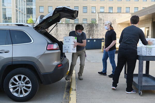
The coronavirus could turn sewage surveillance into a mainstream public health practice.Marc Johnson saw trouble in the water.Dr. Johnson, a virologist at the University of Missouri, had spent much of 2020 studying sewage, collecting wastewater from all over the state and analyzing it for fragments of the coronavirus. People with Covid-19 shed the virus in their stool, and as the coronavirus spread throughout Missouri, more and more of it began to appear in the state’s wastewater.In January, Dr. Johnson spotted something new in his water samples: traces of B.1.1.7, a more contagious variant that was first detected in Britain. Officially, the state had no confirmed cases of B.1.1.7, but the wastewater suggested that the variant had arrived. By the end of the month, the B.1.1.7 levels in Dr. Johnson’s water samples had risen sharply, and in early February, the state finally found its first case. It has since found hundreds more.Using some samples of sewage, Dr. Johnson had been able to peer into Missouri’s coronavirus future. “I can’t believe how well it works,” he said. “I feel like an oracle.”Dr. Johnson is one of many scientists who have been drawn into the once niche field of wastewater epidemiology in the past year. Researchers in 54 countries are now tracking the coronavirus in sewage, according to the Covid19Poops Dashboard, a global directory of the projects.Their work has validated the idea that wastewater surveillance can be a useful way to track infectious disease across entire communities, revealing epidemiological blind spots and yielding actionable public health information.It has also helped push wastewater epidemiology into the mainstream. In March, the European Commission recommended that member states establish systems to monitor sewage for the coronavirus. And last fall, the Centers for Disease Control and Prevention and the Department of Health and Human Services established the National Wastewater Surveillance System to help local officials respond to Covid-19. It is the first system of its kind in the United States.“Wastewater surveillance is not a new idea,” said Amy Kirby, the program lead for the surveillance system. It has been used in low- and middle-income countries in the fight to eradicate polio, for instance, and has been proposed as a way to keep tabs on noroviruses, a common cause of stomach bugs. “But really, the return on investment to build this large new infrastructure was never enough to warrant building the system for any of those other diseases,” Dr. Kirby said. “But Covid and the pandemic really changed the calculus.”The system, and others like it now emerging around the world, could ultimately usher in a new age of wastewater epidemiology, helping officials track not just the coronavirus, but also other outbreaks and diseases. “I think this is really going to be the beginning of a whole new type of data collection for public health disease surveillance,” Dr. Kirby said.Getting their feet wetWastewater samples for processing were unloaded at the Lab Science Center at the University of Missouri.Michael B. Thomas for The New York TimesAlthough Covid-19 is primarily a respiratory disease, research conducted early in the pandemic revealed that people infected with the coronavirus often shed it in their stool. This finding, combined with the scale and urgency of the crisis, spurred immediate interest in tracking the virus by sampling wastewater.By searching for, and then counting, certain coronavirus genes in sewage, researchers hoped to determine whether the virus was present in a particular region and how widespread it was. Before long, wastewater surveillance projects were popping up everywhere from Kansas City, Mo., to Kathmandu, Nepal.The resulting data, now appearing in a flood of scientific papers and preprints, have provided powerful proof of principle. Scientists have detected the virus in all kinds of environments: in treated and untreated water, in sludge and settled solids, in sewers and septic tanks, in pit latrines and open drainage systems. They found it in water flowing into enormous treatment plants and out of schools, dormitories and nursing homes. “It’s just fascinating how robust this tool has become,” said Peter Grevatt, chief executive of the Water Research Foundation.Teams all over the globe — in the United States, France, Portugal, India, Iran, Brazil, Canada and elsewhere — also found that the wastewater data seemed to be an accurate indicator of what was happening in the real world. When the number of diagnosed Covid-19 cases in an area increased, more coronavirus appeared in the wastewater. Levels of the virus fell when areas instituted lockdowns and surged when they reopened.Multiple teams have also confirmed that sewage can serve as an early warning system; Wastewater viral levels often peaked days before doctors saw a peak in official Covid-19 cases.This lead time, which can range from a couple of days to two weeks, depends partly on the robustness of local clinical testing programs, scientists say: When more people are being tested for the virus more frequently, the wastewater data provides less advance warning. The lead time also exists because infected people often begin shedding the virus, SARS-CoV-2, before they feel symptoms and then, once they fall ill, frequently delay seeking medical care.“I think wastewater has proven itself as one of the most, I would say, objective means of understanding what SARS-CoV-2 is doing in our society,” said Gertjan Medema, a microbiologist at KWR Water Research Institute in the Netherlands.It has proved sensitive, too, allowing researchers to detect a single infected student in a dorm or resident in a nursing home.Plugging holesDr. Johnson is one of many scientists newly drawn to the once niche field of wastewater epidemiology. “I can’t believe how well it works,” he said. “I feel like an oracle.”Michael B. Thomas for The New York TimesWastewater surveillance is not a replacement for clinical testing, experts said, but can be an efficient, cost-effective complement. In one study published last August, researchers calculated that they could test the wastewater from every treatment plant in Germany millions of times for less than it would cost them to test every German resident just once. The approach is likely to be especially valuable in low- and middle-income countries, where testing resources are even more limited.“Not every population gets tested, not everyone has access to health care,” Dr. Johnson said. “If there’s groups of people that are asymptomatic, they probably aren’t getting tested either. So you aren’t really getting the full big picture. Whereas for our testing, everyone poops.”Although there is a lot left to learn, even the small pilot projects started over the last year have already helped officials identify hidden viral hot spots and figure out how to target their resources.For instance, a number of U.S. colleges and universities, including the University of Arizona and the University of North Carolina, have used wastewater surveillance of dorms to find asymptomatic, infected students who had otherwise evaded detection. In the Netherlands, health officials have used wastewater data to determine where to send their mobile testing buses, Dr. Medema said.In Australia, where case numbers have been relatively low, the wastewater monitoring has helped reassure authorities that their pandemic controls are working. “Almost all the samples come back with nothing in them,” said Daniel Deere, the project manager for ColoSSoS, a coronavirus sewage surveillance project in Australia. “It’s been good to give confidence to allow the economy to stay open, to allow movement to continue between states.”(On the occasions when a water sample has come back positive, the government has ramped up testing and launched media campaigns to alert people who live in the region, he said.)Wastewater analysis has also allowed scientists to detect the arrival of certain variants in a region weeks before they are found in actual patients — and to identify mutations that have not yet been detected in patients anywhere.Dr. Johnson and his colleague John Dennehy, a virologist at Queens College, recently identified novel combinations of mutations, which might suggest the presence of new variants, in New York City wastewater. “We don’t know what it means yet,” Dr. Johnson said, noting that the new sequences could be coming from animals, not people. “We can see a little bit into the future, but you know, the crystal ball is always a little cloudy.”Waste not, want notWastewater samples being logged at the Lab Science Center.MichaelB Thomas for The New York TimesThis flurry of research and investment has been a boon to wastewater epidemiology. “This has been just an amazingly huge catalyst for the field,” said Tim Julian, who leads the pathogens and human health group at the Swiss Federal Institute of Aquatic Science and Technology.Over the last year, scientists have refined their methods, and water utilities, environmental laboratories and public health agencies have forged new connections. “The big question mark on everyone’s mind is what happens next,” said Christopher Mason, a geneticist at Weill Cornell Medical College who is part of a team tracking the coronavirus in wastewater samples collected from sites around the world. “How long does this go? How do we really sustain it?”The C.D.C.’s new wastewater surveillance system is one answer.“A lot of the initial efforts were coming from academic researchers, commercial laboratories and a few utilities that were able to do the testing themselves,” Dr. Kirby said. “And they have done great work to develop these methods. But routine, long-term surveillance work is not what any of those groups were designed to do.”The National Wastewater Surveillance System provides funding, technical support, a national data repository and other resources that will allow state and local health departments take over this long-term monitoring. Since its founding last year, the system has grown to include 33 states, four cities, one county and three U.S. territories.Some health departments were initially skeptical that wastewater data could be useful, but they have since come to embrace it, Dr. Kirby said, using it to figure out where to target their testing resources or to forecast hospital demand. “When you see an increase in wastewater data, we know that within a couple of weeks, you’re going to start seeing an increase in hospitalizations,” she said.In the next three to five years, the agency plans to expand the system to include monitoring for other pathogens, including antibiotic-resistant bacteria and microbes that frequently contaminate food, Dr. Kirby said.The European Union is also developing a “sewage sentinel system” that aims to monitor the wastewater in roughly 6,000 cities, Dr. Medema said. Although Covid-19 is the immediate priority, researchers are also assessing the feasibility of using such a system “in a post-pandemic E.U. for antibiotic resistance, infectious diseases, use of pharmaceuticals and illicit drugs and maybe more on the horizon,” he said.These systems could ultimately help officials stay ahead of emerging threats, providing early warnings about whatever pathogen is poised to cause the next pandemic. An Italian team recently found that the new coronavirus was already present in the wastewater in northern Italy in mid-December 2019, days before the first Covid-19 cases in Wuhan, China, were publicly reported.“This isn’t the last infectious disease that will come through our water supplies,” said Belinda Sturm, an environmental engineer at the University of Kansas. “This is a tool that we should make sure that we keep sharpened.”

Read more →