Researchers see need for warnings about long-range wildfire smoke

Smoke from local wildfires can affect the health of Colorado residents, in addition to smoke from fires in forests as far away as California and the Pacific Northwest.
Researchers at Colorado State University, curious about the health effects from smoke from large wildfires across the Western United States, analyzed six years of hospitalization data and death records for the cities along the Front Range, which reaches deep into central Colorado from southern Wyoming.
They found that wildfire smoke was associated with increased hospitalizations for asthma, chronic obstructive pulmonary disease and some cardiovascular health outcomes. They also discovered that wildfire smoke was associated with deaths from asthma and cardiovascular disease, but that there was a difference in the effects of smoke from local fires and that from distant ones.
Long-range smoke was associated with expected increases in hospitalizations and increased risk of death from cardiovascular outcomes.
But when the research team separated out health effects of smoke from local wildfires in early summer 2012 from long-range smoke from late summer 2012 and summer 2015, they found that local wildfires were associated with meaningful decreases in hospitalizations, especially for asthma.
The study, “Differential Cardiopulmonary Health Impacts of Local and Long-Range Transport of Wildfire Smoke,” was recently published in GeoHealth, a journal from the American Geophysical Union.

advertisement

Residents protect themselves from local fires
Sheryl Magzamen, lead author of the study and an associate professor in the Department of Environmental and Radiological Health Sciences at CSU, said the team believes that evacuation efforts and related media coverage of local wildfires may have helped protect residents from adverse health effects of smoke exposure as well as direct impacts of the fires.
“There’s a lack of communication about smoke from distant wildfires,” said Magzamen. “Generally when there are local fires, there are advisories in the news that are associated with evacuations and local fire conditions. Due to the presence of the fire, people take measures to protect themselves. This could be why we see this lower risk of health effects from smoke associated with local fires.”
Researchers described the long-range wildfire smoke as resembling fog, which is what Magzamen said she noticed in Fort Collins in August 2015. At the time, she was collaborating on a project with Jeff Pierce, associate professor in the Department of Atmospheric Science.
“I thought it was weird to see fog on that day,” she explained. “Jeff said, ‘That’s actually smoke.’ We all took a step back.”
Smoke changes with age

advertisement

Pierce, a co-author on this study, said researchers don’t really know how harmful smoke is as it gets older, or becomes long-range smoke.
“In Fort Collins, about half the time we had smoke in late August or September 2020, this was smoke from the Cameron Peak Fire,” he explained. “This smoke was only a couple hours old when it got here. At other times, we were getting smoke from California, and the smoke from the Cameron Peak Fire was either going over our heads or further south.”
The Cameron Peak Fire was reported on Aug. 13, 2020, and burned into October, consuming 208,913 acres on the Arapaho and Roosevelt National Forests in Larimer and Jackson Counties and Rocky Mountain National Park. It was the first wildfire in Colorado history to burn more than 200,000 acres.
The average person would not notice a difference in wildfire smoke, Pierce said.
“If the smoke is even two days old, things happen chemically, which changes the smoke a lot,” he explained. “If it didn’t smell like wood burning, it was long-range smoke from California.”
Magzamen said that the team is working to better understand these chemical changes.
“As the small particles found in wildfire smoke age, they can cause more oxidative stress and more respiratory health effects,” she said. “But wildfire smoke itself is a mixture of particles and gases. Teasing apart the effects of all the components of smoke and what happens to the mixture across space and time — and how those changes impact health — is an enormous scientific challenge.”
Better air quality monitoring
Magzamen said the gap in understanding the source of wildfire smoke is because it historically has been measured by land-based sensors, which are primarily located in large urban areas and sparsely located in other regions, even along the Front Range.
“Even over the last five years, our air quality monitoring networks have been enhanced with new technologies and better measurements of real-time smoke effects,” she said.
CSU researchers are now collaborating with local government officials on messaging related to the different types of wildfire smoke, with a specific aim to reach the most vulnerable populations. This includes caretakers of young children, people experiencing homelessness and others who can’t shelter safely in place during wildfire season.
“We want people to be smoke-aware,” she said. “On the Front Range, we have wildfire smoke every summer. We may not get Cameron Peak-size type of fires every year, but we are downwind for pretty much the entire Western United States,” she said. “It’s critical that we keep people healthy and safe.”

Read more →

Immune cell implicated in development of lung disease following viral infection

Scientists at Washington University School of Medicine in St. Louis have implicated a type of immune cell in the development of chronic lung disease that sometimes is triggered following a respiratory viral infection. The evidence suggests that activation of this immune cell — a type of guardian cell called a dendritic cell — serves as an early switch that, when activated, sets in motion a chain of events that drives progressive lung diseases, including asthma and chronic obstructive pulmonary disease (COPD).
The new study, published in The Journal of Immunology, opens the door to potential preventive or therapeutic strategies for chronic lung disease. More immediately, measuring the levels of these dendritic cells in clinical samples from patients hospitalized with a viral infection, such as influenza or COVID-19, could help doctors identify which patients are at high risk of respiratory failure and death.
Studying mice with a respiratory viral infection that makes the animals prone to developing chronic lung disease, the researchers showed that these dendritic cells communicate with the lining of the airway in ways that cause the airway-lining cells to ramp up their growth and inflammatory signals. The inflammation causes airway-lining cells to grow beyond their normal boundaries and turn into cells that overproduce mucus and cause inflammation, which in turn causes cough and difficulty breathing.
“We’re trying to understand how a viral infection that seems to be cleared by the body can nevertheless trigger chronic, progressive lung disease,” said senior author Michael J. Holtzman, MD, the Selma and Herman Seldin Professor of Medicine. “Not everyone experiences this progression. We believe there’s some switch that gets flipped, triggering the bad response. We’re identifying that switch and ways to control it. This work tells us that this type of dendritic cell is sitting right at that switch point.”
Holtzman’s past work had implicated the lining of the airway — where the viral infection takes hold — as the likely trigger for this process.
“But this study suggests that the cascade starts even further upstream,” said Holtzman, also director of the Division of Pulmonary and Critical Care Medicine. “Dendritic cells are telling the cells lining the airway what to do. There’s more work to be done, but this data tells us that the dendritic cells play an important role in getting the airway-lining cells onto the wrong path.”
Holtzman calls this dendritic cell a type of sentinel because its job is to detect an invading virus and trigger the body’s initial immune response against the infection. The problem comes when the cell doesn’t shut down properly after the threat has passed.
“Many people never develop chronic lung disease after a viral infection,” Holtzman said. “But others have a genetic susceptibility to this type of disease. People who are susceptible to virus-triggered disease include patients with asthma, COPD, and viral infections such as COVID-19. It’s really critical to look for ways to fix this disease response and prevent the problems that might occur after the virus has gone.”
In the meantime, Holtzman said, high levels of these dendritic cells and their products in the lungs of hospitalized patients could serve as a warning to doctors that such patients are likely to develop severe disease and should be provided with respiratory interventions and other supportive therapies that are precisely tailored to their disease process.
“Similarly, if this process is not underway, the patient might be more likely to avoid these types of long-term problems,” Holtzman said. “We’re pursuing this line of research to help improve prediction of severe lung disease after infection and to provide companion therapies that could prevent this switch from being flipped or flip it back to reverse the disease.”
This work was supported by the National Institute of Allergy and Infectious Diseases (NIAID), grant number R01 AI130591 and the National Heart, Lung, and Blood Institute (NHLBI), grant number R35 HL145242, both of the National Institutes of Health (NIH); the Cystic Fibrosis Foundation; and the Hardy Trust and Schaeffer Funds.

Story Source:
Materials provided by Washington University School of Medicine. Original written by Julia Evangelou Strait. Note: Content may be edited for style and length.

Read more →

A Trauma Expert Puts the Meghan and Harry Interview in Context

#masthead-section-label, #masthead-bar-one { display: none }The British Royal FamilyInterview and FalloutWhat Meghan and Harry DisclosedWhat We LearnedMemories of DianaAdvertisementContinue reading the main storySupported byContinue reading the main storyA Trauma Expert Puts the Meghan and Harry Interview in ContextCommentators describe the couple’s experience as “trauma.” But strictly speaking, trauma is an event that alters your mind, leaving you helpless and terrified.Prince Harry, Duke of Sussex, and Meghan, Duchess of Sussex, in London last year.Credit…Justin Tallis/Agence France-Presse — Getty ImagesMarch 9, 2021, 3:05 p.m. ETOprah Winfrey’s interview this week with Prince Harry and his wife, Meghan, Duchess of Sussex, revealed simmering divisions within the royal family, and a deep, abiding sense of loss felt by the prince after his mother’s death in 1997 that intensified amid the subsequent tabloid coverage. Prince Harry, who has become a prominent advocate for mental health causes, has frequently spoken about this loss and its effect on his mental health.The couple left Britain in part because they said that the royal family would not provide them support after repeated attacks by the press on the duchess that left her feeling isolated and distraught. Many in the news media and on social platforms are interpreting their decision as a result of the “trauma” that Harry experienced from the loss of his mother, Princess Diana, and that Meghan experienced after she became part of the royal household.But throwing around these mental health terms risks creating a misunderstanding of what Harry and Meghan are talking about and what they are asking for. It’s clear that the couple lived through periods of deep emotional distress and, in Meghan’s case, isolation and perhaps depression. She described “not wanting to live anymore.”But trauma, in the clinical sense, means something different, according to Dr. Bessel van der Kolk, a psychiatrist based in Boston and the author of “The Body Keeps the Score: Brain, Mind, and Body in the Healing of Trauma.” He talked to The New York Times about how trauma differs from distress, even though trauma can be the source of distress.What qualifies as trauma, particularly childhood trauma?The strict definition of childhood trauma does not include the loss of a mother or a father. Strictly speaking, a trauma is different from a rupture in one’s attachment system, though often the two of them go together, as they do in cases of physical abuse or sexual abuse at the hands of one’s caregivers. The attachment system is a different dimension from trauma: Trauma is an event that blows your mind and leaves you helpless and terrified. The attachment system defines who we belong to, who is there for us, who sees and cherishes us.Losing your mother as a boy certainly shapes your identity because a central relationship is ruptured and your core sense of security is affected.Exposure to long periods of parental discord forces a child to take sides and often makes children overprotective of an injured parent. When their parents are distressed, children often feel responsible to manage their parental relationship as well as they can. A little boy seeing his mom being hurt or humiliated may well develop a deep sense of caring, protection and possibly a deep sense of guilt for not having been able to do more.What are the long-term effects of childhood trauma?The terror of being assaulted is quite different from not being seen or noticed — to being made to feel you don’t belong. Feeling unwanted and despised creates a deep sense of feeling godforsaken and tends to make you feel that you may as well be dead.Sexual and physical abuse tends to put you on guard. You automatically recoil from being involved with others; you may feel a deep sense of threat when you get close to other people. It’s very hard to give up that hyper-alertness. It makes a person extremely cagey, careful not to be caught in the same situation ever again. However, after repeated trauma, some people develop a sense that being used is all they are good for, causing them to become compliant with their abusers.Being treated by family members as irrelevant — the attachment trauma, or being a witness to ongoing patterns of abuse — creates another kind of psychological pattern. People’s identity is formed around questions like “What did I do wrong?” or “What could I have done differently?” That becomes the central preoccupation of their lives.The important factors are what those challenges are, and at what age they occur. Character is formed in the first 10 to 14 years of life. These years are the most critical, and the earlier a real trauma occurs, the more lasting impact it usually has. As people grow older, they become more independent agents and can tolerate more rejection, more emotional pain.Don’t most children live through at least one experience that they later consider traumatic or severely challenging?Yes. Most people have very challenging lives, and major conflicts with family members is not at all out of the ordinary. Being rejected by your in-laws — this is not uncommon, of course, and it does not matter how prominent you are or whether you live in a palace. Then a major issue in the couple’s relationships becomes whether one’s spouse chooses to side with you or with their family.Could the same experience that upends one child’s life have a smaller impact on another child’s life?Yes. People have very different impulses, very different reactions to the same kinds of challenges. But your attachment system — who you belong to, who knows you, who loves, who you play with — this is more fundamental than trauma. As long as people feel safe with the people in their immediate environment, in their families, tribes or troops, they are amazingly resilient.Risking or giving up those bonds, as Harry did, is a very profound step. The default position, psychologically, is to adjust your behavior and expectations to fit in with your family of origin. It takes enormous courage to sever those ties and to create new and more fruitful affiliations.If you are having thoughts of suicide, call the National Suicide Prevention Lifeline in the United States at 1-800-273-8255 (TALK). You can find a list of additional resources at SpeakingOfSuicide.com/resources.AdvertisementContinue reading the main story

Read more →

Therapy sneaks into hard layer of pancreatic cancer tumor and destroys it from within

Every 12 minutes, someone in the United States dies of pancreatic cancer, which is often diagnosed late, spreads rapidly and has a five-year survival rate at approximately 10 percent. Treatment may involve radiation, surgery and chemotherapy, though often the cancer becomes resistant to drugs.
Researchers at University of California San Diego School of Medicine and Moores Cancer Center, in collaboration with Sanford-Burnham-Prebys Medical Discovery Institute and Columbia University, demonstrated that a new tumor-penetrating therapy, tested in animal models, may enhance the effects of chemotherapy, reduce metastasis and increase survival.
The study, published online March 9, 2021 in Nature Communications, showed how a tumor-targeting peptide, called iRGD, can sneak inside the armor that the tumor built to protect itself and use the fibrous tissue as a highway to reach deeper inside, destroying the tumor from within.
The pancreas is a large gland located behind the stomach. It makes enzymes that aid digestion and hormones that regulate blood-sugar levels. Pancreatic ductal adenocarcinoma (PDAC) is a subtype of pancreatic cancer that is highly drug-resistant due, in part, by the hard shell-like outer layer surrounding the tumor.
“This type of tumor is made up of a dense fibrous tissue that acts as a barrier to drugs trying to get through. Many drugs can reach the vessels of the tumor, but they are not able to get deep into the tissue, making treatment less effective, and that is one reason why this type of cancer is so challenging to treat,” said Tatiana Hurtado de Mendoza, PhD, first author of the study and assistant project scientist at UC San Diego School of Medicine and Moores Cancer Center.
“Our study found that the tumor-penetrating peptide iRGD is able to use this fibrous network to deliver chemotherapy drugs deep into the tumor and be more effective.”
The research team examined the microenvironment of PDAC tumors in a mouse model. They found that after targeting the tumor blood vessels, iRGD binds to high levels of β5 integrin, a protein produced by cells known as carcinoma-associated fibroblasts (CAFs) that produce much of the tumor’s protective fibrous cover.
“We were able to closely replicate human disease in our mouse model and found that when iRGD was injected with chemotherapy in mice with high levels of β5 integrin, there was a significant increase in survival and a reduction in the cancer spreading to other organs in the body compared to chemotherapy alone. This could be a powerful treatment strategy to target aggressive pancreatic cancer,” said Andrew Lowy, MD, co-corresponding author of the study, professor of surgery at UC San Diego School of Medicine and chief of the Division of Surgical Oncology at Moores Cancer Center at UC San Diego Health.
“What is also exciting about this finding is the iRGD therapy did not produce any additional side effects. This is critically important when considering treatments for patients.”
The researchers said next steps include a national human clinical trial. They estimate the trial could begin in one year.
“The knowledge gained from our study has the potential to be directly applied to patient care. We also believe that the levels of β5 integrin within a pancreatic cancer could tell us which patients would benefit the most from iRGD-combination therapy,” said Lowy.

Story Source:
Materials provided by University of California – San Diego. Note: Content may be edited for style and length.

Read more →

Rare mutations may have big impact on schizophrenia pathology

Schizophrenia is a neurodevelopmental disorder that disrupts brain activity producing hallucinations, delusions, and other cognitive disturbances. Researchers have long searched for genetic influences in the disease, but genetic mutations have been identified in only a small fraction — fewer than a quarter — of sequenced patients. A new study now shows that “somatic” gene mutations in brain cells could account for some of the disease neuropathology.
The study, led by senior author Jeong Ho Lee, MD, PhD, at Korea Advanced Institute of Science and Technology and the team of Stanley Medical Research Institute, appears in Biological Psychiatry, published by Elsevier.
Traditional genetic mutations, called germline mutations, occur in sperm or egg cells and are passed on to offspring by their parents. Somatic mutations, in contrast, occur in an embryo after fertilization, and they can appear throughout the body or in isolated pockets of tissues, making them much harder to detect from blood or saliva samples, which are typically used for such sequencing studies.
Recently, more-advanced genetic sequencing techniques have allowed researchers to detect somatic mutations, and studies have shown that even mutations present at very low levels can have functional consequences. A previous study hinted that brain somatic mutations were associated with schizophrenia (SCZ), but it was not powerful enough to cement such an association.
In the current study, the researchers used deep whole-exome sequencing to determine the genetic code of all exomes, the parts of genes that encode proteins. The scientists sequenced postmortem samples from 27 people with schizophrenia and 31 control participants both from brain and from liver, heart or spleen tissue, allowing them to compare the sequences in the two tissues. Using a powerful analytic technique, the team identified an average of 4.9 somatic single-nucleotide variants (SNV), or mutations, in brain samples from people with SCZ and 5.6 somatic SNVs in brain samples from control subjects.
Although there was no significant quantitative difference in somatic SNVs between SCZ and control tissues, the researchers found that the mutations in SCZ patients were found in genes already associated with SCZ. Of the germline mutations that had previously been associated with schizophrenia, the genes affected encoding proteins associated with synaptic neural communication, particularly in a brain region called the dorsolateral prefrontal cortex.
The researchers then determined which proteins might be affected by the newly identified somatic mutations. Remarkably, a protein called GRIN2B emerged as highly affected, and two patients with SCZ carried somatic mutations on the GRIN2B gene itself. GRIN2B is a protein component of NMDA-type glutamate receptors, which are critical for neural signaling. Faulty glutamate receptors have long been suspected to contribute to SCZ pathology; GRIN2B ranks among the most-studied genes in schizophrenia.
John Krystal, MD, Editor of Biological Psychiatry, said of the work, “The genetics of schizophrenia has received intensive study for several decades. Now a new possibility emerges, that in some cases, mutations in the DNA of brain cells contributes to the biology of schizophrenia. Remarkably this new biology points to an old schizophrenia story: NMDA glutamate receptor dysfunction. Perhaps the path through which somatic mutations contribute to schizophrenia converges with other sources of abnormalities in glutamate signaling in this disorder.”
Dr. Lee and the team next wanted to assess the functional consequences of the somatic mutations. Because of the location of the GRIN2B mutations found in SCZ patients, the researchers hypothesized that they might interfere with the receptors’ localization on neurons. Experiments in cortical neurons from mice showed that the mutations indeed disrupted the receptors’ usual localization to dendrites, the “listening” ends of neurons, which in turn prevented the formation of normal synapses in the neurons. The finding suggests that the somatic mutations could disrupt neural communication, contributing to SCZ pathology.
The somatic mutations identified in the study had a variant allele frequency of only about 1 percent, indicating that the mutations were rare among brain cells as a whole. Nevertheless, they have the potential to create widespread cortical dysfunction.
Dr. Lee said of the findings: “Besides the comprehensive genetic analysis of brain-only mutations in postmortem tissues from schizophrenia patients, this study experimentally showed the biological consequence of identified somatic mutations, which led to neuronal abnormalities associated with SCZ. Thus, this study suggests that brain somatic mutations can be a hidden major contributor to SCZ and provides new insights into the molecular genetic architecture of SCZ.”

Story Source:
Materials provided by Elsevier. Note: Content may be edited for style and length.

Read more →

Deforestation's effects on malaria rates vary by time and distance

Deforestation may cause an initial increase in malaria infections across Southeast Asia before leading to later decreases, a study published today in eLife suggests.
The results may help malaria control programs in the region develop better strategies for eliminating malaria infections and educating residents on how to protect themselves from infection.
Mosquitos spread the malaria parasite to humans causing infections that can be severe and sometimes deadly. In the area along the Mekong river in Southeast Asia, many residents hunt or harvest wood in the surrounding forests, which can increase their risk of infection. Yet recent outbreaks of malaria in the region have also been linked to deforestation.
“As countries in the region focus their malaria control and elimination efforts on reducing forest-related transmission, understanding the impact of deforestation on malaria rates is essential,” says first author Francois Rerolle, Graduate Student Researcher at the University of California San Francisco (UCSF), US, who works within the UCSF Malaria Elimination Initiative.
To better understand the effects of deforestation on malaria transmission, Rerolle and colleagues examined both forest cover data and village-level malaria incidence data from 2013-2016 in two regions within the Greater Mekong Sub-region.
They found that in the first two years following deforestation activities, malaria infections increased in villages in the area, but then decreased in later years. This trend was mostly driven by infections with the malaria parasite Plasmodium falciparum. Deforestation in the immediate 1-10-kilometer radius surrounding villages did not affect malaria rates, but deforestation in a wider 30-kilometer radius around the villages did. The authors say this is likely due to the effect that wider deforestation can have on human behaviour. “We suspect that people making longer and deeper trips into the forest results in increased exposure to mosquitoes, putting forest-goers at risk,” Rerolle explains.
Previously, studies on the Amazon in South America have found increased malaria infections in the first 6-8 years after deforestation, after which malaria rates fall. The difference in timing may be due to regional differences. The previous studies in the Amazon looked at deforestation driven by non-indigenous people moving deeper into the forest, while communities in the current study have long lived at the forest edges and rely on subsistence agriculture.
“Our work provides a more complete picture of the nuanced effects of deforestation on malaria infections,” says senior author Adam Bennett, Program Lead at the UCSF Malaria Elimination Initiative. “It may encourage more in-depth studies on the environmental and behavioural drivers of malaria to help inform strategies for disease elimination.”

Story Source:
Materials provided by eLife. Note: Content may be edited for style and length.

Read more →

Injectable porous scaffolds promote better, quicker healing after spinal cord injuries

Spinal cord injuries can be life-changing and alter many important neurological functions. Unfortunately, clinicians have relatively few tools to help patients regain lost functions.
In APL Bioengineering, by AIP Publishing, researchers from UCLA have developed materials that can interface with an injured spinal cord and provide a scaffolding to facilitate healing. To do this, scaffolding materials need to mimic the natural spinal cord tissue, so they can be readily populated by native cells in the spinal cord, essentially filling in gaps left by injury.
“In this study, we demonstrate that incorporating a regular network of pores throughout these materials, where pores are sized similarly to normal cells, increases infiltration of cells from spinal cord tissue into the material implant and improves regeneration of nerves throughout the injured area,” said author Stephanie Seidlits.
The researchers show how the pores improve efficiency of gene therapies administered locally to the injured tissues, which can further promote tissue regeneration.
Since many spinal cord injuries result from a contusion, the biomaterial implants need to be injected in or near the injured area without causing damage to any surrounding spared tissue. A major technical challenge has been fabricating scaffold materials with cell-scale pore sizes that are also injectable.
In the researchers’ method, they injected beads of material through a small needle into the spinal cord, where the beads stick together to form a scaffold, where cells can crawl in the pore spaces between the beads. The researchers found inclusion of these larger cell-scale pores within biomaterial scaffolds improved cell infiltration, gene delivery, and tissue repair after spinal cord injury, compared to more conventional materials with nanoscale pores.
The researchers made the highly porous scaffolds using two different methods. One was simpler but created a more irregularly sized pore network. The second was more complicated but created a highly regular pore structure.
Even though both materials had the same average pore size and chemical composition, more regenerating nerves were found to infiltrate scaffolds with regularly shaped pores.
“These results inform how to maximize interfacing with the nervous system,” said Seidlits. “This has potential applications not only for developing new therapies for brain and spinal cord repair but also for brain-machine interfaces, prosthetics, and treatment of neurodegenerative diseases.”

Story Source:
Materials provided by American Institute of Physics. Note: Content may be edited for style and length.

Read more →

First infection of human cells during spaceflight

Astronauts face many challenges to their health, due to the exceptional conditions of spaceflight. Among these are a variety of infectious microbes that can attack their suppressed immune systems.
Now, in the first study of its kind, Cheryl Nickerson, lead author Jennifer Barrila and their colleagues describe the infection of human cells by the intestinal pathogen Salmonella Typhimurium during spaceflight. They show how the microgravity environment of spaceflight changes the molecular profile of human intestinal cells and how these expression patterns are further changed in response to infection. In another first, the researchers were also able to detect molecular changes in the bacterial pathogen while inside the infected host cells.
The results offer fresh insights into the infection process and may lead to novel methods for combatting invasive pathogens during spaceflight and under less exotic conditions here on earth.
The results of their efforts appear in the current issue of the Nature Publishing Group journal npj Microgravity.
Mission control
In the study, human intestinal epithelial cells were cultured aboard Space Shuttle mission STS-131, where a subset of the cultures were either infected with Salmonella or remained as uninfected controls.

advertisement

The new research uncovered global alterations in RNA and protein expression in human cells and RNA expression in bacterial cells compared with ground-based control samples and reinforces the team’s previous findings that spaceflight can increase infectious disease potential.
Nickerson and Barrila, researchers in the Biodesign Center for Fundamental and Applied Microbiomics, along with their colleagues, have been using spaceflight as a unique experimental tool to study how changes in physical forces, like those associated with the microgravity environment, can alter the responses of both the host and pathogen during infection. Nickerson is also a professor in the School of Life Sciences at ASU.
In an earlier series of pioneering spaceflight and ground-based spaceflight analogue studies, Nickerson’s team demonstrated that the spaceflight environment can intensify the disease-causing properties or virulence of pathogenic organisms like Salmonella in ways that were not observed when the same organism was cultured under conventional conditions in the laboratory.
The studies provided clues as to the underlying mechanisms of the heightened virulence and how it might be tamed or outwitted. However, these studies were done when only the Salmonella were grown in spaceflight and the infections were done when the bacteria were returned to Earth.
“We appreciate the opportunity that NASA provided our team to study the entire infection process in spaceflight, which is providing new insight into the mechanobiology of infectious disease that can be used to protect astronaut health and mitigate infectious disease risks,” Nickerson says of the new study. “This becomes increasingly important as we transition to longer human exploration missions that are further away from our planet.”
Probing a familiar adversary

advertisement

Salmonella strains known to infect humans continue to ravage society, as they have since antiquity, causing around 1.35 million foodborne infections, 26,500 hospitalizations, and 420 deaths in the United States every year, according to the Centers for Disease Control. The pathogen enters the human body through the ingestion of contaminated food and water, where it attaches and invades into intestinal tissue. The infection process is a dynamic dance between host and microbe, its rhythm dictated by the biological and physical cues present in the tissue’s environment.
Despite decades of intensive research, scientists still have much to learn about the subtleties of pathogenic infection of human cells. Invasive bacteria like Salmonella have evolved sophisticated countermeasures to human defenses, allowing them to flourish under hostile conditions in the human stomach and intestine to stealthily evade the immune system, making them highly effective agents of disease.
The issue is of particular medical concern for astronauts during spaceflight missions. Their immune systems and gastrointestinal function are altered by the rigors of space travel, while the effects of low gravity and other variables of the spaceflight environment can intensify the disease-causing properties of hitchhiking microbes, like Salmonella. This combination of factors poses unique risks for space travelers working hundreds of miles above the earth — far removed from hospitals and appropriate medical care.
As technology advances, it is expected that space travel will become more frequent — for space exploration, life sciences research, and even as a leisure activity (for those who can afford it). Further, extended missions with human crews are on the horizon for NASA and perhaps space-voyaging companies like SpaceX, including trips to the Moon and Mars. A failure to keep bacterial infections at bay could have dire consequences.
Hide and Seq
In the current study, human intestinal epithelial cells, the prime target for invasive Salmonella bacteria, were infected with Salmonella during spaceflight. The researchers were keen to examine how the spaceflight setting affected the transcription of human and bacterial DNA into RNA, as well as the expression of the resulting suite of human proteins produced from the RNA code, products of a process known as translation.
The research involved the close examination of transcriptional profiles of both the pathogenic Salmonella and the human cells they attack, as well as the protein expression profiles of the human cells to gauge the effects of the spaceflight environment on the host-pathogen dynamic.
To accomplish this, researchers used a revolutionary method known as dual RNA-Seq, which applied deep sequencing technology to enable their evaluation of host and pathogen behavior under microgravity during the infection process and permitted a comparison with the team’s previous experiments conducted aboard the Space Shuttle.
The host and pathogen data recovered from spaceflight experiments were compared with those obtained when cells were grown on earth in identical hardware and culture conditions (e.g., media, temperature).
Earth and sky
Earlier studies by Nickerson and her colleagues demonstrated that ground-based spaceflight analogue cultures of Salmonella exhibited global changes in their transcriptional and proteomic (protein) expression, heightened virulence, and improved stress resistance — findings similar to those produced during their experiments on STS-115 and STS-123 Space Shuttle missions.
However, these previous spaceflight studies were done when only the Salmonella were grown in spaceflight and the infections were done when the bacteria were returned to Earth.
In contrast, the new study explores for the first time, a co-culture of human cells and pathogen during spaceflight, providing a unique window into the infection process. The experiment, called STL-IMMUNE, was part of the Space Tissue Loss payload carried aboard STS-131, one of the last four missions of the Space Shuttle prior to its retirement.
The human intestinal epithelial cells were launched into space (or maintained in a laboratory at the Kennedy Space Center for ground controls) in three-dimensional (3-D) tissue culture systems called hollow fiber bioreactors. The hollow fiber bioreactors each contained hundreds of tiny, porous straw-like fibers coated with collagen upon which the intestinal cells attached and grew. These bioreactors were maintained in the Cell Culture Module, an automated hardware system which pumped warm, oxygenated cell culture media through the tiny fibers to keep the cells healthy and growing until they were ready for infection with Salmonella.
Once in orbit, astronauts aboard STS-131 activated the hardware. Eleven days later, S. Typhimurium cells were automatically injected into a subset of the hollow fiber bioreactors, where they encountered their target — a layer of human epithelial cells.
The RNA-Seq and proteomic profiles showed significant differences between uninfected intestinal epithelial cultures in space vs those on earth. These changes involved major proteins important for cell structure as well as genes important for maintaining the intestinal epithelial barrier, cell differentiation, proliferation, wound healing and cancer. Based on their profiles, uninfected cells exposed to spaceflight may display a reduced capacity for proliferation, relative to ground control cultures.
Infections far from home
Human intestinal epithelial cells act as critical sentinels of innate immune function. The results of the experiment showed that spaceflight can cause global changes to the transcriptome and proteome of human epithelial cells, both infected and uninfected.
During spaceflight, 27 RNA transcripts were uniquely altered in intestinal cells in response to infection, once again establishing the unique influence of the spaceflight environment on the host-pathogen interaction. The researchers also observed 35 transcripts which were commonly altered in both space-based and ground-based cells, with 28 genes regulated in the same direction. These findings confirmed that at least a subset of the infection biosignatures that are known to occur on Earth also occur during spaceflight. Compared with uninfected controls, infected cells in both environments displayed gene regulation associated with inflammation, a signature effect of Salmonella infection.
Bacterial transcripts were also simultaneously detected within the infected host cells and indicated upregulation of genes associated with pathogenesis, including antibiotic resistance and stress responses.
The findings help pave the way for improved efforts to safeguard astronaut health, perhaps through the use of nutritional supplements or probiotic microbes. Ongoing studies of this kind, to be performed aboard the International Space Station and other space habitats, should further illuminate the many mysteries associated with pathogenic infection and the broad range of human illnesses for which they are responsible.
“Before we began this study, we had extensive data showing that spaceflight completely reprogrammed Salmonella at every level to become a better pathogen,” Barrila says. “Separately, we knew that spaceflight also impacted several important structural and functional features of human cells that Salmonella normally exploits during infections on earth. However, there was no data showing what would happen when both cell types met in the microgravity environment during infection. Our study indicates that there are some pretty big changes in the molecular landscape of the intestinal epithelium in response to spaceflight, and this global landscape appears to be further altered during infection with Salmonella.”

Read more →

Making the role of AI in medicine explainable

Researchers at Charité — Universitätsmedizin Berlin and TU Berlin as well as the University of Oslo have developed a new tissue-section analysis system for diagnosing breast cancer based on artificial intelligence (AI). Two further developments make this system unique: For the first time, morphological, molecular and histological data are integrated in a single analysis. Secondly, the system provides a clarification of the AI decision process in the form of heatmaps. Pixel by pixel, these heatmaps show which visual information influenced the AI decision process and to what extent, thus enabling doctors to understand and assess the plausibility of the results of the AI analysis. This represents a decisive and essential step forward for the future regular use of AI systems in hospitals. The results of this research have now been published in Nature Machine Intelligence.
Cancer treatment is increasingly concerned with the molecular characterization of tumor tissue samples. Studies are conducted to determine whether and/or how the DNA has changed in the tumor tissue as well as the gene and protein expression in the tissue sample. At the same time, researchers are becoming increasingly aware that cancer progression is closely related to intercellular cross-talk and the interaction of neoplastic cells with the surrounding tissue — including the immune system.
Although microscopic techniques enable biological processes to be studied with high spatial detail, they only permit a limited measurement of molecular markers. These are rather determined using proteins or DNA taken from tissue. As a result, spatial detail is not possible and the relationship between these markers and the microscopic structures is typically unclear. “We know that in the case of breast cancer, the number of immigrated immune cells, known as lymphocytes, in tumor tissue has an influence on the patient’s prognosis. There are also discussions as to whether this number has a predictive value — in other words if it enables us to say how effective a particular therapy is,” says Prof. Dr. Frederick Klauschen of Charité’s Institute of Pathology.
“The problem we have is the following: We have good and reliable molecular data and we have good histological data with high spatial detail. What we don’t have as yet is the decisive link between imaging data and high-dimensional molecular data,” adds Prof. Dr. Klaus-Robert Müller, professor of machine learning at TU Berlin. Both researchers have been working together for a number of years now at the national AI center of excellence the Berlin Institute for the Foundations of Learning and Data (BIFOLD) located at TU Berlin.
It is precisely this symbiosis which the newly published approach makes possible. “Our system facilitates the detection of pathological alterations in microscopic images. Parallel to this, we are able to provide precise heatmap visualizations showing which pixel in the microscopic image contributed to the diagnostic algorithm and to what extent,” explains Prof. Müller. The research team has also succeeded in significantly further developing this process: “Our analysis system has been trained using machine learning processes so that it can also predict various molecular characteristics, including the condition of the DNA, the gene expression as well as the protein expression in specific areas of the tissue, on the basis of the histological images.
Next on the agenda are certification and further clinical validations — including tests in tumor routine diagnostics. However, Prof. Klauschen is already convinced of the value of the research: “The methods we have developed will make it possible in the future to make histopathological tumor diagnostics more precise, more standardized and qualitatively better.”

Story Source:
Materials provided by Charité – Universitätsmedizin Berlin. Note: Content may be edited for style and length.

Read more →

Lung Cancer Scans Are Recommended for People 50 and Older With Shorter Smoking Histories

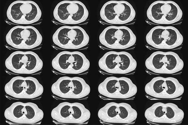
AdvertisementContinue reading the main storySupported byContinue reading the main storyYearly Lung Cancer Scans Are Advised for People 50 and Over With Shorter Smoking HistoriesNew advice from an influential panel will make more women and African-Americans eligible for CT scans, but some who need them most may not be able to afford them.A computed tomography scan of the lungs of a healthy adult man. The panel recommended low-dose CT scans, so-called because they involve a relatively small amount of radiation, and cost about $300.Credit…Alamy Stock PhotoMarch 9, 2021Updated 11:21 a.m. ETNew guidelines from medical experts will nearly double the number of people in the United States who are advised to have yearly CT scans to screen for lung cancer, and will include many more African-Americans and women than in the past.The disease is the leading cause of U.S. cancer deaths, and the goal of the expanded screening is to find it early enough to cure it in more people at high risk because of smoking. In those individuals, annual CT scans can reduce the risk of death from the cancer by 20 to 25 percent, large studies have found.The new recommendations, by the U.S. Preventive Services Task Force, include people ages 50 to 80 who have smoked at least a pack a day for 20 years or more, and who still smoke or have quit within the past 15 years.The advice, published on Tuesday in the medical journal JAMA, differs in two major ways from the task force’s previous guidelines, issued in 2013: It lowers the age when screening should start, to 50 from 55, and it reduces the smoking history to 20 years, from 30.Those changes will add more women and African-Americans to the pool eligible for screening, because they tend to smoke less heavily than the white male study participants on whom earlier guidelines were based. Women and Black Americans also tend to develop lung cancer earlier and from less tobacco exposure than do white men, experts said.Why the risk appears to differ by race and gender is not known.“Some studies have alluded to some hormonal influences in women,” Dr. Mara Antonoff, a lung surgeon at the M.D. Anderson Cancer Center in Houston, said in an interview. “In terms of racial differences, we don’t have an answer. We have population-based data to show they have a tendency to develop lung cancer younger and with less exposure to tobacco, but we don’t have a mechanism.”Under the new criteria, 14.5 million people in the United States will qualify for the screening, an increase of 6.4 million.The task force includes 16 physicians, scientists and public health experts who periodically evaluate screening tests and preventive treatments. Members are appointed by the director of the federal Agency for Healthcare Research and Quality, but the group is independent and its recommendations often help shape U.S. medical practice.The use of chest X-rays to detect lung cancer was largely abandoned decades ago because they could not find the disease early enough to be useful.The CT scans, called low-dose CT — because they involve a relatively small amount of radiation — cost about $300. Patients are advised to stop the screening once they have not smoked for 15 years, or if they develop health problems that would substantially shorten their life expectancy or make them unable to have lung surgery if needed.Patients have not flocked to clinics for this screening. Researchers estimate that only 6 to 18 percent of those who qualify and could be helped by the screening have taken advantage of it. Some cannot afford it.“Part of the low uptake is simply lack of access to care,” said Dr. Robert Smith, a screening expert at the American Cancer Society. “Smoking in general is increasingly concentrated in lower-income populations.”The Affordable Care Act does require that insurers cover any screening broadly recommended by the task force, with no out-of-pocket costs.Bettye Givens, right, of south Kansas City, reviewing lung scans with her doctor in 2017. She received a low-dose CT scan that detected her lung cancer at its earliest stage and was successfully treated.Credit…Andy Marso/Kansas City Star/TNS/Alamy Live NewsBut researchers have found that half the population eligible for lung-cancer screening had either no insurance, or Medicaid, Dr. Smith said. Not all Medicaid plans have covered the screening, according to an editorial in JAMA.“There could be a 15-year period when you might quality for screening and not have any insurance,” Dr. Smith said.He and other researchers also said that patients may be missing out on lung-cancer screening because they just don’t know about it. It has not received as much attention as other cancer screenings, like mammograms, colonoscopies and Pap tests. Some doctors may not encourage it as strongly, and especially with former smokers, may not take the time to calculate a patient’s smoking history to see if it matches the guidelines.The changes in the criteria for smoking history and screening age were based on new data from multiple studies, Dr. Alex H. Krist, the task force chairman and a professor of family medicine and population health at Virginia Commonwealth University, said in an interview.“Lung cancer is the No. 1 cancer killer in America,” Dr. Krist said, adding that with the new data, “we have even more confidence that screening does save lives.”Like other kinds of screenings affected by the pandemic, those for lung cancer remain below 2019 levels, according to an analysis of Medicare data by Avalere Health, a consulting firm, conducted for Community Oncology Alliance, which represents independent cancer specialists.While the number of screenings had started to rebound in the summer, the fresh spike in Covid cases later in the year caused them to fall again. In November, screenings were down by 30 percent, compared to 2019, and the number of lung biopsies had also dropped, indicating cases were not being diagnosed.Using its own grading system, the task force gave its recommendation a B, saying there was “moderate certainty” that annual screening was of “moderate net benefit.”That may not sound like a ringing endorsement, given that a grade of A means “high certainty that the net benefit is substantial.” But anything with an A or B grade should be offered to patients, according to task force rules.“There is building evidence that a pretty simple, five-minute, low-dose, low radiation scan can really save a lot of people’s lives,” said Dr. Bernard J. Park, a lung surgeon and the clinical director of the lung-screening service at Memorial Sloan Kettering Cancer Center in New York. About 75 to 85 percent of the cancers found with this screening are Stage 1, and curable with just surgery or radiation, he estimated.Dr. Park said that many people who signed up for the screening had quit smoking or were trying to stop, but that a few regarded clear scans as a sign that they could keep smoking.Dr. Smith said that the American Cancer Society was due to revise its own guidelines for lung-cancer screening, and that its advice would probably be similar to that of the task force.In 2013, the American Academy of Family Physicians declined to recommend for or against CT screening for lung cancer, saying there was insufficient evidence. But the president, Dr. Ada Stewart, said in an emailed statement on Monday that the academy would review the new task force evidence and decide whether to update its own recommendation to its members.Globally, there were 2.09 million new cases of lung cancer in 2018, and the disease is also the leading cause of cancer deaths, killing 1.76 million people that year, according to the World Health Organization.There were 228,820 new cases of lung cancer in the United States in 2020, and 135,720 people died from it, according to the National Cancer Institute. About 90 percent of cases occur in people who smoke, and current smokers’ risk of developing the disease is about 20 times that of nonsmokers.Only about 20.5 percent of patients survive five years after the diagnosis. Most cases are diagnosed late, after the cancer has begun to spread. But if it can be found and treated early, cure is possible, doctors say.CT screening has risks, and doctors say those must be explained to patients, who may decide to decline the testing. The scans detect tiny nodules in the lungs that may be early cancers — or maybe not. A suspicious-looking spot could be just a minor infection, inflammation or a benign growth, Dr. Park said.Often, the nodules can just be monitored with repeat scans, but it can be nerve-racking for patients to spend months waiting for the next test, knowing there is something in their lung that might be malignant.False positive rates, when something harmless is mistaken for cancer, have ranged from 3.9 to 25 percent and higher in studies, but tend to decrease over time, as the patient has more annual scans.A major concern about false positives is that they can lead to invasive procedures like lung biopsies. One large study found that invasive procedures were performed needlessly in 1.7 percent of the patients who were screened. The task force report said that standards created by radiology societies for evaluating the scans could help to prevent some unnecessary procedures spurred by false positives.Another possible risk from screening is the chance that the cumulative radiation exposure could cause cancer. But the dose is low, and the risk is thought to be small, especially when compared with the risk of lung cancer caused by smoking.In theory, screening could also lead to unneeded invasive tests and treatment for a cancer that would not have progressed or harmed the patient. How often that might occur is not known, but it is considered rare.Reed Abelson contributed to this article.AdvertisementContinue reading the main story

Read more →