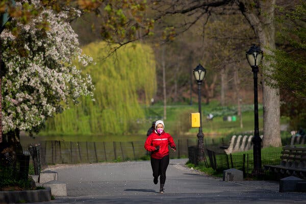
AdvertisementContinue reading the main storySupported byContinue reading the main storyPhys EdGetting to the Bottom of the Runner’s HighFor years we’ve been crediting endorphins, but it’s really about the endocannabinoids.Credit…Kevin Hagen for The New York TimesMarch 10, 2021, 5:00 a.m. ETWe can stop crediting endorphins, the natural opioid painkillers produced by our bodies, for the floaty euphoria we often feel during aerobic exercise, according to a nifty new study of men, women and treadmills. In the study, runners developed a gentle intoxication, known as a runner’s high, even if researchers had blocked their bodies’ ability to respond to endorphins, suggesting that those substances could not be behind the buzz. Instead, the study suggests, a different set of biochemicals resembling internally homegrown versions of cannabis, better known as marijuana, are likely to be responsible.The findings expand our understanding of how running affects our bodies and minds, and also raise interesting questions about why we might need to be slightly stoned in order to want to keep running.In surveys and studies of experienced distance runners, most report developing a mellow runner’s high at least sometimes. The experience typically is characterized by loose-limbed blissfulness and a shedding of anxiety and unease after half an hour or so of striding. In the 1980s, exercise scientists started attributing this buzz to endorphins, after noticing that blood levels of the natural painkillers rise in people’s bloodstreams when they run.More recently, though, other scientists grew skeptical. Endorphins cannot cross the blood-brain barrier, because of their molecular structure. So, even if runners’ blood contains extra endorphins, they will not reach the brain and alter mental states. It also is unlikely that the brain itself produces more endorphins during exercise, according to animal studies.Endocannabinoids are a likelier intoxicant, these scientists believed. Similar in chemical structure to cannabis, the cannabinoids made by our bodies surge in number during pleasant activities, such as orgasms, and also when we run, studies show. They can cross the blood-brain barrier, too, making them viable candidates to cause any runner’s high.A few past experiments had strengthened that possibility. In one notable 2012 study, researchers coaxed dogs, people and ferrets to run on treadmills, while measuring their blood levels of endocannabinoids. Dogs and humans are cursorial, meaning possessed of bones and muscles well adapted to distance running. Ferrets are not; they slink and sprint but rarely cover loping miles, and they did not produce extra cannabinoids while treadmill running. The dogs and people did, though, indicating that they most likely were experiencing a runner’s high and it could be traced to their internal cannabinoids.That study did not rule out a role for endorphins, however, as Dr. Johannes Fuss realized. The director of the Human Behavior Laboratory at the University Medical Center Hamburg-Eppendorf in Germany, he and his colleagues had long been interested in how various activities affect the inner workings of the brain, and after reading the ferret study and others, thought they might look more closely into the runner’s high.They began with mice, which are eager runners. For a 2015 study, they chemically blocked the uptake of endorphins in the animals’ brains and let them run, then did the same with the uptake of endocannabinoids. When their endocannabinoid system was turned off, the animals ended their runs just as anxious and twitchy as they had been at the start, suggesting that they had felt no runner’s high. But when their endorphins were blocked, their behavior after running was calmer, relatively more blissed-out. They seemed to have developed that familiar, mild buzz, even though their endorphin systems had been inactivated.Mice emphatically are not people, though. So, for the new study, which was published in February in Psychoneuroendocrinology, Dr. Fuss and his colleagues set out to replicate the experiment, to the extent possible, in humans. Recruiting 63 experienced runners, male and female, they invited them to the lab, tested their fitness and current emotional states, drew blood and randomly assigned half to receive naloxone, a drug that blocks the uptake of opioids, and the rest, a placebo. (The drug they had used to block endocannabinoids in mice is not legal in people, so they could not repeat that portion of the experiment.)The volunteers then ran for 45 minutes and, on a separate day, walked for the same amount of time. After each session, the scientists drew blood and repeated the psychological tests. They also asked the volunteers whether they thought they had experienced a runner’s high.Most said yes, they had felt buzzed during the run, but not the walk, with no differences between the naloxone and placebo groups. All showed increases, too, in their blood levels of endocannabinoids after running and equivalent changes in their emotional states. Their euphoria after running was greater and their anxiety less, even if their endorphin system had been inactivated.Taken as a whole, these findings are a blow to endorphins’ image. “In combination with our research in mice,” Dr. Fuss says, “these new data rule out a major role for endorphins” in the runner’s high.The study does not explain, though, why a runner’s high exists at all. There was no walker’s high among the volunteers. But Dr. Fuss suspects the answer lies in our evolutionary past. “When the open savannas stretched and forests retreated,” he says, “it became necessary for humans to hunt wild animals by long-distance running. Under such circumstances, it is beneficial to be euphoric during running,” a sensation that persists among many runners today, but with no thanks due, it would seem, to endorphins.AdvertisementContinue reading the main story
Read more →