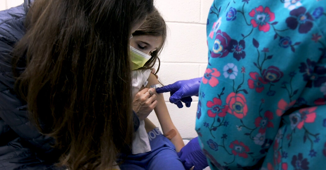
50 new genes for eye color

The genetics of human eye colour is much more complex than previously thought, according to a new study published today.
An international team of researchers led by King’s College London and Erasmus University Medical Center Rotterdam have identified 50 new genes for eye colour in the largest genetic study of its kind to date. The study, published today in Science Advances, involved the genetic analysis of almost 195,000 people across Europe and Asia.
These findings will help to improve the understanding of eye diseases such as pigmentary glaucoma and ocular albinism, where eye pigment levels play a role.
In addition, the team found that eye colour in Asians with different shades of brown is genetically similar to eye colour in Europeans ranging from dark brown to light blue.
This study builds on previous research in which scientists had identified a dozen genes linked to eye colour, believing there to be many more. Previously, scientists thought that variation in eye colour was controlled by one or two genes only, with brown eyes dominant over blue eyes.
Co-senior author Dr Pirro Hysi, King’s College London, said: “The findings are exciting because they bring us to a step closer to understanding the genes that cause one of the most striking features of the human faces, which has mystified generations throughout our history. This will improve our understanding of many diseases that we know are associated with specific pigmentation levels.”
Co-senior author Dr Manfred Kayser, Erasmus University Medical Center Rotterdam, said:
“This study delivers the genetic knowledge needed to improve eye colour prediction from DNA as already applied in anthropological and forensic studies, but with limited accuracy for the non-brown and non-blue eye colours.”

Story Source:
Materials provided by King’s College London. Note: Content may be edited for style and length.

Read more →

Probiotics increase gut bacteria diversity in extremely preterm infants

Extremely preterm infants can suffer from a life-threatening inflammation of the gut. A new clinical study has shown that supplements of a lactic acid bacterium may have positive effects by increasing the diversity of intestinal bacteria in these infants. The study has been led by researchers at Linköping University, Sweden, and published in the scientific journal Cell Reports Medicine.
A litre of milk weighs a kilogram. Most infants who are born extremely prematurely weigh less than that. An infant who should have developed and grown for three more months in the protective environment of the mother’s womb is, of course, extremely vulnerable. As a consequence of advances in neonatal care, many premature infants survive, although one out of four of the extremely premature infants die.
“Preterm infants can be affected by a very severe inflammation of the intestines, which almost only occurs in such infants. The condition, necrotising enterocolitis (or NEC), leads to parts of the intestine dying. One of three infants who contract the infection die, and those who survive often suffer from long-term complications such as short gut syndrome and neurodevelopmental disabilities,” says Thomas Abrahamsson, paediatrician at the neonatal intensive care unit at Crown Princess Victoria Children´s Hospital and associate professor at the Department of Biomedical and Clinical Sciences (BKV) at Linköping University, who has led the study.
The bacteria in the intestine of preterm infants differ from those in full-term infants. This has led many people to investigate whether giving probiotic supplements that contain certain bacteria has a positive effect. One finding is that the lactic acid bacterium Lactobacillus reuteri can reduce the risk of NEC in preterm infants. It is, however, not clear whether this is true also for extremely preterm infants, nor is the mechanism behind the positive effect known.
The study now published is part of a clinical study carried out in Linköping and Stockholm. The researchers looked at 132 infants who had been born extremely prematurely, between week 23 and 28 of pregnancy, i.e. 17 to 12 weeks before the due date. All weighed less than a kilogram at birth. Each infant was randomly assigned to one of two groups: to receive oil drops that contained the probiotic or placebo. The treatment was given daily during the neonatal period. The scientists investigated how the intestinal bacterial flora was influenced by the supplement of L. reuteri, and analysed bacteria in the stools at several time points.
“We see that the composition of bacteria in the intestine differs during the first month of the probiotic treatment. During the first week of life, the bacterial groups Staphylococcus and Klebsiella, which may cause disease, were more common in the group that received placebo,” says Magalí Martí Generó, principal research engineer in BKV, and principal author of the article.
Klebsiella can cause inflammation and has been associated with NEC and sepsis. The present study does not allow any conclusions about whether the probiotic treatment influences the risk of these diseases in these extremely premature infants. Larger studies will be required to determine this.
“The supplemented probiotic L. reuteri survives in the intestine, even though these extremely premature infants are treated with large doses of antibiotics that kill bacteria. The positive effect of the treatment in increasing the diversity of intestinal bacteria may be one mechanism behind the positive effects of this probiotic shown in previous studies,” says Thomas Abrahamsson.
Supplementation with probiotics is used in increasing numbers of neonatal clinics. The scientific evidence that supplements of probiotics to preterm infants have a positive effect and can be used safely is considered to be sufficiently strong.

Story Source:
Materials provided by Linköping University. Original written by Karin Söderlund Leifler. Note: Content may be edited for style and length.

Read more →

Dry eye disease negatively affects physical and mental health as well as vision

Patients suffering from dry eye disease symptoms have a lower quality of life compared to those without symptoms, a new study reports. The findings showed that patients with the condition reported negative effects on visual function, their ability to carry out daily activities and their work productivity.
Dry eye disease is a common condition and a frequent reason for patients to seek medical care. It can affect people of any age but is most prevalent in women and in older people. Symptoms include irritation and redness in the eyes, blurred vision, and a sensation of grittiness or a foreign body in the eye. It has been reported that up to a third of adults over 65 years old have the condition, although the actual number is likely to be higher as there is no established diagnostic test and people with mild symptoms are less likely to report them to their doctor.
Treatment often involves prescriptions of artificial tears, ocular lubricants and astringents, which come at a cost to the NHS; in 2014, 6.4 million items were prescribed at a cost of over £27 million.
This new study, led by the University of Southampton, set out to explore how dry eye disease affects the lives of adults in the UK through an online survey of one thousand patients with the condition and further one thousand without. Participants undertook a questionnaire from the National Eye Institute about their visual function and a EuroQol questionnaire on health-related quality of life. Those who declared that they experienced dry eye disease also answered further questions to assess the severity of their symptoms.
The results, published in the journal BMJ Open, showed that a higher proportion of participants with dry eye disease had problems with mobility and experienced more difficulties in their day-to-day activities than patients without the condition. The surveys also revealed they were more likely to suffer from anxiety and depression.
Those with the most severe symptoms we more likely to experience a negative impact on their social and emotional functioning as well work productivity, including missing more time from work as a result of their symptoms.
Dr Parwez Hossain, Associate Professor in Ophthalmology at the University of Southampton, led the study. He said: “This study provided some very useful information on the burden that dry eye disease places on patients. As well as confirming the impact on work and social lives we also discovered showed that the extent of the effects are consistent with the severity of symptoms. We also found that participants with dry eye disease symptoms were a lot more likely to suffer from other comorbidities, twice as many suffered from arthritis, hearing loss or irritable bowel disease compared to the cohort without symptoms.
“Whilst we cannot draw causal associations through this study, the presence of dry eye disease does appear to impact on an individual’s health and vision related quality of life.”
Although both groups reported similar levels of digital screen use and reading, the cohort with symptoms reported more exposure to environmental factors such as air conditioning, forced heating or air pollution. The research team believe that these factors could either contribute to dry eye disease, or be noticed more by sufferers.

Story Source:
Materials provided by University of Southampton. Note: Content may be edited for style and length.

Read more →

Imaging the human eye: detailed images of rod and cone photoreceptors

Researchers have developed a noninvasive technique that can capture images of rod and cone photoreceptors with unprecedented detail. The advance could lead to new treatments and earlier detection for retinal diseases such as macular degeneration, a leading cause of vision loss.
“We are hopeful that this technique will better reveal subtle changes in the size, shape and distribution of rod and cone photoreceptors in diseases that affect the retina,” said research team leader Johnny Tam from the National Eye Institute. “Figuring out what happens to these cells before they are lost is an important step toward developing earlier interventions to treat and prevent blindness.”
In Optica, The Optical Society’s (OSA) journal for high impact research, the researchers show that their new imaging method overcomes resolution limitations imposed by the diffraction barrier of light. The researchers accomplish this feat while using light that is safe for imaging the living human eye.
“The diffraction limit of light can now be routinely surpassed in microscopy, which has revolutionized biological research,” said Tam. “Our work represents a first step toward routine sub-diffraction imaging of cells in the human body.”
Using less light to see more
Achieving high-resolution images of photoreceptors in the back of the eye is challenging because the eye’s optical elements (such as lens and cornea) distort light in a way that can substantially reduce image resolution. The diffraction barrier of light also limits the ability of optical instruments to distinguish between two objects that are too close together. Although there are various methods for imaging beyond the diffraction limit, most of these approaches use too much light to safely image living human eyes.

advertisement

To overcome these challenges, the researchers improved upon a retinal imaging technique known as adaptive optics scanning light ophthalmoscopy, which uses deformable mirrors and computational methods to correct for optical imperfections of the eye in real time.
“One might think that more light is needed to get a better image, but we demonstrate that we can improve resolution by strategically blocking light in various locations within our instrument,” said Tam. “This approach reduces the overall power of light delivered to the eye, making it ideal for live imaging applications.”
For the new approach, the researchers generated a ring-shaped, or hollow, beam of light. Using this type of beam improved the resolution across the photoreceptors but at the expense of depth resolution. To regain the lost depth resolution, the researchers used a small pinhole called a sub-Airy disk to block light coming back from the eye. They showed that this imaging approach could be used to enhance a microscopy technique called non-confocal split-detection, which is used to acquire complementary views of the photoreceptors.
Testing in the clinic
After demonstrating that imaging resolution was improved in theoretical simulations, the researchers confirmed their simulations using various test targets. They then used the new method to image rod and cone photoreceptors in five healthy volunteers at the National Institutes of Health’s Clinical Center.
The new approach yielded about a 33 percent increase in transverse resolution and 13 percent improvement in axial resolution compared to traditional adaptive optics scanning light ophthalmoscopy. Using their optimized approach, the researchers were able to see a circularly shaped subcellular structure in the center of cone photoreceptors that could not be clearly visualized previously.
“The ability to noninvasively image photoreceptors with subcellular resolution can be used to track how individual cells change over time,” said Tam. “For example, watching a cell begin to degenerate, and then possibly recover, will be an important advance for testing new treatments to prevent blindness.”
The researchers plan to image the eyes of more patients with the new technique and use the images to begin to answer fundamental questions linked to rod and cone health. For example, they are interested in visualizing rod and cone health in people who have rare genetic diseases. They say that their imaging approach could be applied to other point scanning-based microscopy and imaging approaches in which it is important to image with low levels of light.

Story Source:
Materials provided by The Optical Society. Note: Content may be edited for style and length.

Read more →

Scientists move closer to developing 'game-changing' test to diagnose Parkinson's

Results published today show it is possible to identify Parkinson’s based on compounds found on the surface of skin. The findings offer hope that a pioneering new test could be developed to diagnose the degenerative condition through a simple and painless skin swab.
Scientists at The University of Manchester have developed a technique which works by analysing compounds found in sebum — the oily substance that coats and protects the skin — and identifying changes in people with Parkinson’s Disease. Sebum is rich in lipid-like molecules and is one of the lesser studied biological fluids in the diagnosis of the condition. People with Parkinson’s may produce more sebum than normal — a condition known as seborrhoea.
The research has been funded by charities Parkinson’s UK and the Michael J. Fox Foundation as well as The University of Manchester Innovation Factory. The work was originally funded following an observation by Joy Milne, whose husband was diagnosed with Parkinson’s at the age of 45. Working with Dr Tilo Kunath at the University of Edinburgh, Joy demonstrated an incredible ability to distinguish a distinctive Parkinson’s odour in individuals using her sense of smell, even before symptoms emerge in those affected.
The team, led by Professor Perdita Barran, The University of Manchester, and the clinical lead Professor Monty Silverdale at Salford Royal Foundation Trust, recruited 500 people with and without Parkinson’s. Samples of sebum were taken from their upper backs for analysis. Using different mass spectrometry methods, 10 chemical compounds in sebum were identified which are elevated or reduced in people with Parkinson’s. This allows scientists to distinguish people with Parkinson’s with 85 per cent accuracy.
The team confirmed their earlier findings published in ACS Central Science that the volatile compounds on skin can be used to diagnose the condition, increasing the number of people sampled and including participants from the Netherlands, as well as the UK.
In a new study published today in Nature Communications, high resolution mass spectrometry was used to profile the complex chemical signature in sebum of people with Parkinson’s and show subtle but fundamental changes as the condition progresses. Detailed analysis showed changes in people with Parkinson’s in lipid (fat) processing and mitochondria. Problems with mitochondria — the tiny energy-producing batteries that power cells — are one of the hallmarks of Parkinson’s.

advertisement

This means this ‘world first’ testing strategy is not only useful in diagnosing Parkinson’s but also in monitoring the development of the condition. The skin swab could provide an incredibly important new tool in clinical trials helping researchers measure whether new, experimental treatments are able to slow, stop or reverse the progression of Parkinson’s.
The study unveiled novel diagnostic sebum-based biomarkers for Parkinson’s, provides insight into understanding of how the condition develops, and links lipid dysregulation to altered mitochondrial function.
These promising results published today could lead to a definitive test to diagnose Parkinson’s accurately, speedily and cost effectively. The team is now seeking funding to further develop the test and explore the potential for using the test to ‘stratify’ patients.
Working with the University of Manchester Innovation Factory, the team has patents filed for their diagnostic techniques and are planning to create a spin-out company to commercialise the new tests. They are also working to use this approach to develop tests for COVID-19 as shown in research last week in EClinical Medicine as well as other conditions and are actively seeking investors interested in supporting the drive to bring this technology to market.
Professor Perdita Barran, Professor of Mass Spectrometry at The University of Manchester, said: “We believe that our results are an extremely encouraging step towards tests that could be used to help diagnose and monitor Parkinson’s.
“Not only is the test quick, simple and painless but it should also be extremely cost-effective because it uses existing technology that is already widely available.
“We are now looking to take our findings forwards to refine the test to improve accuracy even further and to take steps towards making this a test that can be used in the NHS and to develop more precise diagnostics and better treatment for this debilitating condition.”
Parkinson’s tends to develop gradually and it may be many months, even years, before the symptoms become obvious enough for an individual to visit their GP. A DaTscan is regularly used to help specialists confirm the loss of dopamine-producing cells that cause the development of Parkinson’s. However, similar loss may also occur in some other rarer neurological conditions. With no molecular test for the condition, diagnosis is made by a neurologist based on a combination of symptoms such as tremor, slowness, stiffness and balance issues. However, many of the symptoms of Parkinson’s can overlap with other conditions, especially in the early stages when progression is gradual and symptoms are more subtle.
In a recent survey of more than 2,000 people with Parkinson’s carried out by Parkinson’s UK, more than a quarter (26 per cent) reported they were misdiagnosed with a different condition before receiving the correct Parkinson’s diagnosis.¹

Read more →

Parkinson's skin-swab test 'in sight'

SharecloseShare pageCopy linkAbout sharingimage copyrightPARKINSON’S UKA simple skin-swab test could be used to help diagnose the degenerative brain condition Parkinson’s disease, UK scientists say. Studies with volunteers show it can quickly detect tell-tale compounds in sebum – the oily substance that protects the skin. People with Parkinson’s can have higher than usual concentrations of these.Researchers discovered this after a woman amazed doctors with her ability to detect Parkinson’s through smell. Retired nurse Joy Milne, 68, from Perth, noticed the “musky” smell on her husband, Les, years before his Parkinson’s diagnosis.She has been helping scientists at the University of Manchester explore the link. And a blinded test of her odour-detecting skills found she was 100% accurate.The research team moved on to using a mass-spectrometry machine to detect the compounds.And they now have data from 500 people, showing the skin test can correctly distinguish those with Parkinson’s more than eight out of every 10 times.Investigator Prof Perdita Barran said: “We believe that our results are an extremely encouraging step towards tests that could be used to help diagnose and monitor Parkinson’s. “Not only is the test quick, simple and painless but it should also be extremely cost-effective because it uses existing technology that is already widely available.”We are now looking to take our findings forwards to refine the test to improve accuracy even further and to take steps towards making this a test that can be used in the NHS and to develop more precise diagnostics and better treatment for this debilitating condition.”Disease progression Currently, there is no cure and no definitive test for Parkinson’s.And diagnosis can take years. The researchers are seeking funding to further develop their test, which they believe could also help track disease progression and measure whether treatments are having an impact, at the molecular level. Their latest work, published in the journal Nature Communications, suggests progression of the disease comes with changes in the regulation of lipids in cells.’Enjoy life’Daxa Kalayci, 56, of Leicester, was misdiagnosed several times over four years before finally finding out she had Parkinson’s in 2019. “I was misdiagnosed with anxiety, stress-related tremors and told that my problems stemmed from going through the menopause,” she said.”Despite my diagnosis eventually being confirmed… a quick and simple diagnostic test for Parkinson’s would have given me the chance to start my treatment earlier and enjoy life a lot more. “But instead, I lost so many years not being able to pursue a career as a paramedic or go back to nursing.”This test could be a game-changer for people living with Parkinson’s and searching for answers, like I was.”Related Internet LinksParkinson’s UKThe BBC is not responsible for the content of external sites.

Read more →

Pandemic Raises Concerns About Childhood Lead Poisoning

#masthead-section-label, #masthead-bar-one { display: none }The Coronavirus OutbreakliveLatest UpdatesMaps and CasesRisk Near YouVaccine RolloutGuidelines After VaccinationAdvertisementContinue reading the main storySupported byContinue reading the main storyPandemic Raises Concerns About Childhood Lead PoisoningLead screenings for children plummeted last spring, and stay-at-home orders may have increased household exposure to the toxic metal.A worker scraping off lead paint. The C.D.C. estimates that more than 20 million housing units in the United States contain lead-based paint, which was essentially banned in 1978.Credit…Jamie Hooper/AlamyMarch 11, 2021, 5:00 a.m. ETOver the past half-century, public health officials have made enormous progress in protecting American children from lead poisoning and the irreversible neurological damage it can cause. Since the 1970s, the percentage of children with high levels of lead in their blood has plummeted.But in 2020, a new health threat, the coronavirus, endangered these hard-earned gains.When Covid-19 cases spiked last spring, lockdowns and day care closures confined young children to their homes, where lead exposure can be particularly high. The growing national emergency also delayed lead-removal efforts and disrupted routine childhood lead screenings, leaving health officials unable to identify and treat many children living in lead-laden homes.Last month, the Centers for Disease Control and Prevention estimated that in the early months of the pandemic, roughly 10,000 children with elevated levels of lead in their blood may have gone undetected.“Hundreds of thousands of children have missed their essential tests for lead,” said Joseph Courtney, a senior epidemiologist at the C.D.C.’s lead poisoning prevention and environmental health tracking branch, who conducted the analysis. “And it’s something that has potentially permanent lifetime effects.”There is no safe level of exposure to lead, which can disrupt neurological and cognitive development, causing learning disabilities, behavioral problems and developmental delays.The C.D.C. estimates that more than 20 million housing units in the United States contain lead-based paint, which was banned in 1978. When the paint flakes, cracks or peels, the lead mixes with dust, which children can ingest or inhale. Young children, whose brains are still developing, are at particular risk.“The main route of exposure for most children is lead-contaminated dust, from paint getting on their hands or their toys,” said David Jacobs, chief scientist at the National Center for Healthy Housing. “And then normal child behavior at this age is, everything goes in the mouth.”For that reason, many states recommend or require that children of certain ages have their blood tested for lead. (Federal guidelines require that all children who are enrolled in Medicaid receive blood lead level tests at ages one and two.) Doctors typically perform these tests as part of a child’s regular checkup.But when the pandemic hit last March, government officials issued stay-at-home orders, and many medical offices closed. Others started conducting appointments virtually. “You can’t do a blood lead test by telemedicine,” said Dr. Stephanie Yendell, senior epidemiology supervisor at the Minnesota Department of Public Health. “You can’t get a blood sample by video call.”That month in Minnesota, the number of tests for blood lead level fell to 70 percent of what it had been the previous year, Dr. Yendell said. “And then in April, we bottomed out at 43 percent compared to the year before.”In New York City, which was hit by an early wave of Covid-19 cases, childhood lead screenings declined by 88 percent last April.Anecdotal reports of these declines soon made their way to the C.D.C., which asked state and local health officials to share their lead screening data so the agency could conduct a more formal analysis. Thirty-four health departments — representing 32 states, plus New York City and Washington, D.C. — ultimately did so.Paint is the most common cause of childhood lead poisoning, but lead pipes also pose a threat; the longer that water sits stagnant in such pipes, the more lead leeches into it.Credit…Julio Cortez/Associated PressEvery one of those departments reported a drop-off in testing last spring, Dr. Courtney and his colleagues found. Over the first five months of 2020, roughly 500,000 fewer children had their blood lead levels tested in these districts compared with the same period of 2019. The decline was especially steep in March, April and May, when testing levels fell 50 percent compared to the previous year.The Coronavirus Outbreak

Read more →

Nobody Wants Cancer. But a ‘Big C’ Label Has Surprising Upsides.

AdvertisementContinue reading the main storySupported byContinue reading the main storyVoicesNobody Wants Cancer. But a ‘Big C’ Label Has Surprising Upsides.Classifying a rare blood disorder as a cancer opened new doors for disease investigation, treatment and hope for a cure.Credit…Paper Illustration by Reina Takahashi, photographed by Tony Cenicola/The New York TimesMarch 11, 2021, 5:00 a.m. ETI have a rare blood disorder. When it was diagnosed 25 years ago, it was called an “orphan disease,” meaning the small number of people affected didn’t justify research investments by major pharmaceutical companies.Enter the World Health Organization and its 2008 decision to classify the condition as a blood “cancer.” This opened new doors for disease investigation and understanding. It prompted a growing number of super specialist practitioners, and most important, hope for some 300,000 people living in the United States with what are now called myeloproliferative neoplasms, or MPNs.Nobody likes to hear a cancer diagnosis, particularly for a disease they thought was benign. In fact, when I first saw my blood disorder referred to as a cancer, I wrote to the MPN Research Foundation, a nonprofit research and advocacy group, and suggested they remove the “Big C” word from their website. How, I thought, could they be so misleading?That’s when I discovered I was behind the times. MPNs were indeed reclassified as a malignant condition of the bone marrow, affecting sometimes one, two or all three types of blood cells: white cells, red cells and platelets.The average age people are diagnosed with an MPN is 60-something. My diagnosis came at age 38. For the next 15 years, I was treated for essential thrombocythemia, a type of MPN that caused my bone marrow to produce significantly higher than normal numbers of platelets. The proliferation of platelets put me at risk for dangerous clots and ultimately led to complete blockages of two important veins of the portal system, which carries blood to the liver.The Big C label began to look like good news.Once MPNs were classified as blood cancers — including essential thrombocythemia, polycythemia vera and myelofibrosis — interest grew from research laboratories, major medical centers, and small and mega pharmaceutical companies, which now saw these rare and poorly understood conditions as an opportunity. Perhaps pieces to the MPN puzzle could shed light on more common blood cancers, like leukemia and lymphoma. And perhaps treatments for those widely studied cancers could be used to treat MPNs.“The cancer designation did open up significant new funding opportunities, for example from the National Cancer Institute,” said Barbara Van Husen, board chair of the MPN Research Foundation. “It has definitely accelerated research.” There are more than 200 clinical trials underway for various MPNs, as well as ongoing research into stem cell transplantation, currently the only potential cure for these rare cancers.In my case, for reasons researchers are working to understand, my bone marrow flipped a switch. I was no longer making excessive platelets. Instead I was producing too many red cells and was given a revised diagnosis of polycythemia vera, a distinctly different MPN. I went for regular phlebotomies, the modern version of bloodletting in which pints of my blood were drained into a collection bag to reduce blood volume. Think blood transfusion, reversed.Doctors increased the dose of the 30-year-old chemotherapy drug I had been taking since my blood clots first appeared. The drug is still considered standard of care “if well tolerated.” It effectively adjusted my red counts. Unlike newer, more targeted drugs, however, it does not discriminate, potentially killing off not just red cells but white cells and platelets as well.I continued to live a full and largely unaffected life, though I often suffered symptoms such as debilitating headaches and bone pain.The hallmark fatigue of these blood disorders was not yet on my radar. I hiked. I cycled. I traveled. I was a night owl typically functioning “well” on five to six hours of sleep.Then, in September of 2019, my summer bike rides became an exhausting effort. By October, I was so fatigued that walking up a slightly inclined sidewalk had me leaning on a lamppost to catch my breath. Doctors identified scarring (“fibrosis”) in my bone marrow, elevating my diagnosis once again, to the third evolution of my MPN journey, myelofibrosis.“You’re a poster child for exploring new drug therapies for MPNs,” Dr. Ellen Ritchie, of Weill Cornell Medical Center in Manhattan, recently told me. Just seven months into my participation in a global phase 2 clinical trial for a new combination drug therapy, tests indicated significant, measurable improvement. It appears my disease is beginning to reverse.More than two decades after my first of many bone marrow biopsies, I have more energy than many of the “healthy” people I know.We tend to think of cancer as acute. Get it out quickly, go through treatments, prevent it from spreading. But for those with a chronic cancer like an MPN, there isn’t a distinctive beginning, middle and end. It can be overwhelming, emotionally exhausting, and even as we learn more about the disease, full of uncertainty.“Often, individuals with a chronic illness, like cancer, are more challenged to be hopeful about their future, while also needing to be focused on thriving on a daily basis,” says Laura Kaplan, a licensed clinical social worker whose Connecticut practice helps people navigate serious illness. “It is important for their identity and quality of life to try to keep a good perspective about living with the process of their disease.”For years, I chose not to let my condition define me. I chose not to identify myself as someone living with cancer. I would simply live my life fully, day to day. I would work hard and play hard, enjoy raising my two boys, deal with whatever challenged my health, and keep on moving. That approach continues to serve me well.Ruth Fein Revell, a health and environment writer, serves on a patient advisory board of the MPN Research Foundation.AdvertisementContinue reading the main story

Read more →

Sharp reductions in costs of producing cannabis, fentanyl likely to spur widespread changes in use, dependence

The legalization of cannabis and the arrival of nonmedical fentanyl are fundamentally changing drug markets in North America. A large part of these changes relates to the ability to produce large quantities of the drugs at low costs, which has slashed wholesale prices for both drugs and retail prices for cannabis. A new analysis explores the effects of these changes on use. The analysis concludes that sharp declines in production costs for cannabis and opioids could dramatically reduce the price per dose for consumers in ways that alter patterns of use and dependence.
The analysis, by a researcher at Carnegie Mellon University (CMU), is published in the International Journal of Drug Policy.
“Historical analogies suggest that very large declines in price can have effects on use that go beyond just expanding traditional patterns of consumption,” explains Jonathan Caulkins, professor of operations research and public policy at CMU’s Heinz College, who wrote the analysis. “The overall situation with cannabis and fentanyl may look more different in 2040 compared to today — just as today looks different compared to 2000.”
Caulkins focused on the motivations for use of these drugs, factors that appear ready to change. He also considered market factors, noting that basic relationships among production costs, prices, and consumption have held up in markets over centuries. And he looked at wild cards — cultural, sociological, and political changes that could be equally influential.
Caulkins started with two key economic ideas: First, prices in competitive markets fall to match the marginal cost of production. For example, North American cannabis production costs have decreased as much as 95 percent. Second, when prices fall, consumption rises. This has occurred with cannabis, although as yet, there is no indication that fentanyl production has reduced retail opioid prices — but monitoring retail opioid prices is difficult.
Therefore, falling prices affect consumption, but the effects of precipitous declines may not be simply a larger version of the effects of modest price declines. Among other factors to consider is elasticity of demand, including how the degree of responsiveness to price changes varies from one setting to the next and from one outcome to the next. In short, for many products used widely by society (e.g., lighting and electricity, computers, cigarettes), Caulkins explains, their meaning changed when production costs fell radically.
“Liberalization of cannabis policy and reduced production costs may fundamentally change the place of cannabis in society,” Caulkins notes. Consider, for example, that cannabis operations are listed on the NASDAQ and Toronto stock exchanges, legalization has led to a wide range of products such as edibles and vaping, and advertising for the product has soared. More changes are likely, he suggests.
Significant declines in wholesale opioid prices could also have far-reaching and unexpected effects, Caulkins predicts. Among them: reducing the value of criminal organizations’ cross-border smuggling, making distribution less violent.
“We don’t know what the future holds,” he adds, “but I predict that if someone in 2040 lists the major changes in drug markets, use, and dependence that occurred since 2020, there will be items on that list that pertain to the declines in production costs brought about by cannabis legalization and the spread of synthetic opioids.”
Based on this prediction, Caulkins concludes: “It’s not too soon to invest more in monitoring markets to stay abreast of the diverse ramifications that may flow from these radical reductions in production costs.”

Read more →