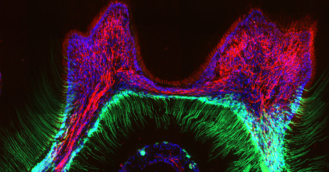
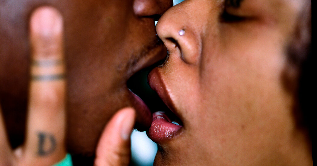
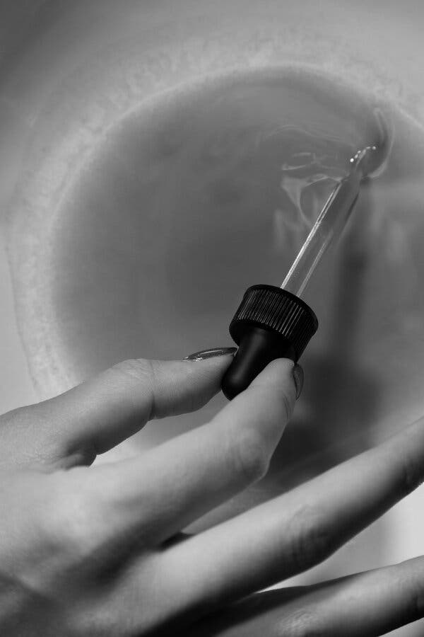
Study uncovers clues to COVID-19 using imaging

Since the pandemic hit, researchers have been uncovering ways COVID-19 impacts other parts of the body, besides the lungs.
Now, for the first time, a visual correlation has been found between the severity of the disease in the lungs using CT scans and the severity of effects on patient’s brains, using MRI scans. This research is published in the American Journal of Neuroradiology. It will be presented at the 59th annual meeting of the American Society of Neuroradiology (ASNR) and has also been selected as a semifinalist for that organization’s Cornelius Dyke Award.
The results show that by looking at lung CT scans of patients diagnosed with COVID-19, physicians may be able to predict just how badly they’ll experience other neurological problems that could show up on brain MRIs, helping improve patient outcomes and identify symptoms for earlier treatment.
CT imaging can detect illness in the lungs better than an MRI, another medical imaging technique. However, MRI can detect many problems in the brain, particularly in COVID-19 patients, that cannot be detected on CT images.
The study was led by Achala Vagal, MD, professor in the department of radiology, and Abdelkader Mahammedi, MD, assistant professor of radiology. Both are UC Health radiologists and members of the UC Gardner Neuroscience Institute.
“We’ve seen patients with COVID-19 experience stroke, brain bleeds and other disorders affecting the brain,” says Mahammedi. “So, we’re finding, through patient experiences, that neurological symptoms are correlating to those with more severe respiratory disease; however, little information has been available on identifying potential associations between imaging abnormalities in the brain and lungs in COVID-19 patients.
“Imaging serves as proof for physicians, confirming how an illness is forming and with what severity and helps in making final decisions about a patient’s care.”
In this study, which was conducted not only at UC, but also at large institutions in Spain, Italy and Brazil, researchers reviewed electronic medical records and images of hospitalized COVID-19 patients from March 3 to June 25, 2020. Patients who were diagnosed with COVID-19, experienced neurological issues and who had both lung and brain images available were included.
Of 135 COVID-19 patients with abnormal CT lung scans and neurological symptoms, 49, or 36%, were also found to develop abnormal brain scans and were more likely to experience stroke symptoms.
Mahammedi says this study will help physicians classify patients, based on the severity of disease found on their CT scans, into groups more likely to develop brain imaging abnormalities. He adds that this correlation could be important for implementing therapies, particularly in stroke prevention, to improve outcomes in patients with COVID-19.
“These results are important because they further show that severe lung disease from COVID-19 could mean serious brain complications, and we have the imaging to help prove it,” says Mahammedi. “Future larger studies are needed to help us understand the tie better, but for now, we hope these results can be used to help predict care and ensure that patients have the best outcomes.”
This research was supported by the National Institutes of Health (the National Institute of Neurological Disorders and Stroke and National Institute on Aging) (NS103824, NS117643, NS100417, 1U01NS100699, U01NS110772). Researchers cite no conflict of interest.

Story Source:
Materials provided by University of Cincinnati. Original written by Katie Pence. Note: Content may be edited for style and length.

Read more →

New review explores effective sampling techniques for collecting airborne viruses and ultrafine particles

As the world continues to grapple with the COVID-19 pandemic, an international team of researchers have published a review of the best techniques to collect airborne aerosols containing viruses.
In the review, which was published by the Science of the Total Environment journal, a team led by the University of Surrey concluded that the most effective way to collect and detect airborne pathogens, particularly viruses, was to use cyclone sampling techniques.
For example, the sampler draws the air through the cyclone separator. It then uses centrifugal forces to collect the particles on a sterile cone containing the liquid collection vessel, such as DMEM (Dulbecco’s minimal essential medium). The collected sample can then be readily used for any analysis for virus detection.
The research team hope that this wide-ranging review can serve as an information hub packed with the best methods and samplers involved in airborne virus collection.
The study is part of the INHALE project — an EPSRC funded project that aims to assess air pollution’s impact on personal health in urban environments. The project involves Imperial College London, the University of Surrey and the University of Edinburgh.
The INHALE team also reviewed effective techniques for capturing fine (PM2.5) and ultrafine (PM0.1) particles to understand their toxicity and their role on reactive oxygen species in cells, their elemental composition and carbon content. The team also set out to find the best solution to prevent samples from being destroyed, a common problem found in toxicological experiments that makes large sample collection challenging. The study concluded that Harvard impactor samplers could be used for both indoor and outdoor environments to effectively collect these fine and ultrafine samples.
Professor Prashant Kumar, lead author of the study and Founding Director of the Global Centre for Clean Air Research at the University of Surrey, said: “The scientific community will have to become more efficient and resourceful if we are to overcome foes such as airborne viruses and air pollution. Knowing the right tools to use — as well as how and where to use them — is crucial in our ongoing fight to make the air we breathe cleaner and safer for all.”
Professor Fan Chung, co-lead of INHALE from Imperial College London, said: “I am pleased that this timely review found support for the techniques that have been adopted in the INHALE research program. The collection of ultrafine particles is of particular importance because of the commonly found difficulties of collecting enough for toxicity studies. Ultimately, the success of INHALE will depend on the ability to capture enough of these fine and ultrafine particles as far as possible in their natural state.”
Professor Chris Pain, co-lead of INHALE from Imperial College London, said: “Understanding the application of these sampling techniques is hugely important for environmental and health research in general and for the INHALE project itself, particularly concerning collecting ultra-fine particles.”
This work was supported by the EPSRC INHALE (Health assessment across biological length scales for personal pollution exposure and its mitigation) project (EP/T003189/1).

Story Source:
Materials provided by University of Surrey. Note: Content may be edited for style and length.

Read more →

Release of serotonin from mast cells contribute to airway hyperresposivness in asthma

In asthma, the airways become hyperresponsive. Researchers from Uppsala University have found a new mechanism that contributes to, and explains, airway hyperresponsiveness. The results are published in the scientific journal Allergy.
Some 10 per cent of Sweden’s population suffer from asthma. In asthmatics, the airways are hyperresponsive (overreactive) to various types of stimuli, such as cold air, physical exertion and chemicals. The airways become constricted, making breathing difficult.
To diagnose asthma, a “methacholine test” is commonly used to determine whether a person is showing signs of airway hyperresponsiveness. Methacholine binds to what are known as muscarinic receptors in the smooth muscle cells lining the inside of the trachea. These muscle cells then begin to contract, causing constriction of the trachea.
In the new study, the scientists show that the airway hyperresponsiveness induced by methacholine is due partly to the body’s mast cells. The research was conducted using a mouse model of asthma, where the mice were made allergic to house dust mites.
Mast cells, which are immune cells of a specific type belonging to the innate immune system, are found mainly in tissues that are in contact with the external environment, such as the airways and the skin. Because of their location and the fact that they have numerous different receptors capable of recognising parts of foreign or pathogenic substances, they react quickly and become activated. In their cytoplasm, mast cells have storage capsules, known as granules, in which some substances are stored in their active form. When the mast cell is activated, these substances can be rapidly released and provoke a physiological reaction. This plays a major part in the body’s defence against pathogens, but in asthma and other diseases where the body starts reacting against harmless substances in the environment, it becomes a problem.
In their study, the researchers were able to demonstrate that the mast cells contribute to airway hyperresponsiveness by having a receptor that recognises methacholine: muscarinic receptor-3 (M3). When methacholine binds M3, the mast cells release serotonin. This then acts on nerve cells, which in turn control the airways. Thereafter, the airways produce acetylcholine, which also acts on M3 in smooth muscle cells and makes the trachea contract even more. A vicious cycle is under way.
The scientists’ discovery also means that drugs like tiotropium, which were previously thought to work solely by blocking M3 in smooth muscle, are probably also efficacious because they prevent activation through M3 in mast cells. Accordingly, the ability of mast cells to rapidly release serotonin in response to various stimuli, thereby contributing to airway hyperresponsiveness, has been underestimated.

Story Source:
Materials provided by Uppsala University. Original written by Linda Koffmar. Note: Content may be edited for style and length.

Read more →

Multiple factors synergistically drive socioeconomic disparities in flu burden

A comprehensive modeling study sheds new light on socioeconomic-based mechanisms that drive disparities in influenza burden across the U.S. Casey Zipfel of Georgetown University in Washington D.C. and colleagues present this analysis in the open-access journal PLOS Computational Biology.
People of lower socioeconomic status experience increased burden of influenza. Past studies have identified various factors that underlie this health inequity, including decreased flu vaccination, lack of access to paid sick leave, lack of healthcare access, increased susceptibility to infection, and different exposure patterns. However, no previous study has considered all of these factors at once.
For the new study, Zipfel and colleagues considered how multiple underlying factors independently and synergistically drive health disparities in influenza burden. They combined large-scale disease datasets and observations from past studies to develop data-driven computational models, enabling them to explore how various factors impact influenza transmission and burden for people of varying socioeconomic status across the U.S.
The analysis showed that people of lower socioeconomic status bear a disproportionate burden of influenza infection in the U.S., and this disparity arises from the synergistic combination of multiple social-economic and healthcare factors. The researchers also identified geographic regions where disparities are most severe and where existing systems to track influenza tend to overlook flu cases among people of low socioeconomic status.
“As the divide in health disparities grows wider across the world, it is imperative that we continue to understand how social determinants impact health, and how this is reflected geographically,” Zipfel says. “Our work spotlights inequities in respiratory disease transmission, currently on display due to the COVID-19 pandemic.”
The new findings could help inform efforts to eliminate public health disparities due to socioeconomic status and systemic racism. Meanwhile, the researchers note the need to collect better data on healthcare access and usage among people of low socioeconomic status in order to validate their model findings and inform future research and public health efforts.

Story Source:
Materials provided by PLOS. Note: Content may be edited for style and length.

Read more →

Treatment for type-2 diabetic heart disease

University of Otago researchers have discovered one of the reasons why more than 50 per cent of people with type 2 diabetes die from heart disease.
And perhaps more significantly, they have found how to treat it.
Associate Professor Rajesh Katare, of the Department of Physiology, says it has been known that stem cells in the heart of diabetic patients are impaired. While stem cell therapy has proved effective in treating heart disease, it is not the case in diabetic hearts.
It has not been known why; until now.
It comes down to tiny molecules called microRNA which control gene expression.
“Based on the results of laboratory testing, we identified the number of microRNAs that are impaired in stem cells of the diabetic heart,” Associate Professor Katare says.

advertisement

“Among several microRNAs we identified that one particular microRNA called miR-30c — which is crucial for the stem cells’ survival, growth and new blood vessel formation — is reduced in the diabetic stem cells. All these functions are required for stem cell therapy to be successful in the heart.
“Importantly, we also confirmed that this microRNA is decreased in the stem cells collected from the heart tissue of the patients undergoing heart surgery at Dunedin Hospital.”
Researchers were able to then increase the level of the lacking miR-30c in the heart by a “simple injection.”
“This resulted in significantly improving the survival and growth of stem cells in the diabetic heart,” Associate Professor Katare says.
“This fascinating discovery has newly identified that impairment in the microRNAs is the underlying reason for the stem cells being not functional in the diabetic heart. More importantly, the results have identified a novel therapy for activation of stem cells in the heart using microRNA, without the need to inject stem cells, which is a time and cost consuming process.”
Associate Professor Katare calls the finding “significant” and says it could help diabetes- sufferers — who are ten per cent of New Zealanders — lead a longer, quality life.
“Apart from identifying the reasons for poor stem cells function in a patient with diabetes, the novel therapy of using microRNA could change the treatment method for heart disease in diabetic individuals.”
Researchers will now undertake more laboratory testing before moving on to humans.
“Our initial analysis revealed that there might be another four potential candidate microRNAs. Therefore, it is essential to test the function of those microRNAs as well. It may be possible that combination therapy with more than one microRNA could further increase the beneficial effects.”

Story Source:
Materials provided by University of Otago. Note: Content may be edited for style and length.

Read more →

Faulty Software Snarls Vaccine Sign-Ups

#masthead-section-label, #masthead-bar-one { display: none }The Coronavirus OutbreakliveLatest UpdatesMaps and CasesRisk Near YouVaccine RolloutGuidelines After VaccinationAdvertisementContinue reading the main storySupported byContinue reading the main storyFaulty Software Snarls Vaccine Sign-UpsHealth departments continue to grapple with delays caused by technical problems with numerous websites used for making appointments.Awaiting shots in Richmond, Va. Health departments around the country continue to face delays giving vaccines, in part because of flawed appointment software like that used in Richmond.Credit…Carlos Bernate for The New York TimesMarch 12, 2021Updated 9:26 a.m. ETWhen coronavirus vaccines first became available, state health officials in Virginia turned to software recommended by the Centers for Disease Control and Prevention to schedule appointments. But people complained that the software, called VAMS, was too confusing for older adults to use.So the state switched to another system, PrepMod — but that had problems, too. Links sent to seniors for their appointments were reusable and found their way to Facebook, leading to one vaccination event in Richmond with dozens of overbookings. Some of those people threatened health care workers when they were turned away.“It was a nightmare scenario,” said Ruth Morrison, the policy director for the Richmond and Henrico County health district. “People showing up confused, irate, thinking they had an appointment.”State and local health departments around the country continue to face delays dispensing shots, in part because flaws remain in the appointment software tools like those used in Richmond. The problems threaten to slow the vaccine rollout even as supplies and distribution are picking up quickly across the country.Large software systems have often been problematic for companies and governments. HealthCare.gov, a site released after the Affordable Care Act, crashed early on. But the issues with the vaccine sites have an added sense of urgency because health officials are trying to vaccinate as many people as possible, as fast as possible.On Thursday, President Biden said that his administration would send out technical teams to help states improve their websites. He also said the federal government would open a website by May 1 that would allow Americans to find out where the vaccine is available.Many state officials have switched software providers, only to see little or no improvement. In California, tech mishaps have allowed ineligible people to snatch up appointments. Massachusetts residents have been stymied by crashing websites. Some North Carolina residents are eschewing online sign-ups entirely, instead engaging in a vaccine free-for-all.PrepMod is being used by 28 states and localities, after many states shunned the $44 million VAMS tool built by Deloitte. Salesforce and Microsoft have developed vaccine software, too, with their customers being similarly frustrated. Smaller tech companies have pitched their own scheduling tools as well.“Some of these systems have strengths, but they all have weaknesses, too,” said Ruth Morrison, the policy director for the Richmond and Henrico County health district.Credit…Carlos Bernate for The New York Times“It’s like a patchwork quilt,” said Ms. Morrison, who decided after the failed PrepMod trial that her county would try something else. “Some of these systems have strengths, but they all have weaknesses, too.”Other health officials have defended the appointment systems, and the developers behind the software said the complaints about their products were overblown.Tiffany Tate, PrepMod’s creator and the executive director of the Maryland Partnership for Prevention, said criticisms of her system largely stemmed from health providers’ lack of knowledge about how to use it, or from the constantly shifting needs of states.“The pandemic’s evolving, and we’ve got to be able to keep up with it,” she said. “We just have to be a very flexible platform.”Deloitte, whose software is used by nine states, said VAMS was originally intended for smaller groups at early stages of states’ vaccine rollouts, so the company was “responding quickly to meet their changing needs” and was updating the system to handle a greater load.Health experts say multiple factors complicated the software rollout. In some cases, developers condensed work that would normally take years into weeks, leading to glitches. In addition, the varied approaches to determining eligibility in the dozens of localities using the software have made it difficult to develop a one-size-fits-all approach.Some states use more than half a dozen appointment scheduling systems, from tools used by federal, state and local agencies to the software employed by private hospitals and pharmacies to rudimentary solutions like SignUpGenius. Some sites do not support appointment scheduling at all, but allow people to browse databases to find available vaccines or get on wait lists. Often, the systems cannot communicate with one another.At one vaccination event in Richmond intended for older adults, dozens of people who had re-used appointment links showed up, overbooking the event.Credit…Carlos Bernate for The New York Times“You’re basically building and testing data systems on the fly as millions of people are trying to find vaccines,” said Claire Hannan, the executive director of the Association of Immunization Managers, which advocates for state health departments.The Coronavirus Outbreak

Read more →

Faulty Software Snarls Coronavirus Vaccine Sign-Ups

#masthead-section-label, #masthead-bar-one { display: none }The Coronavirus OutbreakliveLatest UpdatesMaps and CasesRisk Near YouVaccine RolloutGuidelines After VaccinationAdvertisementContinue reading the main storySupported byContinue reading the main storyFaulty Software Snarls Vaccine Sign-UpsHealth departments continue to grapple with delays caused by technical problems with numerous websites used for making appointments.Awaiting shots in Richmond, Va. Health departments around the country continue to face delays giving vaccines, in part because of flawed appointment software like that used in Richmond.Credit…Carlos Bernate for The New York TimesMarch 12, 2021Updated 9:26 a.m. ETWhen coronavirus vaccines first became available, state health officials in Virginia turned to software recommended by the Centers for Disease Control and Prevention to schedule appointments. But people complained that the software, called VAMS, was too confusing for older adults to use.So the state switched to another system, PrepMod — but that had problems, too. Links sent to seniors for their appointments were reusable and found their way to Facebook, leading to one vaccination event in Richmond with dozens of overbookings. Some of those people threatened health care workers when they were turned away.“It was a nightmare scenario,” said Ruth Morrison, the policy director for the Richmond and Henrico County health district. “People showing up confused, irate, thinking they had an appointment.”State and local health departments around the country continue to face delays dispensing shots, in part because flaws remain in the appointment software tools like those used in Richmond. The problems threaten to slow the vaccine rollout even as supplies and distribution are picking up quickly across the country.Large software systems have often been problematic for companies and governments. HealthCare.gov, a site released after the Affordable Care Act, crashed early on. But the issues with the vaccine sites have an added sense of urgency because health officials are trying to vaccinate as many people as possible, as fast as possible.On Thursday, President Biden said that his administration would send out technical teams to help states improve their websites. He also said the federal government would open a website by May 1 that would allow Americans to find out where the vaccine is available.Many state officials have switched software providers, only to see little or no improvement. In California, tech mishaps have allowed ineligible people to snatch up appointments. Massachusetts residents have been stymied by crashing websites. Some North Carolina residents are eschewing online sign-ups entirely, instead engaging in a vaccine free-for-all.PrepMod is being used by 28 states and localities, after many states shunned the $44 million VAMS tool built by Deloitte. Salesforce and Microsoft have developed vaccine software, too, with their customers being similarly frustrated. Smaller tech companies have pitched their own scheduling tools as well.“Some of these systems have strengths, but they all have weaknesses, too,” said Ruth Morrison, the policy director for the Richmond and Henrico County health district.Credit…Carlos Bernate for The New York Times“It’s like a patchwork quilt,” said Ms. Morrison, who decided after the failed PrepMod trial that her county would try something else. “Some of these systems have strengths, but they all have weaknesses, too.”Other health officials have defended the appointment systems, and the developers behind the software said the complaints about their products were overblown.Tiffany Tate, PrepMod’s creator and the executive director of the Maryland Partnership for Prevention, said criticisms of her system largely stemmed from health providers’ lack of knowledge about how to use it, or from the constantly shifting needs of states.“The pandemic’s evolving, and we’ve got to be able to keep up with it,” she said. “We just have to be a very flexible platform.”Deloitte, whose software is used by nine states, said VAMS was originally intended for smaller groups at early stages of states’ vaccine rollouts, so the company was “responding quickly to meet their changing needs” and was updating the system to handle a greater load.Health experts say multiple factors complicated the software rollout. In some cases, developers condensed work that would normally take years into weeks, leading to glitches. In addition, the varied approaches to determining eligibility in the dozens of localities using the software have made it difficult to develop a one-size-fits-all approach.Some states use more than half a dozen appointment scheduling systems, from tools used by federal, state and local agencies to the software employed by private hospitals and pharmacies to rudimentary solutions like SignUpGenius. Some sites do not support appointment scheduling at all, but allow people to browse databases to find available vaccines or get on wait lists. Often, the systems cannot communicate with one another.At one vaccination event in Richmond intended for older adults, dozens of people who had re-used appointment links showed up, overbooking the event.Credit…Carlos Bernate for The New York Times“You’re basically building and testing data systems on the fly as millions of people are trying to find vaccines,” said Claire Hannan, the executive director of the Association of Immunization Managers, which advocates for state health departments.The Coronavirus Outbreak

Read more →

Study provides insights into architecture of abnormal protein deposits in brain disorders

Scientists at the Case Western Reserve University School of Medicine have determined the structure of protein “fibrils” linked to Lou Gehrig’s disease and other neurodegenerative disorders — findings that provide clues to how toxic proteins clump and spread between nerve cells in the brain.
Their results may also lead to developing drugs to treat diseases such as amyotrophic lateral sclerosis (ALS) and frontotemporal dementia (FTD).
“These devastating brain disorders that affect tens of thousands of Americans?are on the rise worldwide, and there are no effective treatments to stop their progression,” said Witold Surewicz, a professor in the Department of Physiology and Biophysics at the School of Medicine and the study’s senior author.
The study was published March 12 in the online journal Nature Communications. Qiuye Li, a graduate student in the department, was lead author; Case Western Reserve medical student Michael Babinchak contributed.
The study was supported by the National Institutes of Health.
TDP-43 is normally a soluble protein that interacts with nucleic acids. However, in several neurodegenerative disorders, this protein forms large, harmful rope-like clumps that accumulate in brains of afflicted patients.

advertisement

These abnormal structures, known as amyloid fibrils, are a signature of brain pathology in amyotrophic lateral sclerosis (ALS; also called Lou Gehrig’s disease) and FTD, the most common cause of dementia in people at younger ages — in their 50s and 60s.
Similar fibrilar structures of TDP-43 are also linked to other brain disorders, including Alzheimer’s disease and chronic traumatic encephalopathy, a condition caused by repeated brain injuries and often found in athletes who played football and other contact sports.
By using a technique of electron microscopy at very low temperature (cryo-electron microscopy), the authors analyzed thousands of images of fibrils formed in the test tube by the key fragment of TDP-43. They determined the complex architecture of these elongated structure at a resolution close to individual atoms. This structural insight revealed, among other findings, the nature of the template on which more copies of TDP-43 can lock.
Based on this structural model, the researchers also discussed how the fibril structure could be controlled by amino acid mutations in TDP-43 linked to hereditary forms of ALS and FTD, as well as by aging-dependent modifications of the protein.
“This is really an exciting development because it reveals a mechanism for the growth of these toxic aggregates,” Li said.
“This, in turn, provides important clues as to how these aggregates may spread between the cells in affected brains.”
“Detailed knowledge about fibrillar structures formed by TDP-43 may also lead to the development of drugs to treat these devastating brain disorders,” Surewicz said.

Story Source:
Materials provided by Case Western Reserve University. Note: Content may be edited for style and length.

Read more →

Breast cancer: The risks of brominated flame retardants

Brominated flame retardants (BFRs) are found in furniture, electronics, and kitchenware to slow the spread of flames in the event of a fire. However, it has been shown that these molecules may lead to early mammary gland development, which is linked to an increased risk of breast cancer. The study on the subject by Professor Isabelle Plante from the Institut national de la recherche scientifique (INRS) made the cover of the February issue of the journal Toxicological Sciences.
Part of the flame retardants are considered to be endocrine disruptors, i.e. they interfere with the hormonal system. Since they are not directly bound to the material in which they are added, the molecules escape easily. They are then found in house dust, air and food.
This exposure can cause problems for mammary glands because their development is highly regulated by hormones. “BFRs pose a significant risk, particularly during sensitive periods, from intrauterine life to puberty and during pregnancy,” says Professor Plante, co-director of the Intersectoral Centre for Endocrine Disruptor Analysis and environmental toxicologist. Endocrine disruptors, such as BFRs, can mimic hormones and cause cells to respond inappropriately.
The effects of environmental exposure
In their experiments, the research team exposed female rodents to a mixture of BFRs, similar to that found in house dust, prior to mating, during gestation and during lactation. Biologists were able to observe the effects on the offspring at two stages of development and on the mothers.
In pre-pubertal rats, the team noted early development of mammary glands. For pubescent rats, the results, published in 2019, showed a deregulation of communication between cells. Similar consequences were observed in female genitors in a 2017 study. All of these effects are associated with an increased risk of breast cancer.
Professor Isabelle Plante points out that peaks in human exposure to BFRs have been observed in the early 2000s. “Young women exposed to BFRs in utero and through breastfeeding are now in the early stages of fertility. Their mothers are in their fifties, a period of increased risk for breast cancer,” says Professor Plante. This is why the team is currently studying endocrine disruptors related to a predisposition to breast cancer, funded by the Breast Cancer Foundation and the Cancer Research Society.
Debate over legislation
In all three studies, most of the effects were observed when subjects were exposed to the lowest dose, from dust, and not the higher doses. This observation raises questions about the current legislation for endocrine disruptors. “To evaluate the “safe” dose, experts give an increasing dose and then, when they observe an effect, identify it as the maximum dose. With endocrine disruptors, the long-term consequences would be caused by lower doses” reports Professor Plante.
Although counter-intuitive, this observation comes from the fact that high doses trigger a toxic response in the cells. When the body is exposed to lower doses, similar to the concentration of hormones in our body, the consequences rather consist in the deregulation of the hormonal system.

Story Source:
Materials provided by Institut national de la recherche scientifique – INRS. Original written by Audrey-Maude Vézina. Note: Content may be edited for style and length.

Read more →

RNA editing protein ADAR1 protects telomeres and supports proliferation in cancer cells

Scientists at The Wistar Institute identified a new function of ADAR1, a protein responsible for RNA editing, discovering that the ADAR1p110 isoform regulates genome stability at chromosome ends and is required for continued proliferation of cancer cells. These findings, reported in Nature Communications, reveal an additional oncogenic function of ADAR1 and reaffirm its potential as a therapeutic target in cancer.
The lab of Kazuko Nishikura, Ph.D., professor in the Gene Expression & Regulation Program of The Wistar Institute Cancer Center, was one of the first to discover ADAR1 in mammalian cells and to characterize the process of RNA editing and its multiple functions in the cell.
Similar to changing one or more letters in a written word, RNA editing allows cells to make discrete modifications to single nucleotides within an RNA molecule. This process can affect RNA metabolism and how it is translated into proteins and has implications for neurological and developmental disorders and antitumor immunity.
There are two forms of the ADAR1 protein, ADAR1p150 and ADAR1p110. While the RNA editing role of the former, located in the cytoplasm, has been extensively characterized, the function of the nuclear ADAR1p110 isoform remained elusive.
“We discovered that in the nucleus, ADAR1p110 oversees a similar mechanism to ADAR1p150, the better-known cytoplasmic variant, but the editing process in this case targets particular nucleic acid structures called R-loops when formed at the chromosome ends,” said Nishikura. “Through this function, ADAR1p110 seems to be essential for cancer cell proliferation.”
R-loops form during gene transcription when, instead of dissociating from its template DNA strand, the newly synthesized RNA remains attached to it, leading to a stable DNA/RNA hybrid. While these structures can be beneficial for transcriptional regulation in certain conditions, accumulation of R-loops can cause DNA damage, chromosome rearrangements and genomic instability and is linked to neurological disorders and cancer.
Nishikura and colleagues found that ADAR1p110 helps the cells resolve R-loops and prevent their accumulation by editing both the DNA and the RNA strands involved in the structure and facilitating degradation of the RNA strand by the RNase H2 enzyme.
Notably, researchers found that ADAR1p110 depletion results in accumulation of R-loops at the chromosome ends, indicating that ADAR1p110 acts on R-loops formed in the telomeric regions and is required to preserve telomere stability.
Telomeres serve as an internal clock that tells normal cells when it’s time to stop proliferating. Just like the plastic coating on the tips of shoelaces, telomeres protect chromosome ends from the loss of genetic material at each cell division, by their progressive shortening eventually triggers growth arrest or cell death.
Cancer cells bypass this mechanism to become immortal. Researchers found that ADAR1p110 depletion leads to extensive telomeric DNA damage and arrested proliferation specifically in cancer cells.
“It has recently been suggested ADAR1 inhibitors could potentiate tumor response to immunotherapy by interfering with certain cytoplasmic ADAR1p150 functions,” said Nishikura. “Based on our findings on the role of nuclear ADAR1p110 in maintaining telomere stability in cancer cells, we predict that ADAR1 inhibitors would be very effective anticancer therapeutics by interfering with two different and independent pro-oncogenic ADAR1functions exerted by the two isoforms.”
Work supported by: National Institutes of Health (NIH) grants GM040536, CA175058, and GM130716; additional support was provided by the Emerson Collective, the Japan Society for the Promotion of Science (JSPS), and the Uehara Memorial Foundation. Core support for The Wistar Institute was provided by the Cancer Center Support Grant P30CA010815.

Story Source:
Materials provided by The Wistar Institute. Note: Content may be edited for style and length.

Read more →