How to Nudge People Into Getting Tested for the Coronavirus
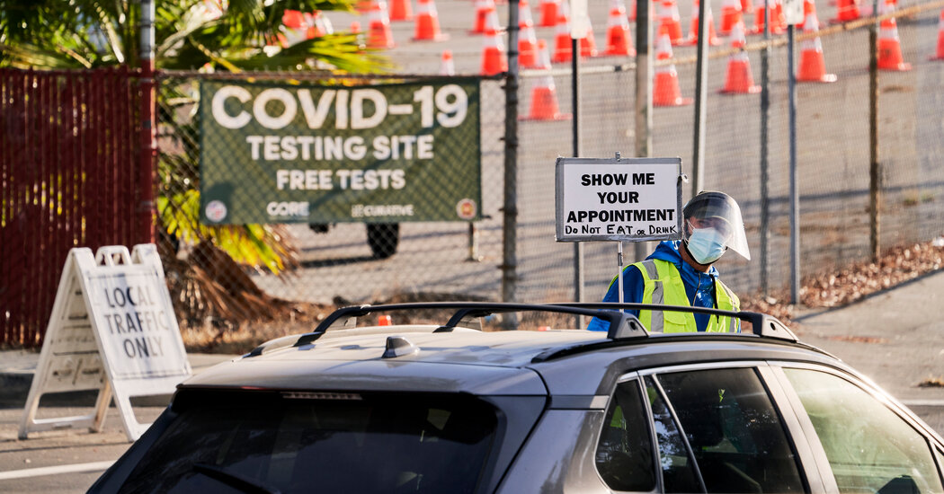
Insights from behavioral economics and other fields suggest strategies that could increase participation in mass screening programs.In October, Dr. Folasade May, an internist and public health researcher at the University of California, Los Angeles, began a new workplace routine. Once a week, she would make the three-minute walk from her office to a campus coronavirus testing site. After having her temperature checked and scanning a bar code on her phone, she would carefully swab the inside of her nose. Within a day or two, an email would arrive with her results.Week after week, her results came back negative. She continued to participate in the testing program even after being vaccinated, and she continued to test negative. And then, two weeks ago, an email landed in her inbox that stunned her: Her sample was positive for the coronavirus.The result — which turned out to be a false positive — briefly threw her life into chaos.“Our whole family was turned upside-down for an afternoon trying to figure out how to separate ourselves in our small house, trying to figure out who’s going to take care of the kids and who else did we expose,” Dr. May said.It is a situation that other families may soon find themselves in as mass coronavirus screening programs ramp up in schools and workplaces. Some of these programs will make testing mandatory, but many others will rely on voluntary participation — or on people regularly self-administering tests in their own homes.The more people who participate, the more effective these programs will be at slowing the spread of the virus, and keeping tabs on it. But there are also clear disincentives to participating, ranging from inconvenience to anxiety over the consequences of a positive result, which can mean missing work, keeping a child home from school or days of quarantine.“There are huge implications of a positive Covid test that a lot of people are very wary of, especially if they’re feeling well,” said Dr. May, who studies health disparities and the barriers to accessing health care. “I don’t know that people are going to come out in droves to get it done.”Although coronavirus screening is new, researchers in several fields — including behavioral economics, implementation science and health psychology — have spent years studying how to encourage people to engage in other kinds of health-related behaviors, including cancer screening, H.I.V. testing and vaccination. Some of these same strategies, they say, could help health officials design coronavirus screening programs, or even vaccination programs, with better participation rates.“The reality is that people are going to have to wake up and decide whether this is something they intend to do on any given day or any given week,” said Harsha Thirumurthy, associate director of the Center for Health Incentives and Behavioral Economics at the University of Pennsylvania. “And we know — from lots of examples of people’s decisions to seek testing for other health conditions, or even people’s decisions to engage in other health-related behaviors — that people are constantly weighing the costs and benefits of engaging in these services.”Here are five ways that health officials say can help shift the calculus.Make it easyHumans have a bias toward the status quo, a tendency to want to keep things as they are rather than shake them up. Many studies have shown that people are more likely to engage in various behaviors, from becoming an organ donor to enrolling in a 401(k) plan, when those behaviors are presented as the default choice.In a randomized clinical trial of nearly 5,000 emergency room patients, researchers found that the share of patients who were willing to take a rapid H.I.V. test climbed to 66 percent from 38 percent when the test was presented as a medical service they had to deliberately decline, rather than one they had to proactively ask for.Similarly, coronavirus screening programs are more likely to see wider participation if they are opt-out rather than opt-in. “The more you ask people to put in their own cognitive efforts and behavioral efforts into this, the less likely they’re going to do it,” said Derek Reed, who directs the applied behavioral economics laboratory at the University of Kansas.And, of course, the actual testing process should be quick and convenient, experts say, with strategically located testing sites and streamlined procedures that allow people to easily incorporate testing into their routines.Ask people to planExperts also suggested asking people to think through the logistics of when and how they plan to get tested. Studies show that people who clearly formulate a plan for how they intend to accomplish something — whether it’s voting in an upcoming election or getting a flu vaccine — are more likely to follow-through.One possibility, Dr. Reed said, would be to text people reminders of their testing appointments, and ask them to reply with, say, a 1 if they plan to walk to the appointment, a 2 if they plan to drive or a 3 if they plan to take the bus. “And then depending on the response, you just automatically ping back Google map directions or a link to campus or community bus system maps or timetables,” he said.These kinds of nudges are likely to be most effective for people who are already motivated to get tested but may have trouble following through. “Often you need to nudge them a little bit by just removing frictions to get rid of these small costs,” said Sebastian Linnemayr, a behavioral economist at the RAND Corporation, a think tank in California.Provide (the right) incentivesHealth officials could also reward people who participate in testing programs. “There probably needs to be some sort of incentive at the patient level,” Dr. May said. “We’ve seen the same thing in cancer screening. We’ve seen health insurers provide incentives to patients to participate in healthy lifestyles, to participate in screening measures.”But the specific incentives matter, and some studies suggest that people are more motivated by an opportunity to win a large reward than the guarantee of a smaller payout.In a November survey of 200 undergraduate students at the University of Kansas, Dr. Reed and his colleagues found that 70 percent of them said they would be willing to get tested if the university gave them a $5 gift card. But if the university instead entered them in a lottery, in which they had a 5 percent chance of winning $100, 88 percent said they would participate, said Dr. Reed, who shared his findings with the university.These kinds of lotteries may be effective because people tend to overestimate their chances of winning them. “The other part of it is that the lottery does introduce an element of fun or engagement in this idea that you’re part of something that other people are also participating in,” Dr. Thirumurthy said.Leverage social tiesPeer pressure can be a powerful public health tool; research has shown, for instance, that college women are more willing to get the human papillomavirus vaccine if they believe that their peers are also doing so.Government, school and workplace leaders can help coronavirus testing become a social norm by making it public how many others are participating. “Then, send out that information over and over again: ‘Don’t be left out, 85 percent already got tested and are making the school a better place,’” Dr. Linnemayr said. He added, “It’s really important that you signal, ‘This is an activity that we, as a community, value and reward.’”Health officials could also consider leveraging social networks more explicitly. In a randomized trial conducted in Kenya, Dr. Thirumurthy found that providing women with two H.I.V. self-testing kits, and encouraging them to give one to their male partners, significantly increased the share of men tested for H.I.V.He is now working with colleagues to evaluate a similar program for the coronavirus, asking people to distribute self-testing kits to others in their social circles.“Who has the strongest ability to persuade you to test?” Dr. Thirumurthy said. “Is it going to be your employer or somebody in the community? Or is it going to be someone who is a close friend or a family member?”Support people who test positiveEven the cleverest nudges, however, will not be enough to convince people to get tested if they think a positive result will have a devastating effect on their lives. A major barrier to H.I.V. testing has been the fear among potential recipients that testing positive would cost them their friends, jobs, housing or health insurance.A positive coronavirus test doesn’t necessarily carry the same stigma, but it can still have serious ripple effects on people’s lives. “If it keeps you out of work and you can’t earn an income to support your family, that’s a non-starter for a lot of people,” said Jennifer Nuzzo, an epidemiologist at Johns Hopkins University.So governments, employers and community organizations should do what they can to reduce the costs associated with testing positive, health experts said. That could mean providing paid leave or allowing employees to work remotely if they must quarantine; offering hotel vouchers so people have a safe place to isolate; or offering financial and food assistance. Such services can “soften the blow” of testing positive, Dr. Thirumurthy said: “There needs to be some type of safety net.”
Read more →