Plan to Ditch the Mask After Vaccination? Not So Fast.

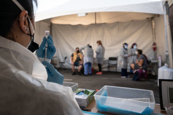
#masthead-section-label, #masthead-bar-one { display: none }The Coronavirus OutbreakliveLatest UpdatesMaps and CasesRisk Near YouVaccine RolloutNew Variants TrackerAdvertisementContinue reading the main storySupported byContinue reading the main storyPlan to Ditch the Mask After Vaccination? Not So Fast.It’s not clear whether vaccinated people may still spread the virus, but the answer to that question is coming soon. Until then, scientists urge caution.A health care worker prepared a dose of the Pfizer-BioNTech vaccine at a vaccination site in San Francisco on Monday.Credit…Mike Kai Chen for The New York TimesMarch 3, 2021, 3:23 p.m. ETWith 50 million Americans immunized against the coronavirus, and millions more joining the ranks every day, the urgent question on many minds is: When can I throw away my mask?It’s a deeper question than it seems — about a return to normalcy, about how soon vaccinated Americans can hug loved ones, get together with friends, and go to concerts, shopping malls and restaurants without feeling threatened by the coronavirus.Certainly many state officials are ready. On Tuesday, Texas lifted its mask mandate, along with all restrictions on businesses, and Mississippi quickly followed suit. Governors in both states cited declining infection rates and rising numbers of citizens getting vaccinated.But the pandemic is not yet over, and scientists are counseling patience.It seems clear that small groups of vaccinated people can get together without much worry about infecting one another. The Centers for Disease Control and Prevention is expected shortly to issue new guidelines that will touch on small gatherings of vaccinated Americans.But when vaccinated people can ditch the masks in public spaces will depend on how quickly the rates of disease drop and what percentage of people remain unvaccinated in the surrounding community.Why? Scientists do not know whether vaccinated people spread the virus to those who are unvaccinated. While all of the Covid-19 vaccines are spectacularly good at shielding people from severe illness and death, the research is unclear on exactly how well they stop the virus from taking root in an immunized person’s nose and then spreading to others.It’s not uncommon for a vaccine to forestall severe disease but not infection. Inoculations against the flu, rotavirus, polio and pertussis are all imperfect in this way.The coronavirus vaccines “are under a lot more scrutiny than any of the previous vaccines ever have been,” said Neeltje van Doremalen, an expert in preclinical vaccine development at the National Institutes of Health’s Rocky Mountain Laboratories in Montana.And now coronavirus variants that dodge the immune system are changing the calculus. Some vaccines are less effective at preventing infections with certain variants, and in theory could allow more virus to spread.The research available so far on how well the vaccines prevent transmission is preliminary but promising. “We feel confident that there’s a reduction,” said Natalie Dean, a biostatistician at the University of Florida. “We don’t know the exact magnitude, but it’s not 100 percent.”Still, even an 80 percent drop in transmissibility might be enough for immunized people to toss their masks, experts said — especially once a majority of the population is inoculated, and as rates of cases, hospitalizations and deaths plummet.A line to register for a vaccination appointment in San Francisco. Experts say that people who have been inoculated should continue to wear masks to protect others.Credit…Mike Kai Chen for The New York TimesBut most Americans are still unvaccinated, and more than 1,500 people are dying every day. So given the uncertainty around transmission, even people who are immunized must continue to protect others by wearing masks, experts said.“They should wear masks until we actually prove that vaccines prevent transmission,” said Dr. Anthony S. Fauci, director of the National Institute for Allergy and Infectious Diseases.The Coronavirus Outbreak

Read more →
New, highly precise 'clock' can measure biological age

Using the model organism Caenorhabditis elegans, researchers at the University of Cologne have developed an ‘aging clock’ that reads the biological age of an organism directly from its gene expression, the transcriptome. Bioinformatician David Meyer and geneticist Professor Dr Björn Schumacher, director of the Institute for Genome Stability in Aging and Disease at the CECAD Cluster of Excellence in Aging Research and the Center for Molecular Medicine Cologne (CMMC), describe their so-called BiT age (binarized transcriptomic aging clock) in the article ‘BiT age: A transcriptome based aging clock near the theoretical limit of accuracy’ in Aging Cell.
We are all familiar with chronological age — our age since birth. But biological age can differ from it, at times significantly. Everyone ages differently. Scientists can use aging clocks to determine an organism’s biological age. Until now, aging clocks such as Horvath’s epigenetic clock have been based on the pattern of methylations, small chemical groups that attach to DNA and change with age. Using the transcriptome, the new clock takes into consideration the set of genes that are read from DNA (messenger RNA) to make proteins for the cell.
Until now, the transcriptome was considered too complex to indicate age. Sometimes genes transcribe a particularly large amount of mRNA, sometimes less. Hence, so far it has not been possible to develop precise aging clocks based on gene activity. Meyer and Schumacher’s new approach uses a mathematical trick to eliminate the differences in gene activity. The binarized transcriptome aging clock divides genes into two groups — ‘on’ or ‘off’ — thus minimizing high variation. This makes aging predictable from the transcriptome. ‘Surprisingly, this simple procedure allows very accurate prediction of biological age, close to the theoretical limit of accuracy. Most importantly, this aging clock also works at high ages, which were previously difficult to measure because the variation in gene activity is particularly high then,’ said Meyer.
BiT age is based exclusively on approximately 1,000 different transcriptomes of C. elegans, for which the lifespan is precisely known. Model organisms such as the nematode provide a controllable view of the aging process, allowing biomarkers to be discovered and the effects of external influences such as UV radiation or nutrition on longevity to be studied.
The new aging clock allows researchers to accurately predict the pro- and anti-aging effects of gene variants and various external factors in the nematode at a young age. The aging clock also showed that genes of the immune response as well as signalling in neurons are significant for the aging process. ‘BiT age can also be applied to predict human age quickly and with very high accuracy. Measuring biological age is important to determine the influence of environment, diet or therapies on the aging process and the development of age-related diseases. This clock could therefore find wide application in aging research. Since BiT age is based purely on gene activity, it can basically be applied to any organism,’ Schumacher explained.

Story Source:
Materials provided by University of Cologne. Note: Content may be edited for style and length.

Read more →

Color blindness-correcting contact lenses

Imagine seeing the world in muted shades — gray sky, gray grass. Some people with color blindness see everything this way, though most can’t see specific colors. Tinted glasses can help, but they can’t be used to correct blurry vision. And dyed contact lenses currently in development for the condition are potentially harmful and unstable. Now, in ACS Nano, researchers report infusing contact lenses with gold nanoparticles to create a safer way to see colors.
Some daily activities, such as determining if a banana is ripe, selecting matching clothes or stopping at a red light, can be difficult for those with color blindness. Most people with this genetic disorder have trouble discriminating red and green shades, and red-tinted glasses can make those colors more prominent and easier to see. However, these lenses are bulky and the lens material cannot be made to fix vision problems. Thus, researchers have shifted to the development of special tinted contact lenses. Although the prototype hot-pink dyed lenses improved red-green color perception in clinical trials, they leached dye, which led to concerns about their safety. Gold nanocomposites are nontoxic and have been used for centuries to produce “cranberry glass” because of the way they scatter light. So, Ahmed Salih, Haider Butt and colleagues wanted to see whether incorporating gold nanoparticles into contact lens material instead of dye could improve red-green contrast safely and effectively.
To make the contact lenses, the researchers evenly mixed gold nanoparticles into a hydrogel polymer, producing rose-tinted gels that filtered light within 520-580 nm, the wavelengths where red and green overlap. The most effective contact lenses were those with 40 nm-wide gold nanoparticles, because in tests, these particles did not clump or filter more color than necessary. In addition, these lenses had water-retention properties similar to those of commercial ones and were not toxic to cells growing in petri dishes in the lab. Finally, the researchers directly compared their new material to two commercially available pairs of tinted glasses, and their previously developed hot-pink dyed contact lens. The gold nanocomposite lenses were more selective in the wavelengths they blocked than the glasses. The new lenses matched the wavelength range of the dyed contact lenses, suggesting the gold nanocomposite ones would be suitable for people with red-green color issues without the potential safety concerns. The researchers say that the next step is to conduct clinical trials with human patients to assess comfort.

Story Source:
Materials provided by American Chemical Society. Note: Content may be edited for style and length.

Read more →

Study reveals details of immune defense guidance system

At the beginning of an immune response, a molecule known to mobilize immune cells into the bloodstream, where they home in on infection sites, rapidly shifts position, a new study shows. Researchers say this indirectly amplifies the attack on foreign microbes or the body’s own tissues.
Past studies had shown that the immune system regulates the concentration of the molecule, sphingosine 1 phosphate (S1P), in order to draw cells to the right locations. The targeted cells have proteins on their surface that are sensitive to levels of this molecule, enabling them to follow the molecule’s “trail,” researchers say. S1P concentration gradients, for instance, can guide immune T cells to either stay in lymph nodes, connected glands in which these cells mature, or move into blood vessels.
For the first time, researchers at NYU Grossman School of Medicine showed in mice experiments that S1P levels in lymph nodes increase as the immune response mounts. Such activation of immune cells can cause inflammation, swelling, and/or death of targeted cells.
While past work had shown that S1P is produced by cells attached to lymph nodes, the new study found that monocytes, circulating immune cells, also produced it when mice were infected with a virus. This in turn may influence the migration of T cells, a set of white blood cells that expands rapidly in response to infection, say the study authors.
Publishing in the journal Nature online March 3, study results showed that T cells left mouse lymph nodes less than half as fast when S1P levels rose, while mostly immature cells escaped when S1P levels were not spiking.
“Our research shows a larger role for sphingosine 1 phosphate in coordinating immune defenses in response to infection and inflammation,” says study lead investigator Audrey Baeyens, PhD, a postdoctoral fellow at NYU Langone and its Skirball Institute of Biomolecular Medicine. “While further testing is needed, our findings raise the prospect of controlling levels of S1P to either boost or diminish the body’s immune response, as needed.”
Moreover, the researchers found that when lymph node levels of S1P went up, it signaled T cells to remain in lymph nodes. Such “trapped” T cells, with longer time to mature and become fully armed in the node, increase in their toxicity. These mature T cells can attack cells infected by viruses, or healthy cells as part of autoimmune diseases.
Indeed, medications that block S1P, preventing immune cells from leaving the lymph nodes, are used to curb unwanted and autoimmune inflammation related to inflammatory bowel disease, psoriasis, and multiple sclerosis, a disease for which fingolimod (Gilenya) is one of the few approved treatments.
Researchers say their findings could also explain why multiple sclerosis patients can experience severe disease relapse immediately after ceasing fingolimod treatment, as T cells held long in lymph nodes are then freed to attack the body’s nerves, a key trait of the disease.
“Now that we have a better understanding of sphingosine 1 phosphate inhibition, we can work on finding new uses for this class of medications, perhaps by manipulating the time T cells spend in the lymph nodes,” says study senior investigator Susan Schwab, PhD. Schwab is an associate professor in the Department of Pathology at NYU Langone and Skirball.
For the study, S1P levels were measured in mice bred to develop symptoms of multiple sclerosis, a disease involving severe inflammation of the brain and spine. They also measured S1P levels in mice exposed to viral genetic material to mimic the inflammation that occurs in infection.
Schwab says the team next plans to study how different S1P levels affect T cell maturation, and how these different maturation times strengthen or weaken the overall immune response to infection.

Read more →

Genomics study identifies routes of transmission of coronavirus in care homes

Care homes are at high risk of experiencing outbreaks of COVID-19, the disease caused by SARS-CoV-2. Older people and those affected by heart disease, respiratory disease and type 2 diabetes — all of which increase with age — are at greatest risk of severe disease and even death, making the care home population especially vulnerable.
Care homes are known to be high-risk settings for infectious diseases, owing to a combination of the underlying vulnerability of residents who are often frail and elderly, the shared living environment with multiple communal spaces, and the high number of contacts between residents, staff and visitors in an enclosed space.
In research published in eLife, a team led by scientists at the University of Cambridge and Wellcome Sanger Institute used a combination of genome sequencing and detailed epidemiological information to examine the impact of COVID-19 on care homes and to look at how the virus spreads in these settings.
SARS-CoV-2 is an RNA virus and as such its genetic code is prone to errors each time it replicates. It is currently estimated that the virus mutates at a rate of 2.5 nucleotides (the A, C, G and U of its genetic code) per month. Reading — or ‘sequencing’ — the genetic code of the virus can provide valuable information on its biology and transmission. It allows researchers to create ‘family trees’ — known as phylogenetic trees — that show how samples relate to each other.
Scientists and clinicians in Cambridge have pioneered the use of genome sequencing and epidemiological information to trace outbreaks and transmission networks in hospitals and community-based healthcare settings, helping inform infection control measures and break the chains of transmission. Since March 2020, they have been applying this method to SARS-CoV-2 as part of the COVID-19 Genomics UK (COG-UK) Consortium.
In this new study, researchers analysed samples collected from 6,600 patients between 26 February and 10 May 2020 and tested at the Public Health England (PHE) Laboratory in Cambridge. Out of all the cases, 1,167 (18%) were care home residents from 337 care homes, 193 of which were residential homes and 144 nursing homes, the majority in the East of England. The median age of care home residents was 86 years.

advertisement

While the median number of cases per care home was two, the ten care homes with the largest number of cases accounted for 164 cases. There was a slight trend for nursing homes to have more cases per home than residential homes, with a median of three cases.
Compared with non-care home residents admitted to hospital with COVID-19, hospitalised care home residents were less likely to be admitted to intensive care units (less than 7% versus 21%) and more likely to die (47% versus 20%).
The researchers also explored links between care homes and hospitals. 68% of care home residents were admitted to hospital during the study period. 57% were admitted with COVID-19, 6% of cases had suspected hospital-acquired infection, and 33% were discharged from hospital within 7 days of a positive test. These findings highlight the ample opportunities for SARS-CoV-2 transmission between hospital and care home settings.
When the researchers examined the viral sequences, they found that for several of the care homes with the highest number of cases, all of the cases clustered closely together on a phylogenetic tree with either identical genomes or just one base pair difference. This was consistent with a single outbreak spreading within the care home.
By contrast, for several other care homes, cases were distributed across the phylogenetic tree, with more widespread genetic differences, suggesting that each of these cases was independent and not related to a shared transmission source.

advertisement

“Older people, particularly those in care homes who may be frail, are at particular risk from COVID-19, so it’s essential we do all that we can to protect them,” said Dr Estée Török, an Honorary Consultant at Addenbrooke’s Hospital, Cambridge University Hospitals (CUH), and an Honorary Senior Visiting Fellow at the University of Cambridge.
“Preventing the introduction of new infections into care homes should be a key priority to limit outbreaks, alongside infection control efforts to limit transmission within care homes, including once an outbreak has been identified.”
The team found two clusters that were linked to healthcare workers. One of these involved care home residents, a carer from that home and another from an unknown care home, paramedics and people living with them. The second involved several care home residents and acute medical staff at Cambridge University Hospitals NHS Foundation Trust who cared for at least one of the residents. It was not possible to say where these clusters originated from and how the virus spread.
“Using this technique of ‘genomic surveillance’ can help institutions such as care homes and hospitals better understand the transmission networks that allow the spread of COVID-19,” added Dr William Hamilton from the University of Cambridge and CUH. “This can then inform infection control measures, helping ensure that these places are as safe as possible for residents, patients, staff and visitors.”
The absolute number of diagnosed COVID-19 cases from care home residents declined more slowly in April than for non-care home residents, increasing the proportion of cases from care homes and contributing to the slow rate of decline in total case numbers during April and early May 2020.
“Our data suggest that care home transmission was more resistant to lockdown measures than non-care home settings. This may reflect the underlying vulnerability of the care home population, and the infection control challenges of nursing multiple residents who may also share communal living spaces,” said Gerry Tonkin-Hill from the Wellcome Sanger Institute.
The team found no new viral lineages from outside the UK, which may reflect the success of travel restrictions in limiting new viral introductions into the general population during the first epidemic wave and lockdown period.
This work was funded by COG-UK, Wellcome, the Academy of Medical Sciences, the Health Foundation and the NIHR Cambridge Biomedical Research Centre.

Read more →

Tackling tumors with two types of virus

An international research group led by the University of Basel has developed a promising strategy for therapeutic cancer vaccines. Using two different viruses as vehicles, they administered specific tumor components in experiments on mice with cancer in order to stimulate their immune system to attack the tumor. The approach is now being tested in clinical studies.
Making use of the immune system as an ally in the fight against cancer forms the basis of a wide range of modern cancer therapies. One of these is therapeutic cancer vaccination: following diagnosis, specialists set about determining which components of the tumor could function as an identifying feature for the immune system. The patient is then administered exactly these components by means of vaccination, with a view to triggering the strongest possible immune response against the tumor.
Viruses that have been rendered harmless are used as vehicles for delivering the characteristic tumor molecules into the body. In the past, however, many attempts at creating this kind of cancer therapy failed due to an insufficient immune response. One of the hurdles is that the tumor is made up of the body’s own cells, and the immune system takes safety precautions in order to avoid attacking such cells. In addition, the immune cells often end up attacking the “foreign” virus vehicle more aggressively than the body’s own cargo. With almost all cancer therapies of this kind developed so far, therefore, the desired effect on the tumor has failed to materialize. Finding the appropriate vehicle is just as relevant in terms of effectiveness as the choice of tumor component as the point of attack.
Arenaviruses as vehicles
The research group led by Professor Daniel Pinschewer of the University of Basel had already discovered in previous studies that viruses from the arenavirus family are highly suitable as vehicles for triggering a strong immune response. The group now reports in the journal Cell Reports Medicine that the combination of two different arenaviruses produced promising results in animal experiments.
The researchers focused on two distantly related viruses called Pichinde virus and Lymphocytic choriomeningitis virus, which they adapted via molecular biological methods for use as vaccine vectors. When they took the approach of administering the selected tumor component first with the one virus and then, at a later point, with the other, the immune system shifted its attack away from the vehicle and more towards the cargo. “By using two different viruses, one after the other, we focus the triggered immune response on the actual target, the tumor molecule,” explains Pinschewer.
Tumor eliminated or slowed down
In experiments with mice, the researchers were able to measure a potent activation of killer T cells that eliminated the cancer cells. In 20% to 40% of the animals — depending on the type of cancer — the tumor disappeared, while in other cases the rate of tumor growth was at least temporary slowed.
“We can’t say anything about the efficacy of our approach in humans as yet,” Pinschewer points out. However, ongoing studies with a cancer therapy based on a single arenavirus have already shown promising results. The effects on tumors in animal experiments cannot be assumed to translate directly into the effect on corresponding cancer types in humans. “However, since the therapy with two different viruses works better in mice than the therapy with only one virus, our research results make me optimistic,” Pinschewer adds.
The biotech company Hookipa Pharma, of which Pinschewer is one of the founders, is now investigating the efficacy of this novel approach to cancer therapy in humans. “We are currently exploring what our approach by itself can actually achieve,” the researcher says. “If it proves successful, a wide range of combinations with existing therapies could be envisaged, in which the respective mechanisms would join forces to eliminate tumors even better.”

Story Source:
Materials provided by University of Basel. Note: Content may be edited for style and length.

Read more →

Accelerating gains in abdominal fat during menopause tied to heart disease risk

Women who experience an accelerated accumulation of abdominal fat during menopause are at greater risk of heart disease, even if their weight stays steady, according to a University of Pittsburgh Graduate School of Public Health-led analysis published today in the journal Menopause.
The study — based on a quarter century of data collected on hundreds of women — suggests that measuring waist circumference during preventive health care appointments for midlife women could be an early indicator of heart disease risk beyond the widely used body mass index (BMI) — which is a calculation of weight vs. height.
“We need to shift gears on how we think about heart disease risk in women, particularly as they approach and go through menopause,” said senior author Samar El Khoudary, Ph.D., M.P.H., associate professor of epidemiology at Pitt Public Health. “Our research is increasingly showing that it isn’t so important how much fat a woman is carrying, which doctors typically measure using weight and BMI, as it is where she is carrying that fat.”
El Khoudary and her colleagues looked at data on 362 women from Pittsburgh and Chicago who participated in the Study of Women’s Health Across the Nation (SWAN) Heart study. The women, who were an average age of 51, had their visceral adipose tissue — fat surrounding the abdominal organs — measured by CT scan and the thickness of the internal carotid artery lining in their neck measured by ultrasound, at a few points during the study. Carotid artery thickness is an early indicator of heart disease.
The team found that for every 20% increase in abdominal fat, the thickness of the carotid artery lining grew by 2% independent of overall weight, BMI and other traditional risk factors for heart disease.
They also found that abdominal fat started a steep acceleration, on average, within two years before the participants’ last period and continued a more gradual growth after the menopausal transition.
Saad Samargandy, Ph.D., M.P.H., who was a doctoral student at Pitt Public Health at the time of the research, explained that fat that hugs the abdominal organs is related to greater secretion of toxic molecules that can be harmful to cardiovascular health.
“Almost 70% of post-menopausal women have central obesity — or excessive weight in their mid-section,” said Samargandy, also the first author of the journal article. “Our analysis showed an accelerated increase of visceral abdominal fat during the menopausal transition of 8% per year, independent of chronological aging.”
Measuring abdominal fat by CT scan is expensive, inconvenient and could unnecessarily expose women to radiation — so El Khoudary suggests that regularly measuring and tracking waist circumference would be a good proxy to monitor for accelerating increases in abdominal fat. Measuring weight and BMI alone could miss abdominal fat growth because two women of the same age may have the same BMI but different distribution of fat in their body, she added.
“Historically, there’s been a disproportionate emphasis on BMI and cardiovascular disease,” said El Khoudary. “Through this long-running study, we’ve found a clear link between growth in abdominal fat and risk of cardiovascular disease that can be tracked with a measuring tape but could be missed by calculating BMI. If you can identify women at risk, you can help them modify their lifestyle and diet early to hopefully lower that risk.”
Late last year, El Khoudary led a team in publishing a new scientific statement for the American Heart Association that calls for increased awareness of the cardiovascular and metabolic changes unique to the menopausal transition and the importance of counseling women on early interventions to reduce cardiovascular disease risk factors.
El Khoudary noted that more research is needed to determine if certain diet, exercise or lifestyle interventions are more effective than others, as well as whether there is a clear cut-off point for when growth in waist circumference becomes concerning for heart disease risk.

Story Source:
Materials provided by University of Pittsburgh. Note: Content may be edited for style and length.

Read more →

Researchers illuminate potential precursors of blood cancers

Utah researchers report significant new insights into the development of blood cancers. In work published today in Blood Cancer Discovery, a journal of the American Association for Cancer Research, scientists describe an analysis of published data from more than 7,000 patients diagnosed with leukemia and other blood disorders. Their findings provide new clues about mutations that may initiate cancer development and those that may help cancer to progress.
The researchers sought to identify mutation hotspots, or frequent changes in specific locations of the cancer patients’ genetic information. The researchers then used these hotspots to look for whether the same mutations were present in the DNA data of more than 4,500 people who were not known to have a cancer diagnosis. They found that approximately 2 percent of these presumably healthy participants had, at low levels, mutations identical to those frequently observed in the cancer patients.
“Understanding how a disease develops is greatly benefited by studying persons who are currently healthy but are on a trajectory for disease onset,” said Clint Mason, PhD, assistant professor of pediatrics at the U of U, who specializes in cancer genomics and bioinformatics and led the study.
Mutations occur throughout the 3 billion bases of DNA that are present in human cells. Many will have no impact on health, yet certain mutations may cause or support development of diseases, including cancer. Therefore, scientists are working to discern which mutations have a significant impact on health. In this study, the researchers sought to understand which mutations are most frequently present in adults and children both with and without blood cancers. They hope this information may illuminate an understanding of these cancers, and further, may potentially be used to identify people who are progressing towards cancer.
The team of researchers consisted nearly entirely of University of Utah (U of U) faculty and students. Mason was joined by co-first authors Julie Feusier, PhD, a trainee in the department of human genetics and now a postdoctoral fellow at Huntsman Cancer Institute, and Sasi Arunachalam, PhD, a postdoctoral fellow at Huntsman Cancer Institute and now a research associate at St. Jude Children’s Research Hospital, in performing analyses along with other co-authors.
For the first part of the study, the researchers completed a large data mining analysis to examine published data from 48 cancer studies that reported mutations present in persons who were being diagnosed with leukemia or other hematologic malignancies. Across the 7,430 pediatric and adult cancer patients of those studies, 434 DNA locations were identified as frequently mutated. Then, in a subsequent analysis of many terabytes of publicly available genetic data, the researchers identified these same cancer-relevant mutations at low levels among 83 of the 4,538 persons who were presumably free from cancer.
“When identical mutations are found to be present in a small percentage of blood cells of a healthy person, it may indicate that something abnormal has begun to occur,” explained Mason.
This abnormal process (known as clonal hematopoiesis) has previously been found to be increasingly common as people age. As a result, researchers generally focus on the study of adults when investigating this phenomenon. Yet in this study, in addition to adults, the researchers analyzed data from 400 children who were likely to be free from cancer. They found preliminary evidence that early cancer mutations could be detected in this age group.
In an accompanying commentary, to be published by Blood Cancer Discovery in its “In the Spotlight” section, Barbara Spitzer, MD, and Ross Levine, MD, of the Memorial Sloan Kettering Cancer Center, wrote, “This work demonstrates the first evidence that [clonal hematopoiesis] is observed in children outside of those with advanced malignancies.” They further commented, “This expansion of the range of hematologic malignancy hotspot mutations goes beyond an improved understanding of the molecular repertoire of hematologic malignancies… More complete knowledge of relevant mutations may increase our detection of patients at highest risk for malignant transformation.”
Finding a low-level mutation identical to one that is frequently present at cancer diagnoses could be alarming to a currently healthy person screened for such events. But fortunately, because cancer frequently requires multiple mutations to be present in a sizeable fraction of cells, most persons with a single low-level mutation are not likely to develop cancer for many years or decades — if they develop cancer at all. Large longitudinal studies are needed to identify the timeframe over which specific mutations or combinations of mutations accelerate progression to cancer.
The Utah researchers next hope to determine the stability of mutations identified at younger ages. This will give a preliminary glimpse into their potential to serve as biomarkers for those having greatest risk of cancer. Future intervention studies will require such an identification.
“Our goal has been to help fill in gaps in the understanding of cancer development so that future prevention work can take place more quickly and effectively,” said Mason. “We are grateful to have been able to contribute a few puzzle pieces to that monumental effort.”

Read more →

Los Angeles County finds fewer cases among health care workers as more get vaccinated.

#masthead-section-label, #masthead-bar-one { display: none }The Coronavirus OutbreakliveLatest UpdatesMaps and CasesRisk Near YouVaccine RolloutNew Variants TrackerAdvertisementContinue reading the main storyCovid-19 Live Updates: Biden’s Call for Vaccinating Educators Is Part of Ambitious Schools PlanLos Angeles County finds fewer cases among health care workers as more get vaccinated.March 3, 2021, 2:20 p.m. ETMarch 3, 2021, 2:20 p.m. ETNew virus cases among health care workers in the Los Angeles County fell to 69 for the week of Feb. 14, from more than 1,800 cases during the week of Nov. 29, the county said.Credit…Isadora Kosofsky for The New York TimesWhat was once a flood of health care workers catching the coronavirus in Los Angeles County has now slowed to a trickle, in large part because the vast majority of them have been vaccinated, local public health officials said. Reports of new virus cases among health care workers in the county have fallen by 94 percent since late November, just before vaccination began.The statistics are encouraging, both in Los Angeles County and across the country. Some health care workers initially expressed reluctance to get a Covid-19 vaccine shot, often out of fear about the safety of the vaccines, which were hurried into use under emergency authorizations from the Food and Drug Administration.Workers in nursing homes and long-term care facilities, which have been hot spots during the pandemic, have been of special concern: At one point, those workers accounted for one-quarter of all cases among health care workers in Los Angeles County.But by the end of February, the county said, 69 percent of health care workers in those facilities — including 78 percent of nursing home and long-term care facilities staffs — had received at least one shot of vaccine.The results have been stark: 434 new virus cases were reported in the county among nursing-home health care workers during the week of Nov. 29, but for the week of Feb. 14, there were 10 cases, according to county data.The same has happened with the county’s health care workers in general: New cases fell to 69 for the week of Feb. 14, from more than 1,800 cases during the week of Nov. 29, the county said.“High rates of vaccination are correlated with the lowest rates of cases and deaths among health care workers at nursing homes,” the county public health department wrote in a statement on Monday, “and we are grateful to everyone that got vaccinated and to the teams that coordinated vaccinations at each site.”The county as a whole made major progress over the same period, with new cases overall down 71 percent. But even so, the risk of getting the virus there remains high.A recent survey by the Kaiser Family Foundation found that Los Angeles County is ahead of most of the country in getting health care workers immunized. The nationwide survey, conducted between Feb. 15 and Feb. 23, found that 54 percent of health care workers had already received at least one dose of vaccine by then, and 10 percent more said they planned to get a shot as soon as they could. Some 15 percent said they would “definitely not” get the vaccine.AdvertisementContinue reading the main story

Read more →

When I Lost My Sense of Taste to Covid, Anorexia Stepped In

#masthead-section-label, #masthead-bar-one { display: none }At HomeWatch: ‘WandaVision’Travel: More SustainablyFreeze: Homemade TreatsCheck Out: Podcasters’ Favorite PodcastsAdvertisementContinue reading the main storySupported byContinue reading the main storyVoicesWhen I Lost My Sense of Taste to Covid, Anorexia Stepped InWith flavor gone, my old eating disorder came roaring back.Credit…Nathalie LeesMarch 3, 2021, 11:22 a.m. ETThe day after my family and I were diagnosed with Covid-19 last September, I made myself a cup of coffee. I had been awake most of the night with chills and hoped I’d find comfort in its familiar aroma and warmth.I lowered my face to the surface of my mug and inhaled. Nothing. I started searching for smell wherever I could. In the bathroom, I untwisted the cap on one of my perfume bottles and couldn’t detect its jasmine fragrance. I brought a candle up to my nose, but it was scentless.When I sipped my coffee, all I could sense was its warmth. I started to make breakfast for my 4-year-old daughter and my 3-year-old son ­— maybe there I could find something with taste. I put a strawberry in my mouth and could feel its seeds but couldn’t detect its sweetness. I bit down on an almond-butter granola bar, sinking my teeth into the sadness of a reality I didn’t want to face.I was diagnosed with anorexia at age 12, the year after my mom died. She’d been sick with metastatic breast cancer for three years, and even when it spread to her bone marrow, her liver and her brain, I was still convinced she’d get better. It’s what my family had told me, and so I believed it to be true. Until it wasn’t.When she died, I felt as though life had become out of control. Pretty quickly, I realized that I could not impose order on the larger world, but I could control something that had always been in my life and always would be: food. And so began a three-year stretch of multiple hospitalizations and a 17-month-long stay at a residential treatment facility.Now, at 35, after 20 years in recovery, I’m far better than I’d ever thought I’d be. But some days, my mind still flirts with anorexia. The disorder secretly seduces me, satisfying my affinity for control and order. It always lurks in the background and I have to make a concerted effort to keep it cornered.Without taste, I was triggered. Anorexia beckoned me, reminding me that I could shed even more weight off my already slender frame if I skimped here and slacked there. When I would make my breakfast in the mornings after losing my taste, I’d forgo frothed milk in my coffee, opting to drink it black instead. I’d put one and a half slices of cheese on my grilled cheese sandwich instead of two and a half. I’d start to place granola on top of my yogurt, but uncomfortably familiar questions would stop me.Do you really need to eat that? Why waste the calories?Without taste, food became a formality. It was merely sustenance, and so I settled for the bland, bare minimum. Chewing felt like a chore, and every bite took effort I didn’t want to expend.I was craving comfort. After days of not eating enough, I decided to seek it in a food that I used to love eating with my mom: ice cream. I ordered a pint of Ben and Jerry’s Glampfire Trail Mix and as soon as it arrived I dug my spoon into satisfying chunks of pretzel, chewy marshmallows and crunchy fudge-covered almonds. I couldn’t taste a thing, but I detected texture. I liked the act of digging my teeth into something that took work to chew. I liked hearing the crunch of the almonds, and swirling the softness of marshmallow in my mouth.I found myself relating to one of Ben & Jerry’s founders, Ben Cohen, who has very little sense of taste and no sense of smell. When he and his partner, Jerry Greenfield, were developing their signature ice cream in the 1970s, anosmia-stricken Ben advocated for chunks. He became the texture taster, the one who would determine if teeth could be satisfied even when the tongue could not. After three small spoonfuls, I put the ice cream back in the freezer, not allowing myself to have any more.There are often competing forces at play in my recovery; the healthy side of me that recognizes I need to eat more and wants to indulge in foods I enjoy, and the old eating disorder that tells me I shouldn’t.The next day, family friends dropped off a homemade broccoli and cheese casserole, coloring books for my kids and a dozen bags of groceries filled with food we like to eat: cinnamon raisin bagels, red grapes, smoothie mixes and more. I wanted nothing more than to enjoy the home-cooked meal, which looked like something my mom would have made. I ate some of it, but not enough.As our symptoms subsided and our two-week quarantine ended, I started to see the effects of eating too little. I could see it in my slightly sunken-in cheeks, could feel it in the contours of my hip bone, could hear it in my stomach, which groaned in the dark of night. I took a photo of myself and recognized I was too thin. My husband noticed, too. He reassured me that my taste would come back, and he reminded me of how much traction I’d lose if I let myself get stuck in the setback.Over the years, I’ve had to change my perspective on what it means to be in recovery. I used to strive for “full recovery” — a life without slip-ups or setbacks — and would always feel like I had failed whenever I faltered. Now I frame my thinking around what I call “the middle place,” that sticky space between sickness and full recovery. I make it my goal to continuously progress through that space — for myself, for my family. Recovery is about recognizing that I’m in control of my choices, even when anorexia comes knocking, pleading for another chance. During Covid, I opened the door a crack, but eventually closed it.My sense of taste was gone for about five weeks, and once it came back I started to regain my footing and, eventually, the pounds I had lost. Taste first showed up one morning when I was eating a banana; soon more flavors re-emerged.And then one Sunday afternoon, I ate creamy tomato bisque and felt and smelled and tasted every single spoonful. There was the warmth, the savory tomatoes, the bliss of basil.I finished the soup and was still hungry. So I got myself a generous side of crackers and Gouda cheese, which I ate with unfettered enjoyment. For the first time in five weeks, I finished that meal feeling full.If you need help with an eating disorder, the National Eating Disorders Association helpline can be reached at 800-931-2237. For crisis situations, text “NEDA” to 741741 to be connected with a trained volunteer at Crisis Textline.Mallary Tenore Tarpley teaches journalism at the University of Texas at Austin, where she is the associate director of the Knight Center for Journalism in the Americas. She is writing a memoir about her childhood experiences with anorexia.AdvertisementContinue reading the main story

Read more →