Separating beer waste into proteins for foods, and fiber for biofuels

Home brewing enthusiasts and major manufacturers alike experience the same result of the beer-making process: mounds of leftover grain. Once all the flavor has been extracted from barley and other grains, what’s left is a protein- and fiber-rich powder that is typically used in cattle feed or put in landfills. Today, scientists report a new way to extract the protein and fiber from brewer’s spent grain and use it to create new types of protein sources, biofuels and more.
The researchers will present their results today at the spring meeting of the American Chemical Society (ACS). 
“There is a critical need in the brewing industry to reduce waste,” says Haibo Huang, Ph.D., the project’s principal investigator. His team partnered with local breweries to find a way to transform leftover grain into value-added products.
“Spent grain has a very high percentage of protein compared to other agricultural waste, so our goal was to find a novel way to extract and use it,” says Yanhong He, a graduate student who is presenting the work at the meeting. Both Huang and He are at Virginia Polytechnic and State University (Virginia Tech).
Craft brewing has become more popular than ever in the U.S. This increased demand has led to an increase in production, generating a major uptick in waste material from breweries, 85% of which is spent grain. This byproduct comprises up to 30% protein and up to 70% fiber, and while cows and other animals may be able to digest spent grain, it is difficult for humans to digest it because of its high fiber content.
In order to transform this waste into something more functional, Huang and He developed a novel wet milling fractionation process to separate the protein from the fiber. Compared to other techniques, the new process is more efficient because the researchers do not have to dry the grain first. They tested three commercially available enzymes — alcalase, neutrase and pepsin — in this process and found that alcalase treatment provided the best separation without losing large amounts of either component. After a sieving step, the result was a protein concentrate and a fiber-rich product.
Up to 83% of the protein in the spent grain was recaptured in the protein concentrate. Initially the researchers proposed using the extracted protein as a cheaper, more sustainable replacement for fishmeal to feed farmed shrimp. But more recently, Huang and He have started to explore using the protein as an ingredient in food products, catering to the consumer demand for alternate protein sources.
However, that still left the remaining fiber-rich product without a specific use. Last year, Huang’s postdoctoral researcher Joshua O’Hair, Ph.D., reported finding a new species of Bacillus lichenformis in a spring at Yellowstone National Park. In the paper, they noted that the bacteria could convert various sugars to 2,3-butanediol, a compound that is used to make many products, such as synthetic rubber, plasticizers and 2-butanol, a fuel. So, He pretreated the extracted fiber with sulfuric acid, then broke it down into sugars from cellulose and hemicellulose. She then fed the sugars to the microbe, producing 2,3-butanediol.
Next, the team plans to work on scaling up the process of separating the protein and fiber components in order to keep up with the volume of spent grain generated at breweries. They are also working with colleagues to determine the economic feasibility of the separation process, as the enzymes currently used to separate the protein and fiber components are expensive. Huang and He hope to find suitable enzymes and green chemicals to make this process even more sustainable, scalable and affordable.
Story Source:
Materials provided by American Chemical Society. Note: Content may be edited for style and length.

Read more →

Cannabis legalization and link to increase in fatal collisions

Legalization of recreational cannabis may be associated with an increase in fatal motor vehicle collisions based on data from the United States, and authors discuss the implications for Canada in an analysis in CMAJ (Canadian Medical Association Journal).
“Analyses of data suggest that legalization of recreational cannabis in United States jurisdictions may be associated with a small but significant increase in fatal motor vehicle collisions and fatalities, which, if extrapolated to the Canadian context, could result in as many as 308 additional driving fatalities annually,” says Ms. Sarah Windle, Lady Davis Institute/McGill University, Montreal, Quebec, with coauthors.
In Canada, the number of people reporting cannabis consumption increased from 14% in 2018 (before legalization) to 17% in 2019 (after legalization). Among cannabis users with a driver’s licence, 13% reported driving within 2 hours of cannabis consumption, with the number of individuals who reported driving after recent cannabis use increasing from 573,000 to 622,000. An analysis of 2012 data estimated the cost of cannabis-related collisions in Canada to be $1.1 billion annually in societal and economic costs, with drivers aged 34 years and younger responsible for the bulk of the costs.
Health care providers can play a role in educating patients, and the authors suggest resources to help.
“Health care professionals have an opportunity to educate patients about the safer use of cannabis products, including advising against cannabis use and driving (especially in combination with alcohol), with a suggested wait time of at least 6 hours before driving,” the authors say.
Government regulation and public awareness could also help reduce the risk of injuries and deaths from driving after cannabis use.
“Implementation of impaired driving regulations and educational campaigns, including federal THC driving limits and public awareness of these limits, may contribute to the prevention of potential increases in cannabis-impaired driving in Canada,” the authors conclude.
Story Source:
Materials provided by Canadian Medical Association Journal. Note: Content may be edited for style and length.

Read more →

Tattoo made of gold nanoparticles revolutionizes medical diagnostics

The idea of implantable sensors that continuously transmit information on vital values and concentrations of substances or drugs in the body has fascinated physicians and scientists for a long time. Such sensors enable the constant monitoring of disease progression and therapeutic success. However, until now implantable sensors have not been suitable to remain in the body permanently but had to be replaced after a few days or weeks. On the one hand, there is the problem of implant rejection because the body recognizes the sensor as a foreign object. On the other hand, the sensor’s color which indicates concentration changes has been unstable so far and faded over time. Scientists at Johannes Gutenberg University Mainz (JGU) have developed a novel type of implantable sensor which can be operated in the body for several months. The sensor is based on color-stable gold nanoparticles that are modified with receptors for specific molecules. Embedded into an artificial polymeric tissue, the nanogold is implanted under the skin where it reports changes in drug concentrations by changing its color.
Implant reports information as an “invisible tattoo”
Professor Carsten Soennichsen’s research group at JGU has been using gold nanoparticles as sensors to detect tiny amounts of proteins in microscopic flow cells for many years. Gold nanoparticles act as small antennas for light: They strongly absorb and scatter it and, therefore, appear colorful. They react to alterations in their surrounding by changing color. Soennichsen’s team has exploited this concept for implanted medical sensing.
To prevent the tiny particles from swimming away or being degraded by immune cells, they are embedded in a porous hydrogel with a tissue-like consistency. Once implanted under the skin, small blood vessels and cells grow into the pores. The sensor is integrated in the tissue and is not rejected as a foreign body. “Our sensor is like an invisible tattoo, not much bigger than a penny and thinner than one millimeter,” said Professor Carsten Soennichsen, head of the Nanobiotechnology Group at JGU. Since the gold nanoparticles are infrared, they are not visible to the eye. However, a special kind of measurement device can detect their color noninvasively through the skin.
In their study published in Nano Letters, the JGU researchers implanted their gold nanoparticle sensors under the skin of hairless rats. Color changes in these sensors were monitored following the administration of various doses of an antibiotic. The drug molecules are transported to the sensor via the bloodstream. By binding to specific receptors on the surface of the gold nanoparticles, they induce color change that is dependent on drug concentration. Thanks to the color-stable gold nanoparticles and the tissue-integrating hydrogel, the sensor was found to remain mechanically and optically stable over several months.
Huge potential of gold nanoparticles as long-lasting implantable medical sensors
“We are used to colored objects bleaching over time. Gold nanoparticles, however, do not bleach but keep their color permanently. As they can be easily coated with various different receptors, they are an ideal platform for implantable sensors,” explained Dr. Katharina Kaefer, first author of the study.
The novel concept is generalizable and has the potential to extend the lifetime of implantable sensors. In future, gold nanoparticle-based implantable sensors could be used to observe concentrations of different biomarkers or drugs in the body simultaneously. Such sensors could find application in drug development, medical research, or personalized medicine, such as the management of chronic diseases.
Interdisciplinary team work brought success
Soennichsen had the idea of using gold nanoparticles as implanted sensors already in 2004 when he started his research in biophysical chemistry as a junior professor in Mainz. However, the project was not realized until ten years later in cooperation with Dr. Thies Schroeder and Dr. Katharina Kaefer, both scientists at JGU. Schroeder was experienced in biological research and laboratory animal science and had already completed several years of research work in the USA. Kaefer was looking for an exciting topic for her doctorate and was particularly interested in the complex and interdisciplinary nature of the project. Initial results led to a stipend awarded to Kaefer by the Max Planck Graduate Center (MPGC) as well as financial support from Stiftung Rheinland-Pfalz für Innovation. “Such a project requires many people with different scientific backgrounds. Step by step we were able to convince more and more people of our idea,” said Soennichsen happily. Ultimately, it was interdisciplinary teamwork that resulted in the successful development of the first functional implanted sensor with gold nanoparticles.
Story Source:
Materials provided by Johannes Gutenberg Universitaet Mainz. Note: Content may be edited for style and length.

Read more →

The ‘Joy and Envy’ of Vaccine FOMO

As some people start to shake off coronavirus precautions, those who are waiting their turn for a vaccine say the FOMO is real. “It’s like when every friend is getting engaged before you.”At the start of the year, Shay Fan felt relief: Vaccinations were on their way. Her relief turned to joy when her parents and in-laws got their shots.Three months later, Ms. Fan, a 36-year-old freelance marketer and writer in Los Angeles, is still waiting for hers, and that joy is gone.“I want to be patient,” she said.But scrolling through Instagram and seeing photos of people, she said, “in Miami with no masks spraying Champagne into another person’s mouth,” while she sits in her apartment, having not had a haircut or been to a restaurant in more than a year, has made patience hard to practice. “It’s like when every friend is getting engaged before you, and you’re like, ‘Oh, I’m happy for them, but when is it my turn?’”For much of the pandemic, the same rules applied: Stay at home, wear a mask, wash your hands.But now, with vaccine distribution ramping up in some areas while others face a shortage, amid a third wave of coronavirus cases, or even warnings of a fourth, the rules are diverging around the world, and even within the same country.In Britain, people are cautiously emerging from more than three months of lockdown, and 47 percent of the population has had at least one vaccine dose. In New York, where at least 34 percent of people in the state have had at least one vaccine dose, there is talk about life feeling almost normal.However, France, where only 14 percent of the population has received at least one vaccine dose, just entered its third lockdown. And Brazil, which has given at least one dose to 8 percent of the population, is reporting some of the world’s highest numbers of new cases and deaths per day. There are dozens of countries — including Japan, Afghanistan, Kenya, the Philippines — that have given only a single dose to less than 1 percent of their populations.Shay Fan’s husband and daughter opening Christmas presents during a Zoom call with family.Shay FayJuliette Kayyem, 51, a professor at Harvard’s John F. Kennedy School of Government, said the wait was even more difficult because she kept hearing about acquaintances, who she did not think were members of priority groups, getting vaccinated before she did.“Is there a word for joy and envy simultaneously?” Ms. Kayyem said.Ms. Kayyem received her first dose at the end of March. But, instead of relief, she felt a renewed bout of pandemic stress, since her husband and teenagers were still not vaccinated.Tristan Desbos, a 27-year-old pastry chef living in London, received his first shot recently, but said his family in France has not been able to get vaccinated, even though many of them are in a high-risk category. “They don’t understand why they cannot get the vaccine in France,” he said.In the European Union, the main problem is vaccine supply. Amid a new deadly wave of cases, Germany imposed a partial lockdown, Italy barred most of its population from going outside except for essential reasons, and Poland closed nonessential shops.Agnès Bodiou, a 60-year-old nurse in France said she waited weeks for her first shot, despite the government’s promise to prioritize health workers. “The Americans succeeded in vaccinating, the English as well,” she said. “We’re still waiting.”The end of the pandemic also feels far away in the Canadian province of Ontario, which on Saturday entered a four-week state of emergency amid a record number of patients in intensive care. Massimo Cubello, a 28-year-old who lives in Toronto, said he is happy for his vaccinated friends in the United States and Britain, but his Zoom fatigue is setting in, and driveway visits with members of his family have not been that easy because of the cold weather.“It’s good to see people getting vaccinated because that’s all part of the process of getting to where we need to get to, but it definitely does make you a little bit envious and anxious about when we, as Canadians, are going to be able to experience that ourselves,” said Mr. Cubello, who works in marketing.Robbie Bell, 75, left, joking with her friends Loretta McNeir, 68, and Anita McGruder, 72, at a restaurant in Miami last month.Scott McIntyre for The New York TimesIn the United States, this dichotomy has played out mostly along generational or racial lines. Older people, who make up the majority of those vaccinated, have been dining indoors, hugging grandchildren and throwing parties, while many younger people are still ineligible or repeatedly finding the “no appointments” message when they have tried to book.Dr. Lynn Bufka, a psychologist and senior director at the American Psychological Association, said the pandemic has weighed heavily on teenagers, and a long wait for vaccines to be distributed to them could add to the stress.“Children are in many ways those individuals whose lives have been disrupted as much as anyone but with less life experience on how to adapt to these kinds of disruptions,” Dr. Bufka said.For American adults, at least, the fear of missing out should not last for much longer. President Biden has promised enough doses by the end of next month to immunize all of the nation’s roughly 260 million adults. In fact, the pace of vaccinations is quickening to such an extent that Biden administration officials anticipate the supply of coronavirus vaccines to outstrip demand by the middle of next month if not sooner.Ms. Fan, the freelance writer and marketer in Los Angeles, will be eligible to book a vaccine appointment in mid-April. She does not plan to do anything wild — the basics are what she is looking forward to most. “I just need a haircut,” she said.Constant Méheut

Read more →

Why I Gave My Mosaic Embryo a Chance

‘It was like rolling the dice, except for someone you’ve never met.’My husband and I were sitting in an Upper East Side office with deep-toned velvet couches and fluffy throw pillows, surrounded by photos of smiling babies, as the fertility doctor gave his spiel. He told us that after age 35, a woman’s chances of getting pregnant drop. Older women produce few normal embryos even with fertility treatment. But we’d have a healthy baby in our arms within a year — if we tested the embryos.By testing the chromosomes in my embryos, he said, we could weed out the abnormal embryos that may lead to miscarriage or a child with disabilities and only use viable ones.I’ve always been a late bloomer — I met my husband at 37 and married at 39. I was in good health but pushing 40, with diminishing egg count and quality. After six months of trying to conceive on our own, we wanted all the help we could get. My husband and I jumped at the embryo testing suggestion.After two long rounds of in vitro fertilization, we had five embryos, but the genetic testing deemed four of them “abnormal,” meaning they contained extra or missing chromosomes. Our fifth embryo, a girl, was what our genetic counselor called “mosaic,” meaning it had both abnormal and normal cells.Starting in the late 1990s, doctors testing fertilized eggs classified them as normal or abnormal, then added the classification “mosaic” in 2015. Mosaic embryos can be either low- or high-level, depending on the number of abnormal cells. Twenty percent of tested embryos are mosaic.Ours was a low-level mosaic embryo, with a few cells having an extra 22nd chromosome. Scientists are still trying to understand mosaicism, but this meant our embryo could be normal and lead to a healthy baby; she could have genetic abnormalities that would lead to miscarriage; or she could be born with congenital heart defects, asymmetrical development (meaning one side of her body could look like it was melting while the opposite side looked normal) or other disabilities that would cause her to use a wheelchair for life. It was like rolling the dice, except for someone you’ve never met.It turns out there are a lot of online communities for mosaic kids and their families, including one on Facebook dedicated specifically to mosaics with an extra 22nd chromosome. Some adults lived normal lives and only find they have mosaic +22 later in life. Some women who were pregnant with babies with mosaic +22 miscarried. Children — ranging from newborns to young adults — had varying developmental challenges.What scared me most was that in girls, the extra 22nd chromosome could cause infertility. I felt selfish for wanting her so desperately that I would allow her into the world without this same opportunity.We had to make a fast choice: do a third cycle of I.V.F., hoping to get a normal embryo, or risk transferring the mosaic. Should we first try the mosaic embryo or risk having more nonviable embryos to agonize over? Because of the risks to the fetus and the developmental challenges our baby might face, the genetic counselor advised us to not transfer.I had always hoped my future children wouldn’t be short like me. My husband, who sprouts freckles in the sun, hoped they would inherit my darker skin. Otherwise, we had no lofty dreams of them going to Harvard or making any “world’s most beautiful baby” list. We picked a dog that was the runt of the litter, with a lopsided face, because we thought she was modern art. But that’s a lot different from bringing a child into the world knowing it had a risk of living a difficult life.It was a lot to take in. I wasn’t scared that my life would be curtailed if I brought up a child with special needs — I was ready to dedicate myself to a child. But I worried that my wanting a child was blinding me to some of my potential shortcomings. Was I capable of giving up everything to concentrate on this person who would need me in ways I couldn’t even fathom yet? I was terrified that I couldn’t handle having a child with special needs and would take it out on her.I was also a little embarrassed that I cared so much about having a “perfect” baby that fit the standard 46-chromosome human body. Who was I to make this life and death decision for another human?But it turns out that I didn’t know as much as I thought I did. Because genetic tests of I.V.F. embryos are far from perfect.“Labs only test five cells from around 150 that make up the fertilized egg,” said Dr. Hugh Taylor, chairman of the Department of Obstetrics, Gynecology and Reproductive Sciences at the Yale School of Medicine. “We’re fooling ourselves if we think we have full information on an embryo based on those few cells.”A recently published study of 1,000 mosaic embryos found those that progressed into a late-term pregnancy and full term birth had similar odds of being born without any discernible genetic differences to a normal embryo. But there were no guarantees.I didn’t want to try another I.V.F. cycle. In late February 2020, we decided to transfer the embryo into my uterus — just in time for New York City to shut down during the pandemic.Five months later, I got a call from a physician who was filling in for my doctor; she canceled my appointment, claiming she was uncomfortable transferring a mosaic embryo. I was livid and overcome with grief.“The larger question that emerges with embryo testing is who gets to take on the risk of possibly bringing a child with potential disabilities into the world,” Dr. Taylor said. “The decision should not be left to physicians. Patients should be given the freedom to decide, and properly counseled in cases where there are abnormalities that will inevitably lead to death.”Parents I had met online described wheeling or driving their frozen abnormal and mosaic embryos in unwieldy metal tanks to other clinics when their physicians refused to transfer. Fortunately, my regular doctor came back and scheduled a new appointment for the following month.My husband and I got lucky. Our beautiful, imperfect embryo attached to the uterine wall, mesmerizing us with her wild beating heart at biweekly ultrasounds. As each week brought on fresh worries — that I could miscarry, that the baby might have other abnormalities not caught at embryo testing — I found comfort in Dr. Taylor’s words: “Mosaicism is more common than we think. Many of us are mosaic without knowing it.”At three months, my doctor recommended a blood test that checked the baby’s DNA fragments in my blood to see if she was at risk for genetic abnormalities. At this point, my husband and I had begun to notice families in the dog park whose children had genetic disabilities. We quietly found acceptance that we would add variety to the families in our community and decided that we wouldn’t terminate the baby — no matter the result.They came back as normal. But like embryo testing, the blood test couldn’t diagnose a fetus’s genetic condition with certainty. Our doctor offered a more accurate amniocentesis test, but we had already made our decision. I decided to leave it there.Now, during ultrasounds, our daughter hides her face behind her hands or presses hard against the placenta, as if asking us to let her grow in privacy. The last time I glimpsed her full profile, at five months gestation, her nose, long and sharp, was prominent and unmistakable. I wondered if it was one of the characteristics of the extra 22nd chromosome or if she’d simply inherited my husband’s nose. As my due date draws nearer, her genetic profile is less of a concern. I’m thrilled we’ve made it this far.Jacquelynn Kerubo is a writer and public health communicator.

Read more →

‘There Was Nothing Anybody Could Do for These Patients.’ Now There Is.

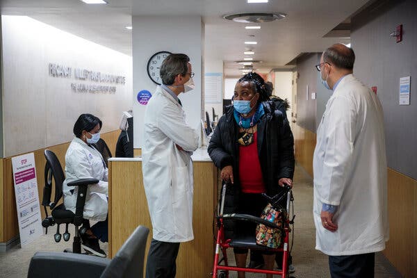
“I can’t do this anymore,” Sonia Sein told herself, her family and her doctor.For six years, she had endured a tube inserted in her windpipe, or trachea, to keep her alive, but her discomfort and distress were becoming more and more unbearable.Largely confined to her Bronx apartment, she needed home health aides and had to quit her career as a social worker for pregnant women. If she talked for over five minutes, she had to stop “because I couldn’t breathe.”The tube was necessary because her trachea — the airway leading to the lungs — had become damaged after she spent weeks on a ventilator for a severe asthma attack in 2014. She had subsequently undergone six major surgeries and more than 10 smaller procedures, but with all conventional approaches to address her condition exhausted, she made plans to have the tube removed and receive only palliative care. “I don’t want to live like this,” she concluded.Today, Ms. Sein, 56, dances and plays tag with her grandchildren and plans to resume working, possibly as an acupuncturist. She says she feels she has been given a “chance of being alive one more time.”Her transformation follows a groundbreaking experimental procedure she underwent in January: the first time, medical experts believe, a donor trachea has been successfully directly transplanted into another person.The 18-hour procedure, conceived and led by Dr. Eric M. Genden Sr., chairman of otolaryngology-head and neck surgery at Mount Sinai Health System in New York, is a milestone because — unlike kidneys, hearts and lungs — the trachea has defied decades of transplantation attempts.“It’s very exciting,” said Dr. G. Alexander Patterson, a professor of surgery at Washington University in St. Louis, who was not involved in the case.Thousands of people in the United States alone develop trachea problems each year from burns, birth defects, tumors and extended intubation on ventilators. The coronavirus pandemic will most likely create more cases because many Covid-19 patients have needed weeks on ventilators.Hundreds of Americans are estimated to die each year because techniques like stents, surgery or lasers cannot heal their damaged tracheas, and they suffocate when airways narrow dangerously or collapse.“There was nothing anybody could do for these patients,” said Dr. Genden, who became captivated by the problem in medical school 30 years ago after a patient with a tracheal tumor died. Guided by several mentors, he delved into research and animal experiments, developing a transplant approach.Because immunosuppressant drugs are required to prevent rejection of the transplant, cancer patients would be eligible only if free of cancer for five years, Dr. Genden said. For other cases, though, doctors say the approach seems promising.“It’s very significant,” said Dr. Pierre Delaere, a professor of head and neck surgery at University Hospital Gasthuisberg in Belgium, a trachea specialist not involved in the case. Still, noting that previous attempts didn’t show documented success, he cautioned that longer-term results were needed before the technique should be embraced, adding, “Let’s see how it works and how you can do it in more patients.”The apparent success of Ms. Sein’s operation is also notable because the trachea field has been rocked for years by a sensational scandal.Ms. Sein speaking with Dr. Eric M. Genden Sr., left, who developed the pioneering procedure, and Dr. Sander S. Florman, director of Mount Sinai’s Transplantation Institute.Sarah Blesener for The New York TimesThat drama began about a decade ago when Dr. Paolo Macchiarini, working at Sweden’s famed Karolinska Institute, garnered headlines and accolades for replacing damaged tracheas with plastic tubes seeded with patients’ stem cells that he cultivated in devices called bioreactors. The transplants, performed on patients from the United States and other countries, were heralded as inaugurating a regenerative medicine revolution.But of 20 patients, including children, most ultimately died, and scientists said Dr. Macchiarini misrepresented data and exaggerated his technique’s effectiveness.A Swedish filmmaker’s investigative documentary raised further questions, as did a Vanity Fair article detailing how the beguiling surgeon became romantically involved with an NBC producer working on a feature about him and apparently conned her into believing he was divorced and would marry her in a ceremony officiated by the pope and attended by the Clintons, Obamas, John Legend and Elton John.The Karolinska Institute, alleging scientific misconduct, dismissed Dr. Macchiarini, who has long denied wrongdoing. Journals retracted several of his studies. In 2019, an Italian court said he had forged documents and abused his position, charges unrelated to his trachea work. In September, a Swedish prosecutor indicted him on aggravated assault charges related to three trachea transplants. The case is pending.Dr. Genden said Dr. Macchiarini’s rise and fall profoundly affected his own path.“Here’s this handsome Italian surgeon at the finest institution in the world, the Karolinska, and he’s everything I’m not: He’s got a beautiful head of hair, he drives a motorcycle, he’s got an accent, he’s incredibly charismatic and dynamic,” Dr. Genden said. “He says, ‘I’ve created this bioreactor and it’s stem cells and it makes tracheas.’ And it’s huge.”Dr. Genden said that when he and colleagues questioned Dr. Macchiarini at a conference early on, “in his bigger-than-life way he says, ‘This is ridiculous, you don’t know what you’re talking about, it functions beautifully.’”Dr. Genden thought his work had “become obsolete, so you basically shut down the lab,” he said. “You can’t justify doing experimental surgery and immunosuppression when you see something else that looks perfect, so you realize, wow, we’re out of business.”As Dr. Macchiarini’s work drew criticism, Dr. Genden revived his idea, but was uncertain about trying it. The scandal meant “there’s an amazing amount of scrutiny,” he said. “We’re going to show up and say, ‘As a student, I had this idea on the back of a napkin and now we’re ready to go’ — and if it fails, the patient dies and it becomes yet another example of some surgeon who thought he could solve a problem and he’s created, instead, just the opposite.”There was another reason to be daunted too: historical assumptions that tracheas weren’t transplantable.“The trachea has been characterized as a simple tube, but it’s very complex,” Dr. Delaere said. About 11 centimeters (4⅓ inches) long, one side curves like a halfpipe, composed of cartilage rings and ligaments. The other side is flat and mobile to move air to the lungs.Video by Mount Sinai HospitalAny replacement trachea must be rigid or “it’ll collapse like a straw in a McDonald’s milkshake,” Dr. Genden said. It must be lined with cilia, hairlike projections “like shag carpeting” that move and clean the air we breathe in, he said. And it needs a blood supply to connect to the patient’s vascular system.Other trachea replacement attempts include transplanting part of a donor’s frozen, preserved aorta, the body’s main artery, and fabricating tracheas from patients’ own chest muscles and rib cartilage.“Some of them have been successful, but they’re cumbersome in different ways and they’re not a trachea,” Dr. Patterson said. “It’s kind of a marginal substitute, and many patients need further interventions to maintain their airways.”Dr. Delaere developed another innovative method, performed on nine patients so far: implanting a section of donor trachea in a patient’s forearm for weeks until it develops blood vessels and can replace part of the patient’s trachea. An advantage is that immunosuppressants are necessary only for several months, he said.But, he added, “It’s an indirect technique.” It takes time and can replace only the trachea’s halfpipe side, “so it’s not a complete tube.”Dr. Genden said some scientists told him “‘You’re out of your mind’” because trying a transplant would risk his reputation. But when Dr. Sander S. Florman, director of Mount Sinai’s Transplantation Institute, and LiveOnNY, a nonprofit organ donation organization, agreed to collaborate, his wife, Audrey, encouraged him.“My wife was the one who said, ‘If you truly believe that people are dying from this and that you could maybe make a difference, you can’t just fold up and go home,’” he said.The “secret sauce” in his approach, he said, is transplanting not just the donor trachea but also its attached esophagus (food tube), thyroid gland and thyroid arteries. That meant the donor trachea was accompanied by a blood supply that Dr. Genden connected to Ms. Sein’s blood vessels.Guided by a high-powered microscope, he used surgical thread half the diameter of a human hair. He opened and cleaned out the donor’s esophagus, laying it against Ms. Sein’s esophagus.“That was a very smart idea to help maintain the fragile blood supply of the airway by leaving the esophagus in place behind it,” Dr. Patterson said.Dr. Genden also transplanted the cricoid, cartilage cuffing the trachea, supplanting Ms. Sein’s completely destroyed cricoid. The nine-centimeter transplant replaced all but two centimeters of her trachea.The donor was a young man (identifying details are being withheld to protect privacy). The different gender was important, allowing Dr. Genden to use chromosomal analysis to detect whether Ms. Sein’s cells populated the new trachea. As of late March, 6.5 percent of cells in the donor trachea were hers, with the proportion increasing, he said.He hopes the immunosuppressant drugs, which can create health risks, can be reduced or even stopped “if the entire graft becomes filled with Sonia’s cells.”Ms. Sein’s trachea before, left, and her new transplanted trachea.Mount SinaiFor Ms. Sein, the procedure was a long-sought dream. In 2017, after another hospital said everything possible had been tried, she felt desperate.“I thought, ‘If they do a transplant for everything, they should do a transplant for trachea,’” she recalled. “I Googled ‘trachea transplant’ and Dr. Genden popped up. So I called and called till I got an appointment.”Dr. Genden said Ms. Sein beseeched him for the transplant, saying, “‘You need to do this for me or I’m going to end my life.’”After realizing other treatment methods wouldn’t help, he concluded that Ms. Sein had a “logical, thoughtful” grasp of transplantation’s risks and of her health condition. He understood how difficult her life had become.At night, when she connected her tracheal tube to a ventilator, “the alarm kept going off to let people know that I stopped breathing,” said Ms. Sein, who hated causing her family concern.Because mucus would clog her tube, “she was constantly suctioning, constantly worried about her airway, and it becomes overwhelming,” Dr. Genden said, like being held down in a swimming pool. “You start to panic because you can’t breathe.”By early 2020, ethical approvals were in place, but the coronavirus pandemic held things up. By this year, Dr. Genden said, her condition had deteriorated so that “if we delay further, it’s not going to work out.”On Jan. 12, Dr. Genden got a call: An appropriate donor had died. The next morning, with the donor and Ms. Sein in adjacent rooms, a team of over 50 medical personnel assembled.“I had a brave face, but I was scared,” Ms. Sein said. She considered backing out.“You got this?” she asked Dr. Genden.“He said ‘yeah,’ so I said ‘Let’s go for it.’”After extracting the donor’s organs, “you go next door and you open up Sonia and you remove the diseased trachea, and there is no going back,” Dr. Genden said. “It’s like when you get on a rocket ship.”At times, he felt “in a dreamlike state,” he said. When the blood vessels were finally connected, the donor trachea began bleeding. “This thing came to life,” Dr. Genden marveled, “and we were like, ‘Holy smokes, we’ve done it.’”Photographs document Ms. Sein’s trachea transformation: Her old windpipe looks raw and red, her new one smooth as porcelain.“I can breathe,” she was amazed to discover. “I could feel it in my lungs.”After several weeks in recovery, she is home. She visits Mount Sinai weekly for blood work. Dr. Genden examines the new trachea by inserting a scope in a hole he left in her neck, which he’ll eventually close.Recently, Ms. Sein, covering the hole with her hand to speak, enthused about having the energy to cook sesame chicken for the first time and how she aims to visit relatives in Puerto Rico. Next month, she will turn 57.“We would have been planning my funeral,” she said, “but now we’re planning a birthday party.”

Read more →

U.S. excess deaths rose nearly 23 percent in 2020, study finds

Extended surges in the South and West in the summer and early winter of 2020 resulted in regional increases in excess death rates, both from COVID-19 and from other causes, a 50-state analysis of excess death trends has found. Virginia Commonwealth University researchers’ latest study notes that Black Americans had the highest excess death rates per capita of any racial or ethnic group in 2020.
The research, publishing Friday in the Journal of the American Medical Association, offers new data from the last 10 months of 2020 on how many Americans died during 2020 as a result of the effects of the pandemic — beyond the number of COVID-19 deaths alone — and which states and racial groups were hit hardest.
The rate of excess deaths — or deaths above the number that would be expected based on averages from the previous five years — is usually consistent, fluctuating 1% to 2% from year to year, said Steven Woolf, M.D., the study’s lead author and director emeritus of VCU’s Center on Society and Health. From March 1, 2020, to Jan. 2, 2021, excess deaths rose a staggering 22.9% nationally, fueled by COVID-19 and deaths from other causes, with regions experiencing surges at different times.
“COVID-19 accounted for roughly 72% of the excess deaths we’re calculating, and that’s similar to what our earlier studies showed. There is a sizable gap between the number of publicly reported COVID-19 deaths and the sum total of excess deaths the country has actually experienced,” Woolf said.
For the other 28% of the nation’s 522,368 excess deaths during that period, some may actually have been from COVID-19, even if the virus was not listed on the death certificates due to reporting issues.
But Woolf said disruptions caused by the pandemic were another cause of the 28% of excess deaths not attributed to COVID-19. Examples might include deaths resulting from not seeking or finding adequate care in an emergency such as a heart attack, experiencing fatal complications from a chronic disease such as diabetes, or facing a behavioral health crisis that led to suicide or drug overdose.

Read more →

Sharing Message of Hope on CBS Sunday Morning

For Easter Sunday, I shared a message of hope with viewers of CBS Sunday Morning. My message: We are at a “love your neighbor” moment with the COVID-19 pandemic, a time when Americans can get vaccinated to help protect others from severe illness and death. The vaccines are a gift to all of us. Do your part. Unwrap the gift, roll up your sleeve, and save lives.

Read more →

New method expands the world of small RNAs

A team led by a biomedical scientist at the University of California, Riverside, has developed a new RNA-sequencing method — “Panoramic RNA Display by Overcoming RNA Modification Aborted Sequencing,” or PANDORA-seq — that can help discover numerous modified small RNAs that were previously undetectable.
RNA plays a central role in decoding the genetic information in DNA to sustain an organism’s life. It is generally known as the intermediate molecule used to synthesize proteins from DNA. Cells are full of RNA molecules in complex and diverse forms, two main types being ribosomal RNA, or rRNA; and transfer RNA, or tRNA; which are involved in the synthesis of proteins.
Small RNAs play essential roles in health and diseases, including cancer, diabetes, neurological diseases, and infertility. Examples of small RNAs are microRNA; piwi-interacting RNA, or piRNA; and tRNA-derived small RNA, or tsRNA. Small RNAs can get modified by chemical groups and thus acquire new functions.
The development of high-throughput RNA sequencing technologies — useful for examining the quantity and sequences of RNA in a biological sample — has uncovered an expanding repertoire of small RNA populations that fine-tune gene expression and protect genomes.
“PANDORA-seq can be widely used to profile small RNA landscapes in various physiological and disease conditions to facilitate the discovery of key regulatory small RNAs involved in these conditions,” said Qi Chen, an assistant professor of biomedical sciences in the UCR School of Medicine, who led the study published today in Nature Cell Biology. “Modified small RNAs wear an ‘invisibility cloak’ that prevents them from being detected by traditional RNA-sequencing methods. How many such modified RNAs are there? What is the origin of their sequences? And what exactly is their biological function? These are questions PANDORA-seq may be able to answer.”
PANDORA-seq employs a stepwise enzymatic treatment to remove key RNA modifications, which then takes off the invisibility cloak used by the modified small RNAs.

Read more →

Masks, ventilation stop COVID spread better than social distancing, study shows

A new study from the University of Central Florida suggests that masks and a good ventilation system are more important than social distancing for reducing the airborne spread of COVID-19 in classrooms.
The research, published recently in the journal Physics of Fluids, comes at a critical time when schools and universities are considering returning to more in-person classes in the fall.
“The research is important as it provides guidance on how we are understanding safety in indoor environments,” says Michael Kinzel, an assistant professor in UCF’s Department of Mechanical and Aerospace Engineering and study co-author.
“The study finds that aerosol transmission routes do not display a need for six feet social distancing when masks are mandated,” he says. “These results highlight that with masks, transmission probability does not decrease with increased physical distancing, which emphasizes how mask mandates may be key to increasing capacity in schools and other places.”
In the study, the researchers created a computer model of a classroom with students and a teacher, then modeled airflow and disease transmission, and calculated airborne-driven transmission risk.
The classroom model was 709 square feet with 9-foot-tall ceilings, similar to a smaller-size, university classroom, Kinzel says. The model had masked students — any one of whom could be infected — and a masked teacher at the front of the classroom.

Read more →