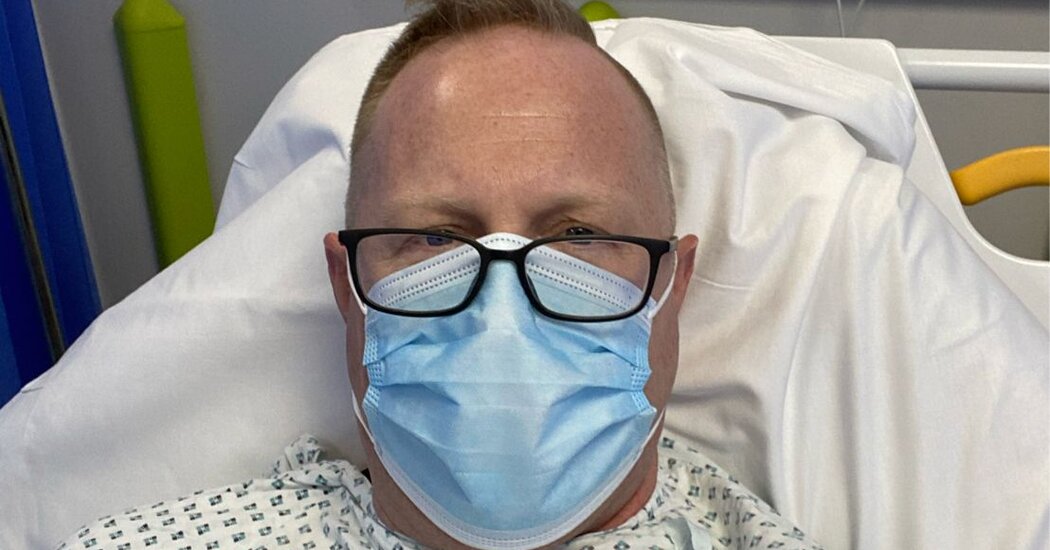
An unvaccinated worker set off an outbreak at a U.S. nursing home where most residents were immunized.

An unvaccinated health care worker set off a Covid-19 outbreak at a nursing home in Kentucky where the vast majority of residents had been vaccinated, leading to dozens of infections, including 22 cases among residents and employees who were already fully vaccinated, a new study reported Wednesday.Most of those who were infected with the coronavirus despite being vaccinated did not develop symptoms or require hospitalization, but one vaccinated individual, who was a resident of the nursing home, died, according to the study released by the Centers for Disease Control and Prevention.Altogether, 26 facility residents were infected, including 18 who had been vaccinated, and 20 health care personnel were infected, including four who had been vaccinated. Two unvaccinated residents also died.The report underscores the importance of vaccinating both nursing home residents and health care workers who go in and out of the sites, the authors said. While 90 percent of the 83 residents at the Kentucky nursing home had been vaccinated, only half of the 116 employees had been vaccinated when the outbreak was identified in March of this year.The study, released in tandem with one involving Chicago nursing homes, underscored the importance of maintaining measures like use of protective gear, infection control protocols and routine testing, no matter the level of vaccination rates. The rise of virus variants also has increased concerns.Resistance to vaccines has been steep among nursing home staffs nationwide, and the low acceptance rates of vaccination increase the likelihood of outbreaks in facilities, according to the authors, a team of investigators from the C.D.C. and Kentucky’s public health department.“To protect skilled nursing facility residents, it is imperative that health care providers, as well as skilled nursing facility residents, be vaccinated,” the authors of the Kentucky study wrote.The outbreak involved a variant of the virus that has multiple mutations in the spike protein, of the kind that make the vaccines less effective. Vaccinated residents and health care workers at the Kentucky facility were less likely to be infected than those who had not been vaccinated, and they were far less likely to develop symptoms. The study estimated that the vaccine, identified as Pfizer-BioNTech, showed effectiveness of 66 percent for residents and 75.9 percent for employees, and were 86 percent to 87 percent effective at protecting against symptomatic disease.In the Kentucky outbreak, the virus variant is not on the C.D.C.’s list of those considered variants of concern or interest. But, the study authors note, the variant does have several mutations of importance: D614G, which demonstrates evidence of increased transmissibility; E484K in the receptor-binding domain of the spike protein, which is also seen in B.1.351, the variant first recognized in South Africa, and P.1. of Brazil; and W152L, which might reduce effectiveness of neutralizing antibodies.In Chicago, meanwhile, routine screening of nursing home residents and staff members identified 627 coronavirus infections in 78 skilled nursing facilities in the city in February, but only 22 were found in individuals who were already fully vaccinated. Two-thirds of the cases in the vaccinated individuals were asymptomatic, the report found, but two residents were hospitalized, and one died.The authors of the Chicago study said their findings demonstrate that nursing homes should continue to follow recommended infection control practices, such as isolation and quarantine, use of personal protective equipment and doing routine testing, regardless of vaccination status.They also emphasized the importance of “maintaining high vaccination coverage among residents and staff members” in order to “reduce opportunities for transmission within facilities and exposure among persons who might not have achieved protective immunity after vaccination.”

Read more →
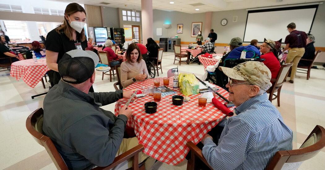
F.D.A. Cites Failures at Plant Where J.&J. Doses Ruined

Federal regulators on Wednesday issued highly critical findings from their inspection of a Baltimore plant that was forced to throw out up to 15 million doses of Johnson & Johnson’s coronavirus vaccine and ordered to temporarily stop all production.The Food and Drug Administration cited a series of shortcomings at the massive plant, which is operated by Emergent BioSolutions. The inspection was triggered by reports that Emergent workers had contaminated a batch of Johnson & Johnson doses with the harmless virus that is used to deliver AstraZeneca’s vaccine, which is also manufactured at the plant.The violations included failure to properly disinfect the factory and its equipment, as well as failure to institute and follow proper procedures designed to prevent contamination of doses and to ensure the strength and purity of the vaccine manufactured there. “There is no assurance that other batches have not been contaminated,” the inspectors wrote.Their 12-page report cited nine violations, ranging from the design of the building to improperly trained employees. The inspection was finished on Tuesday.In a statement, the F.D.A. noted that it has not authorized Emergent to distribute any doses of Johnson & Johnson vaccine, and that no vaccine manufactured at the plant has been released for use in the United States.AstraZeneca’s vaccine is not yet authorized for use in the United States, and all the Johnson & Johnson doses that have been administered in the country so far were manufactured overseas. At the agency’s request, all production at the factory has been halted.“We will not allow the release of any product until we feel confident that it meets our expectations for quality,” the statement from Dr. Janet Woodcock, the F.D.A.’s acting commissioner, and Dr. Peter Marks, the agency’s top vaccine regulator, said.The agency said it was working with Emergent to fix the problems.Emergent is a longtime government contractor that has spent much of the last two decades cornering a lucrative market in federal spending on biodefense. The company’s Baltimore plant is one of two federally designated “Centers for Innovation in Advanced Development and Manufacturing” that were supposed to be at the ready in the event of a pandemic.The New York Times reported earlier this month that the Trump administration awarded a $628 million contract to the company about ten months ago despite a history of performance problems. The contract mainly allowed the government to reserve manufacturing space in the plant for vaccine production. On Tuesday, the House Select Subcommittee on the Coronavirus Crisis, announced that it will investigate that contract award and other issues involving Emergent, saying the firm had a track record of failing to meet contract requirements.The inspectors castigated Emergent’s response to the discovery last month that Johnson & Johnson doses had been contaminated with the benign virus used to create AstraZeneca’s vaccine. The incident “has not been fully investigated,” they wrote.They said Emergent did not thoroughly review whether the cross-contamination was caused by one or more workers who move between AstraZeneca’s and Johnson & Johnson’s manufacturing zones or whether it could be related to how raw materials used in the production of both vaccines are handled.The inspectors found that workers frequently moved between the manufacturing zones without documenting that they had showered and changed their gowns as required. In one ten-day period in February, for instance, 13 employees moved from one zone to another on the same day, but only one documented having showered, they said. After the Johnson & Johnson doses were found to be contaminated, the report said, only routine cleaning was performed.Workers also failed to properly handle manufacturing waste, creating risks of contamination in the warehouse where raw materials are stored, the inspectors found. They also cited peeling paint, crowded equipment and other issues with the building. Overall, it “is not maintained in a clean and sanitary condition,” they wrote.Emergent said in a statement on Wednesday that “while we are never satisfied to see shortcomings in our manufacturing facilities or process, they are correctable and we will take swift action to remedy them.”In its own statement, Johnson & Johnson said it had already stepped up its oversight of Emergent, its subcontractor, and that it would “ensure that all of F.D.A.’s observations are addressed promptly and comprehensively.”One major change has already been made: AstraZeneca will no longer be manufactured at the plant, a move that federal officials insisted upon earlier this month to limit the chance of cross-contamination between two vaccines.Dr. Jose Romero, the Arkansas health secretary and chairman of the expert panel advising the Centers for Disease Control and Prevention on the future of the Johnson & Johnson vaccine, said in an interview that he was dismayed by the F.D.A.’s findings. The panel is meeting on Friday on whether to lift, modify or retain a pause in the administration of Johnson & Johnson’s vaccine that was instituted last week for an entirely different issue: the discovery that eight U.S. residents developed a rare but dangerous blood clotting disorder after they got shots.“I’m shocked” Dr. Romero said. “I can’t put it any other way. Inappropriate disinfection, the prevention of contamination — those are significant and serious violations, at least in my mind, and do of course need to be remedied.“I would not have expected that, given the stringency that we have in this country for good manufacturing practices in these vaccine plants,” he said.

Read more →

Wildfire smoke linked to skin disease

Wildfire smoke can trigger a host of respiratory and cardiovascular symptoms, ranging from runny nose and cough to a potentially life-threatening heart attack or stroke. A new study suggests that the dangers posed by wildfire smoke may also extend to the largest organ in the human body, and our first line of defense against outside threat: the skin.
During the two weeks in November 2018 when wildfire smoke from the Camp Fire choked the San Francisco Bay Area, health clinics in San Francisco saw an uptick in the number of patients visiting with concerns of eczema, also known as atopic dermatitis, and general itch, compared to the same time of the year in 2015 and 2016, the study found.
The findings suggest that even short-term exposure to hazardous air quality from wildfire smoke can be damaging to skin health. The report, carried out by physician researchers at the University of California, San Francisco, in collaboration with researchers at the University of California, Berkeley, appears on April 21 in the journal JAMA Dermatology.
“Existing research on air pollution and health outcomes has focused primarily on cardiac and respiratory health outcomes, and understandably so. But there is a gap in the research connecting air pollution and skin health,” said study lead author Raj Fadadu, a student in the UC Berkeley-UCSF Joint Medical Program. “Skin is the largest organ of the human body, and it’s in constant interaction with the external environment. So, it makes sense that changes in the external environment, such as increases or decreases in air pollution, could affect our skin health.”
Air Pollutants Can Slip through Skin Barriers
Air pollution from wildfires, which consists of fine particulate matter (PM2.5), polycyclic aromatic hydrocarbons (PAHs), and gases, can impact both normal and eczema-prone skin in a variety of ways. These pollutants often contain chemical compounds that act like keys, allowing them to slip past the skin’s outer barrier and penetrate into cells, where they can disrupt gene transcription, trigger oxidative stress or cause inflammation.

Read more →

Changing views on atherosclerosis

Peter Libby, MD, cardiovascular medicine specialist at Brigham and Women’s Hospital and the Mallinckrodt Professor of Medicine at Harvard Medical School; author of a new review paper published in Nature.
Atherosclerosis — hardening of the arteries — is now involved in the majority of deaths worldwide, and advances in our understanding of the biology of the disease are changing traditional views and opening up new avenues for treatment.
The picture of who may be at risk for a heart attack has evolved considerably in recent decades. At one time, a heart attack might have conjured up the image of a middle-aged white man with high cholesterol and high blood pressure who smoked cigarettes. Today, traditional concepts of what contributes to risk have changed. These updated views include new thinking around: Global disease burden: Atherosclerotic cardiovascular disease is now the leading cause of death worldwide. Clinical profile: Women, younger individuals, and people of diverse backgrounds bear an increasing burden of atherosclerotic cardiovascular disease. Role of “good cholesterol”: The protective role of HDL cholesterol (so-called “good cholesterol”) has been called into question and triglycerides have emerged as a promising target for reducing heart disease risk. Inflammation drives atherosclerosis: New data suggests that inflammation may be a critical link between traditional risk factors such as abnormal lipids, smoking, and diabetes and complications of atherosclerosis including heart attack and stroke.”Advances in our understanding of the biology of atherosclerosis have opened avenues to therapeutic interventions that promise to improve the prevention and treatment of now-ubiquitous atherosclerotic diseases,” writes Libby. “From a therapeutic perspective, we have reason for optimism in addressing the growing burden of atherosclerotic risk.”
Story Source:
Materials provided by Brigham and Women’s Hospital. Note: Content may be edited for style and length.

Read more →

SARS-CoV-2: Infection induces antibodies capable of killing infected cells regardless of disease severity

Drawing on epidemiological field studies and the FrenchCOVID hospital cohort coordinated by Inserm, teams from the Institut Pasteur, the CNRS and the Vaccine Research Institute (VRI, Inserm/University Paris-Est Créteil) studied the antibodies induced in individuals with asymptomatic or symptomatic SARS-CoV-2 infection. The scientists demonstrated that infection induces polyfunctional antibodies. Beyond neutralization, these antibodies can activate NK (natural killer) cells or the complement system, leading to the destruction of infected cells. Antibody levels are slightly lower in asymptomatic as opposed to symptomatic individuals, but polyfunctional antibodies were found in all individuals. These findings show that infection induces antibodies capable of killing infected cells regardless of the severity of the disease. The research was published in the journal Cell Reports Medicine on April 21, 2021.
Nearly half of those infected with SARS-CoV-2 do not develop symptoms. Yet, the immune response induced by asymptomatic forms of COVID-19 remains poorly characterized. The extent of the antiviral functions of SARS-CoV-2 antibodies is also poorly characterized. Antibodies are capable of both neutralizing the virus and activating “non-neutralizing” functions. The latter include antibody-dependent cellular cytotoxicity (ADCC) and complement activation, which are major components of the immune response and play a key role in the efficacy of some vaccines. ADCC is a two-stage process in which infected cells are first recognized by antibodies, then destroyed by NK cells. The complement system consists of a series of plasma proteins that also enable the elimination of cells targeted by antibodies. The ability of antibodies to activate these non-neutralizing functions has been little described for SARS-CoV-2 infection so far.
The teams from the Institut Pasteur, the CNRS and the VRI (Inserm/University Paris-Est Créteil) initially developed new assays to measure the various antibody functions. They produced assays to study cell death induced by NK cells or by complement in the presence of antibodies. By analyzing cultures in real time using video microscopy, the scientists showed that NK cells kill infected cells in the presence of antibodies, demonstrating new antiviral activity employed by SARS-CoV-2 antibodies.
The scientists then examined the serum of patients with symptomatic or asymptomatic forms of COVID-19 with their new assays. They also used methods previously developed at the Institut Pasteur, such as the S-Flow assay, to detect SARS-CoV-2 anti-spike antibodies, and the S-Fuse assay, to measure the neutralization capacity of these antibodies.
“This study demonstrated that individuals infected with SARS-CoV-2 have antibodies that are capable of attacking the virus in different ways, by preventing it from entering cells (neutralization) or by activating NK cells to kill infected cells (via ADCC). We therefore use the term polyfunctional antibodies,” explains Timothée Bruel, co-last author of the study and a scientist in the Institut Pasteur’s Virus & Immunity Unit and at the VRI.
By comparing different groups of patients, the scientists then showed that asymptomatic individuals also have polyfunctional antibodies and that their response is slightly weaker than those of patients with moderate forms of COVID-19.
“The study reveals new mechanisms of action of SARS-CoV-2 antibodies and suggests that the protection induced by an asymptomatic infection is very close to that observed after a symptomatic infection,” concludes Olivier Schwartz, co-last author of the study, head of the Virus & Immunity Unit and at the VRI.
Story Source:
Materials provided by Institut Pasteur. Note: Content may be edited for style and length.

Read more →

Prevalence of COVID-19 symptoms among high-risk children

Children with weakened immune systems have not shown a higher risk of developing severe COVID-19 infection despite commonly displaying symptoms, a new study suggests.
During a 16-week period which covered the first wave of the pandemic, researchers from Southampton carried out an observational study of nearly 1500 immunocompromised children — defined as requiring annual influenza vaccinations due to underlying conditions or medication. The children, their parents or guardians completed weekly questionnaires to provide information about any symptoms they had experienced, COVID-19 test results and the impact of the pandemic on their daily life.
The results, published in BMJ Open, showed that symptoms of COVID-19 infection were common in many of the children — with over two thirds of participants reporting at least one symptom and one third experiencing three or more symptoms simultaneously. One hundred and ten patients with symptoms undertook viral PCR tests, none of whom tested positive.
Dr Hans de Graaf from the University of Southampton who led the research said, “Whilst we cannot be certain of the prevalence of COVID-19 amongst the children who took part, because testing was only done when patients were admitted and these children were told to adhere to strict shielding measures, we can assume that any infections would have been mild cases since none of these high risk patients required hospital admissions.”
More than half of the patients or parents reported high levels of anxiety at the start of the study and despite the absence of severe symptoms, these scores remained consistently high throughout the study period.
The researchers believe that these results show that widespread symptom screening for early detection of COVID-19 in not going to be useful in these cases as the children may have frequent upper respiratory tract symptoms likely to be unrelated to COVID-19.
Dr de Graaf continued, “This study was the first to observe the impact of the pandemic on children with compromised immune systems. During the first wave of the pandemic, many may have been shielding so our results suggest that either the shielding measures were effective or that immunocompromised children are less affected by COVID-19 than adults, just like healthy children.”
The report also concludes that the continuous high level of anxiety among participants highlights the need to clearly define and communicate the risk of COVID-19 in children and young people, particularly as lockdown restrictions ease.
Story Source:
Materials provided by University of Southampton. Note: Content may be edited for style and length.

Read more →

Improved management of farmed peatlands could cut 500 million tons CO2

Substantial cuts in global greenhouse gas emissions could be achieved by raising water levels in agricultural peatlands, according to a new study in the journal Nature.
Peatlands occupy just three per cent of the world’s land surface area but store a similar amount of carbon to all terrestrial vegetation, as well as supporting unique biodiversity.
In their natural state, they can mitigate climate change by continuously removing CO2 from the atmosphere and storing it securely under waterlogged conditions for thousands of years.
But many peatland areas have been substantially modified by human activity, including drainage for agriculture and forest plantations. This results in the release, from drained peatlands, of the equivalent of around 1.5 billion tonnes of carbon dioxide (CO2) into the atmosphere each year — which equates to three per cent of all global greenhouse gas (GHG) emissions caused by human activities.
A team of scientists, led by the UK Centre for Ecology and Hydrology (UKCEH), estimated the potential reduction in emissions by restoring all global agricultural peatlands. However, because large populations rely on these areas for their livelihoods, it may not be realistic to expect all agricultural peatlands to be fully rewetted and returned to their natural condition in the near future.
The team therefore also analysed the impact of halving current drainage depths in croplands and grasslands on peat — which cover over 250,000km2 globally — and showed that this could still bring significant benefits for climate change mitigation. The study estimates this could cut emissions by around 500 million tonnes of CO2 a year, which equates to 1 per cent of all global GHG emissions caused by human activities.

Read more →

How SARS coronaviruses reprogram host cells to their own benefit

Coronavirus researchers led by Professor Rolf Hilgenfeld of the University of Luebeck and PD Dr. Albrecht von Brunn of the Ludwig-Maximilian Universitaet (LMU) in Munich have discovered how SARS viruses enhance the production of viral proteins in infected cells, so that many new copies of the virus can be generated. Notably, coronaviruses other than SARS-CoV and SARS-CoV-2 do not use this mechanism, which may therefore provide a possible explanation for the much higher pathogenicity of the SARS viruses. The findings appear in the EMBO Journal.
Coronaviruses that cause harmless colds in humans were discovered more than 50 years ago. When it emerged in 2002/2003, the SARS coronavirus was the first coronavirus found to cause severe pneumonia in infected people. Comparisons of the RNA genomes of innocuous coronaviruses with those of the SARS coronavirus permitted researchers to identify a region that only occurred in the latter, and was called the “SARS-unique domain” (SUD). Such genomic regions and their protein products might be linked to the extraordinary pathogenicity of SARS coronavirus and its cousin, the COVID-19 virus SARS-CoV-2.
The research groups led by Hilgenfeld and von Brunn showed that the SUD proteins of these two viruses interact with a human protein called Paip-1, which is involved in the first steps of protein synthesis. Together with Paip-1 and other proteins in human cells, SUD apparently binds to the ribosomes, the molecular machines that are responsible for protein synthesis in cells. This would lead to an enhancement of the production of all proteins, both those of the host cell and those of the virus. However, in cells infected with SARS-CoV or SARS-CoV-2, the messenger RNA molecules that code for host proteins are selectively destroyed by a viral protein named Nsp1. As a result of this complicated process, the infected cell predominantly produces viral proteins, so that many new copies of the virus can be created.
Albrecht von Brunn’s research group discovered the interaction between the proteins SUD and Paip-1 several years ago. “Being an experienced coronavirologist, I knew that one has to inspect the special regions of the SARS genome when trying to understand this virus,” he says.
The discovery made by the Munich researchers was of great interest to Hilgenfeld, whose research group had already elucidated the three-dimensional structure of the SUD protein some years previously. The two research groups teamed up. Dr. Jian Lei in Hilgenfeld’s group, meanwhile a group leader at Sichuan University in Chengdu (China), succeeded in crystallizing the complex formed by SUD and Paip-1 and determining its three-dimensional structure by X-ray crystallography. And co-first author Dr. Yue “Lizzy” Ma-Lauer of von Brunn’s group characterized the complex of the two proteins and its function using a variety of cell-biological and biophysical methods.
“Interaction studies of this kind between coronavirus proteins and proteins of the infected human cell will help us understand how the viruses change key functions of the cell to their own benefit,” says Hilgenfeld. The project was supported by the German Federal Ministry of Education and Research (BMBF) and by the German Center for Infection Research (DZIF).
Story Source:
Materials provided by Ludwig-Maximilians-Universität München. Note: Content may be edited for style and length.

Read more →

Bypassing broken genes

A new approach to gene editing using the CRISPR/Cas9 system bypasses disease-causing mutations in a gene, enabling treatment of genetic diseases linked to a single gene, such as cystic fibrosis, certain types of sickle cell anemia, and other rare diseases. The method, developed and tested in mice and human tissue cultures by researchers at Penn State, involves inserting a new, fully functional copy of the gene that displaces the mutated gene.
A proof-of-concept for the approach is described in a paper appearing online April 20 in the journal Molecular Therapy.
The CRISPR/Cas9 system has allowed promising new gene therapies that can target and correct disease-causing mutations in a gene. In this process, Cas9 — a bacterial protein — cuts DNA at a specific location, where the genetic sequence can then be edited, trimmed, or a new sequence inserted before the DNA is repaired. However, there are two main limitations to current repair strategies. First, the common repair strategy, called “homology-directed repair,” requires using specific proteins within the cell that are only present during cell division, which means the gene repair process cannot be used in most adult tissues where cell division occurs rarely.
“The second challenge stems from the fact that even when a disease is caused by a single gene, it can result from a variety of different mutations within that gene,” said Douglas Cavener, professor of biology at Penn State and senior author of the paper. “With homology-directed repair, we’d need to design and test the strategy for each and every one of those mutations, which can be expensive and time-intensive. In this study, we designed an approach called Co-opting Regulation Bypass Repair (CRBR), which can be used in both dividing and non-dividing cells and tissues and for a spectrum of mutations within a gene. This approach is especially promising for rare genetic diseases caused by a single gene, where limited time and resources typically preclude design and testing for the many possible disease-causing mutations.”
CRBR takes advantage of the CRISPR/Cas9 system and a cellular repair pathway called “non-homologous end joining” to insert a genetic sequence between a mutated gene’s promoter region — the genetic sequence that controls when and where the gene is functional — and the mutated portion of the gene. The newly inserted sequence contains a condensed version of the normal gene that is used in place of the mutated version. A terminator sequence at the end of the inserted sequence prevents the remaining downstream mutated gene from being used. Because CRBR does not rely on the proteins required by homology-directed repair, it can be used in all types of adult tissues.
“Our approach co-opts the native promoter for a gene,” said Jingjie Hu, a graduate student at Penn State and first author of the paper. “This means that the newly inserted gene will be expressed at the same times and at appropriate levels within the cell as the gene it is replacing. This is an advantage to other types of gene therapies, which rely on an external promoter to drive high levels of expression of the gene that could lead to negative effects if too much is produced or if essential regulation response is missing under certain physiological conditions.”
The research team conducted a series of proof-of-concept experiments to demonstrate the utility of this method. They first focused on the PERK gene, mutations in which can lead to a rare disease called Wolcott-Rallison syndrome. The syndrome results when copies of the gene inherited from both parents have mutations — it is a “recessive” disease — and can cause neonatal diabetes, skeletal problems, growth delay, and other symptoms.

Read more →