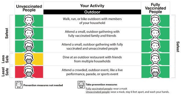
Brain clots 'more likely' with Covid infection than vaccine

SharecloseShare pageCopy linkAbout sharingimage copyrightGetty ImagesThe risk of developing a serious brain clot – known as a cerebral venous sinus thrombosis (CVST) – is 8 to 10 times higher in people with Covid than those who get a vaccine, a study suggests.Based on US data, the Oxford research team says people being vaccinated should be reassured by the findings.It follows investigations into links between the AstraZeneca vaccine and rare blood clots.The study only looked at those who had had a Pfizer or Moderna vaccine.The research, which involved electronic health records of 81 million people in the US, looked at the number of CVST cases seen in the two weeks following a diagnosis of coronavirus and the number of cases occurring in the two weeks after people had their first coronavirus vaccine.It estimates that while these blood clots are uncommon after Covid – with 39 in every million people developing one within two weeks of being ill – they are much rarer still after a vaccine.’Work in progress’But researchers say their study – which has not been through a formal review and is separate from the Oxford vaccine group – is still a work in progress and must be interpreted cautiously because it is difficult to calculate with certainty how common CVSTs are in the general population, partly because of just how rare they are. The study also found:Clots were more common in people who already had cardiovascular disease80% of people who have the clots surviveSome cases were seen in under-30s, showing they are not immune to serious complications from coronavirusIn those who had an mRNA vaccine – such as the Pfizer or Moderna jab – they estimate CVSTs occurred in around four in a million people. Scientists say their study cannot identify whether vaccines are linked to these clots and much larger studies are needed to address this. They say a more complete database would be needed because as in cases it was unclear exactly which mRNA vaccine had been given There are no directly comparable figures for the AZ vaccine because this jab has not been used in the USThe European Medicines Agency says a particular type of CVST occurs in around five in a million people after the first dose of the Oxford-AstraZeneca vaccine – but the populations vaccinated were not the same as in the US and the rates cannot be compared.Paul Harrison, professor of psychiatry at the University of Oxford, said their study had important conclusions for people deciding on vaccines.He said: “Firstly, Covid-19 markedly increases the risk of CVST, adding to the list of blood clotting problems this infection causes. “Secondly, the Covid-19 risk is higher than seen with the current vaccines, even for those under 30; something that should be taken into account when considering the balances between risks and benefits for vaccination.”Rare blood clots – what you need to knowWhere is there surge testing for Covid variants?Is the Oxford-AstraZeneca vaccine safe?From their database they were unable to investigate whether the CVST clots they were seeing had similar features to those seen in rare cases after vaccines – which all had a peculiar pattern of blood cells associated with them, where certain cells called platelets were diminished.Prof Beverley Hunt, of Thrombosis UK, said the mechanisms behind people getting clots after Covid and those experiencing clots after vaccines were likely to be different. She said: “Patients who are hospitalised with Covid-19 have very pro-thrombotic (sticky) changes in their blood, which persist after they have been discharged. This will lead to an increased rate of blood clots. “The mechanism for the very rare blood clots and low platelet counts seen after the AstraZeneca vaccine is different. It is associated with an immune response.”Related Internet LinksOSF COVID-CVT-paper.pdf.websiteThe BBC is not responsible for the content of external sites.

Read more →

Impacts of coronavirus lockdowns: New study collects data on pollutants in the atmosphere

One consequence of the coronavirus pandemic has been global restrictions on mobility. This, in turn, has had an effect on pollution levels in the atmosphere. Researchers from across the world are using this unique opportunity to take measurements, collect data, and publish studies. An international team led by Forschungszentrum Jülich’s Institute of Climate and Energy Research — Troposphere has now published a comprehensive review providing an overview of results up to September 2020. The study also has its own dedicated website, where additional measurement data can be added to supplement and refine existing research results. At the same time, this collection of data allows scientifically substantiated predictions to be made about the pollution levels of future mobility scenarios.
The meta-analysis was coordinated by Prof. Astrid Kiendler-Scharr, director at Jülich’s Institute of Climate and Energy Research — Troposphere. The analysis covers the measurement data of around 200 studies from the first seven months following the onset of the pandemic. It focuses on the following air pollutants: nitrogen dioxide, particulate matter, ozone, ammonia, sulfur dioxide, black carbon, volatile organic compounds (VOCs), and carbon monoxide. A third of the studies take into account the prevailing meteorological situation when calculating the influence of lockdowns on the air composition. The Government Stringency Index (SI) — summarizing the severity of local shutdown measures in a number that can be compared at international level — acted as a reference value.
A key finding of the analysis is that lockdowns, which have the sole aim of slowing down the infection rate, are also reducing the global pollution of the atmosphere with nitrogen dioxide and particulate matter — the higher the SI, the greater this impact. However, this only applies to pollutants that primarily have an anthropogenic origin, i.e. are directly emitted by humans, especially in the field of mobility. In contrast, ozone levels increased. This increase was a result of atmospheric chemical processes caused by reduced nitrogen oxide levels in the air.
The study also highlights current gaps in the data collection and the need for further research. The authors are therefore of the opinion that the period of analysis should be extended to cover the entire year of 2020. The scientists place a particular emphasis on hydrocarbons, which have so far only been examined sporadically in studies, and on extended analyses looking at the impact of emission changes on the climate.
An important addition to the meta-analysis is a database that can be accessed via a website (COVID-19 Air Quality Data Collection — https://covid-aqs.fz-juelich.de/). It contains all data from the study on pollution levels, including data on pollutions levels in individual countries. Researchers can also find a list of publications to date and thus obtain a quick overview of previous studies.
The website also invites scientists to present data from their new studies and to thus become part of the reference system. It therefore acts as a “living version,” with the presentation of collected results being constantly refined. Similarly, there are plans to further develop the data collection to include measurement results and the analysis of other pollutants that are not part of the current canon, for example hydrocarbons.
The important data could also form the basis for better assessments of the impacts on atmospheric chemistry in future scenarios. This includes a considerable, long-term reduction in pollution levels for a comprehensive transition to electromobility.
Story Source:
Materials provided by Forschungszentrum Juelich. Note: Content may be edited for style and length.

Read more →

In surprising twist, some Alzheimer's plaques may be protective, not destructive

One of the characteristic hallmarks of Alzheimer’s disease (AD) is the buildup of amyloid-beta plaques in the brain. Most therapies designed to treat AD target these plaques, but they’ve largely failed in clinical trials. New research by Salk scientists upends conventional views of the origin of one prevalent type of plaque, indicating a reason why treatments have been unsuccessful.
The traditional view holds that the brain’s trash-clearing immune cells, called microglia, inhibit the growth of plaques by “eating” them. The Salk scientists show instead that microglia promote the formation of dense-core plaques, and that this action sweeps wispy plaque material away from neurons, where it causes cell death. The research, which was published in Nature Immunology on April 15, 2021, suggests that dense-core plaques play a protective role, so treatments to destroy them may do more harm than good.
“We show that dense-core plaques don’t form spontaneously. We believe they’re built by microglia as a defense mechanism, so they may be best left alone,” says Greg Lemke, a professor in Salk’s Molecular Neurobiology Laboratory. “There are various efforts to get the FDA to approve antibodies whose main clinical effect is reducing dense-core plaque formation, but we make the argument that breaking up the plaque may be doing more damage.”
Alzheimer’s disease is a neurological condition that results in memory loss, impairment of thinking, and behavioral changes, which worsen as we age. The disease seems to be caused by abnormal proteins aggregating between brain cells to form the hallmark plaques, which interrupt activity that keeps the cells alive.
There are numerous forms of plaque, but the two most prevalent are characterized as “diffuse” and “dense-core.” Diffuse plaques are loosely organized, amorphous clouds. Dense-core plaques have a compact center surrounded by a halo. Scientists have generally believed that both types of plaque form spontaneously from excess production of a precursor molecule called amyloid precursor protein (APP).
But, according to the new study, it is actually microglia that form dense-core plaques from diffuse amyloid-beta fibrils, as part of their cellular cleanup.

Read more →

Estrogen status – not sex – protects against heightened fear recall

A new study shows that markers of fear recall differ between men and women, but in a hormone-dependent manner.
Aberrant fear-memory processing in the brain is thought to underlie anxiety disorders, which affect hundreds of millions of people worldwide. The neurobiological mechanisms underlying these disorders remain poorly understood, but recent studies suggest that neural oscillations in the prefrontal cortex can reflect the strength of fear recall activity, providing a physiological measure.
Women suffer from anxiety disorders at twice the rate of men and indeed the literature shows that there are sex differences in fear recall behaviors, but this area of study has not been extended to neural oscillations. Additional studies suggest a modulatory role for the female sex hormone estradiol (E2) for fear recall and extinction recall.
The new study led by Ursula Stockhorst, PhD, at the University of Osnabrück, Germany, specifically shows that peripheral and brain markers of fear recall differ in a hormone-dependent manner between men and women.
The work appears in Biological Psychiatry: Cognitive Neuroscience and Neuroimaging, published by Elsevier.
Cameron Carter, MD, Editor of Biological Psychiatry: Cognitive Neuroscience and Neuroimaging, said of the work: “This study sheds light on the well-known differences between men and women in their vulnerability to anxiety disorders and shows that aspects of fear learning and extinction that contribute to vulnerability in women are related to differences in estrogen levels.”
For the study, first author and PhD student Philipp Bierwirth, MSc, and colleagues examined 20 men, 20 women using oral hormonal contraceptives (OC) and 20 free-cycling women during their mid-cycle (MC). Women taking OC have suppressed and thus low endogenous E2 levels, whereas free-cycling MC women have higher levels of E2.
Participants underwent a fear-conditioning paradigm in which two of four photographs of neutral male faces were paired with a loud burst of white noise. Following the conditioning, the subjects underwent fear extinction, in which two photographs — only one of which had been paired with the noise — were again presented, but this time with no noise stimulus. The next day, subjects were shown all four photographs again, including the two that had been presented in the extinction phase and thus ready for extinction recall, and two of them without previous extinction — thus subject to fear recall. During all presentations, the researchers measured skin conductance responses (SCR), a peripheral readout of fear expression, and brain oscillations measured by electroencephalography.
Mr. Bierwirth said: “We found stronger peripheral fear expression (via SCR) during fear recall and extinction recall under low-E2 conditions, that is, in men and in OC women, compared to mid-cycle women with higher E2 levels. Most importantly, we also observed enhanced theta oscillations in the medial prefrontal cortex and especially in the dorsal anterior cingulate cortex (dACC), in men and OC women compared to MC women.”
Importantly, the authors also point out that subjects were examined during their natural E2 status. They were not randomized to experimentally manipulated estrogen levels and so causal inferences about estrogen cannot be drawn.
Fear recall-related dACC theta oscillations were attenuated in women with higher E2 levels, which, importantly, supports previous findings suggesting a protective role for E2 against fear overexpression during the recall of fear and extinction memories. The data demonstrate that peripheral and brain oscillatory correlates of fear memory recall do not differ between the sexes per se but vary with E2 status, even among women.
Story Source:
Materials provided by Elsevier. Note: Content may be edited for style and length.

Read more →

Patients who are overweight or obese at risk of more severe COVID-19, study finds

Patients who are overweight or obese have more severe COVID-19 and are highly likely to require invasive respiratory support, according to a new international study.
The research, led by the Murdoch Children’s Research Institute (MCRI) and The University of Queensland and published in Diabetes Care, found obese or overweight patients are at high risk for having worse COVID-19 outcomes. They are also more likely to require oxygen and invasive mechanical ventilation compared to those with a healthy weight.
MCRI researcher Dr Danielle Longmore said the findings, which highlighted the relationship between obesity and increased COVID-19 disease burden, showed the need to urgently introduce strategies to address the complex socio-economic drivers of obesity, and public policy measures such as restrictions on junk food advertising.
“Although taking steps to address obesity in the short-term is unlikely to have an immediate impact in the COVID-19 pandemic, it will likely reduce the disease burden in future viral pandemics and reduce risks of complications like heart disease and stroke,” she said.
The study looked at hospitalised SARS-CoV-2 patients from 18 hospitals in 11 countries including China, America, Italy, South Africa and The Netherlands.
Among the 7244 patients aged 18 years and over, 34.8 per cent were overweight and 30.8 per cent were obese.

Read more →

The architect of genome folding

The DNA molecule is not naked in the nucleus. Instead, it is folded in a very organized way by the help of different proteins to establish a unique spatial organization of the genetic information. This 3D spatial genome organization is fundamental for the regulation of our genes and has to be established de novo by each individual during early embryogenesis. Researchers at the MPI of Immunobiology and Epigenetics in Freiburg in collaboration with colleagues from the Friedrich Mischer Institute in Basel now reveal a yet unknown and critical role of the protein HP1a in the 3D genome re-organization after fertilisation. The study published in the scientific journal Nature identifies HP1a as an epigenetic regulator that is involved in establishing the global structure of the genome in the early Drosophila embryo.
The information of the human genome is encoded by approximately 3 billion DNA base pairs and packaged into 23 pairs of chromosomes. If all chromosomes could be disentangled and linearly aligned, they would be a thin thread of about 2 meters. The DNA molecule must be extensively packaged to fit inside the nucleus, the size of which is in the micrometer range. “The DNA thread is not simply stuffed into the cell nucleus. Instead, it is folded in a very organized way to ensure that different parts of the genome, sometimes several thousand base pairs away from each other, can intercommunicate for appropriate gene functions,” says Nicola Iovino, group leader at the MPI of Immunobiology and Epigenetics in Freiburg.
Part of this packaging are histone proteins acting as spools around which DNA is winded and thereby compacted. This complex of DNA and proteins is called chromatin. As such, chromatin is the fundament for further packaging of the genetic material into chromosomes whose structure is mostly known for its characteristic cross shape. The chromosomes themself occupy distinct positions within the nucleus, known as chromosome territories, that also enable efficient packaging and organization of the genome.
The full machinery contributing to this 3D chromatin organization remains unexplored. Now the lab of Nicola Iovino at the MPI in Freiburg, in collaboration with Luca Giorgetti from the Friedrich Miescher Institute in Basel (Switzerland), was able to show the fundamental role of the heterochromatin protein 1a (HP1a) in the reorganization of the 3D chromatin structure after fertilization. By combining powerful Drosophila genetics with 3D genome modeling, they discovered that HP1a is required to establish a proper chromatin 3D structure at multiple hierarchical levels during early embryonic development.
Early embryos as a model to study chromatin reprogramming
The degree of packaging as well as the corresponding gene activity is influenced by epigenetic modifications. These are small chemical groups that are installed on the histones. “Proteins that carry out these epigenetic modifications can be thought of as being either writers, erasers or reader of the given epigenetic modification. We discovered that the reader protein HP1a is required to establish chromatin structure during early embryonic development in Drosophila,” says Fides Zenk, first-author of the study.

Read more →

New 'time machine' technique to measure cells

Using a new single-cell technique, WEHI researchers have uncovered a way to understand the programming behind how stem cells make particular cell types.
The research uncovered 30 new genes that program stem cells to make the dendritic cells that kick-start the immune response.
By uncovering this process, the researchers hope they will be able to find new immunotherapy treatments for cancer, and plan to expand this technique in other areas such as discovering new drug targets in tumour initiation.
At a glance WEHI researchers have developed a new single cell method to understand the programming behind what causes stem cells to make particular cell types. By testing daughters of a single stem cell in different parallel tests, researchers found 500 genes that predicted dendritic cell fate. Using a CRISPR screen, they discovered 30 key genes amongst the 500 that program dendritic cell production. Researchers intend to expand use of this technique to find the ‘big bang’ moment in cancer development to identify new drug targets to fight cancer.Studying ‘sister’ cells
Led by Dr Shalin Naik, Dr Luyi Tian, Ms Sara Tomei and Mr Jaring Schreuder and published in Immunity, the research outlined the processes involved in kick-starting the generation of dendritic cells driven by the hormone Flt3 ligand, which is used in immunotherapy.

Read more →

Objective analysis of stress in the classroom

Is it the difficulty of a task that determines whether or not students are stressed when working on it? Biologists working in biology didactics set out to find out the answers; to this end, the team used questionnaires and measured the heart rate in 209 test participants.
“This enables us to contrast the subjective perception of stress with an objective measurement method and compare the two,” explains Nina Minkley. Contrary to expectations, it turned out that the effort invested in the task does not increase with its difficulty, nor does the stress level. The study was featured in the journal Frontiers in Education.
Simple questionnaire surveys criticised
To date, the stress experience of students has mostly been surveyed with questionnaires. But this approach has been criticised, because many factors have an effect on one’s own perception that have nothing to do with the task. “For example, women often report higher stress levels than men,” points out Nina Minkley. The researchers can only speculate why this is the case. In the current study, they used an objective method of measuring stress levels.
They equipped 209 secondary school students who were working on biology tasks with chest straps that monitor the heart rate. They also had the participants fill out several questionnaires on their self-concept, their interest in biology and their perception of the tasks. “When we are relaxed, the individual heartbeats differ slightly, whereas when we are stressed, they are less variable,” explains Nina Minkley. Thus, the change in heart rate variability is an objective measurement of the stress level.
Mental effort causes stress
Comparing the questionnaire answers with the measured heart rates revealed that it was mainly mental engagement, i.e. the effort the students invested in solving the tasks, that correlated with the objective stress level. Contrary to expectations, however, more difficult tasks did not increase stress. “Perhaps some tasks can be so difficult that students don’t even try to work on them,” concludes Nina Minkley. “Such objective measures could be used in future studies primarily to survey subjective cognitive stress dimension.”
Story Source:
Materials provided by Ruhr-University Bochum. Note: Content may be edited for style and length.

Read more →

Keeping fit with HIIT really does work

High intensity interval training has become increasingly popular as it’s a quick and effective way to improve health. This is all the more important as countries around the world emerge from lockdowns due to coronavirus and are looking for quick and easy way to exercise again.
Recently, researchers have been studying whether shorter variations of HIIT, involving as little as 4-min of high intensity exercise per session (excluding a warm up and cool down), also improve health.
A new review paper published in the Journal of Physiology collates a decade’s worth of research on the topic of this so-called low-volume high HIIT for health.
The current World Health Organisation (WHO) physical activity guidelines (150-300 min of moderate activity/week or 75-100min of vigorous activity/week) may be unattainable for a large portion of the population who are time poor due to family or work commitments.
This hypothesis is supported by the increasing rates of physical inactivity amongst adults in high income countries.
The findings of this study show that low-volume HIIT (typically involving less than ~20 mins total exercise time — inclusive of warm up and cool down) yields comparable improvements to interventions meeting the current guidelines despite requiring significantly less time.

Read more →

One year of SARS-CoV-2 evolution

A number of SARS-CoV-2 variants have emerged from immunocompromised hosts, research has identified. It is thought that variants of concern — including B.1.1.7, a variant first identified in Kent — were a result of long-term infection in people with a weakened immune system.
Persistent infections in immunocompromised people could cause the virus to mutate more frequently because the person’s immune system cannot clear the virus as quickly as the immune system of a healthy person.
Authors Professor Wendy Barclay, Dr Thomas Peacock, Professor Julian Hiscox and Rebekah Penrice-Randal explain the importance of monitoring genetic changes in SARS-CoV-2 for future control of the virus: “As more and more variants appear, we are getting a better picture of their shared similarities and differences and can better predict what other new variants will look like. Putting all this information together will also help us design booster vaccines that protect against as many variants as possible or design targeted diagnostics” they said.
Their review discusses where mutations have occurred, what part of the virus they affect and how the resulting variants could impact vaccination efforts. According to the authors, mutations in SARS-CoV-2 are expected, as the virus is adapting to humans. “Sequencing of human seasonal coronaviruses has not been done on a scale like SARS-CoV-2, particularly when they would have initially spread into humans. SARS-CoV-2 is at the start of its journey in humans whereas other human coronaviruses have been around, in some cases, for many decades” they said.
Variants with the same or similar mutations have emerged independently in different countries: “SARS-CoV-2 is probably still finding its way in humans in terms of optimal infection and transmission. The scale of the outbreak and the massive sequencing efforts will identify concurrent mutations; basically, the virus is undergoing the same types of selection pressures wherever you are in the world, and the outbreak was all seeded by the same original virus,” explained the authors.
Mutations of particular interest include those in the spike protein. This protein allows the virus to enter host cells and is the main target of the immune system, including immunity generated by all current SARS-CoV-2 vaccines.

Read more →