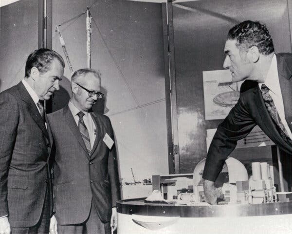
SARS-CoV-2 variants from minks evade inhibition by antibodies

It has been known for about a year that minks can become infected with SARS-CoV-2. The virus had been transmitted from humans to farmed mink and mutated in infected animals. Mutations were acquired in the spike protein, which is crucial for the entry of the virus into host cells and represents the central point of attack for antibodies. These SARS-CoV-2 variants from mink were transmitted back to humans, raising concerns that minks could be a continuing source of infection of humans with SARS-CoV-2 variants with altered biological properties.
Researchers at the German Primate Center (DPZ) — Leibniz Institute for Primate Research in Göttingen, Germany, have now shown that an antibody used for COVID-19 therapy is unable to effectively inhibit SARS-CoV-2 harboring a spike mutation acquired in minks. In addition, the mutation reduced the inhibition of the virus by antibodies produced in SARS-CoV-2 infected humans. These results show that SARS-CoV-2 can acquire mutations in minks that may reduce control of the virus by the human immune system (Cell Reports).
More than three million people have died worldwide from the pandemic spread of SARS coronavirus-2 and its associated disease COVID-19, according to the World Health Organization (WHO). Animal-to-human transmission of the virus is believed to be the origin of the pandemic, which began in Wuhan, China, in December 2019. In April 2020, mink in Dutch mink farms developed a respiratory disease due to infection with SARS-CoV-2, which was transmitted from infected farm workers to the animals. The virus mutated in mink and different virus variants emerged, which were transmitted back to farm workers and then also transmitted from human to human. This observation was also made in Denmark and millions of minks were killed to prevent the transmission of new viral variants to humans.
The spike protein in the envelope of SARS-CoV-2 is responsible for the entry of the virus into cells in which it replicates. Researchers led by Markus Hoffmann and Stefan Pöhlmann of the German Primate Center have studied mutations detected in the spike protein of SARS-CoV-2 from mink, including mutation Y453F. The researchers wanted to find out whether this mutation affects inhibition of the virus by antibodies that are used for COVID-19 therapy or are produced in COVID-19 patients.
“Our results show that one of two antibodies from an antibody cocktail used for COVID-19 therapy no longer efficiently inhibits the viral variant with the Y453F mutation. Furthermore, our study demonstrates that the Y453F mutation reduces inhibition of the virus by antibodies produced by COVID-19 patients. This means that people who were infected with SARS-CoV-2 may have reduced protection against mink variants of the virus,” says Markus Hoffmann, first author of the study. In sum, SARS-CoV-2 can mutate in minks in a way that reduces immune control by antibodies. Whether this is also possible in other animals to which the virus can be transmitted by infected individuals is currently unclear. “In the meantime, the Y453F mutation has also occurred in humans, but not through infection with a mink variant. When the virus replicates for a long time in people who are immunocompromised, resistant variants can emerge. In this case, the resistance-mediating mutation was identical to the one observed in mink,” says Stefan Pöhlmann.
Story Source:
Materials provided by Deutsches Primatenzentrum (DPZ)/German Primate Center. Note: Content may be edited for style and length.

Read more →

Two blood thinners at once: More risk with the same reward

More blood thinners aren’t automatically better, another study confirms.
A new publication in JAMA Internal Medicine focuses on the minimal pros and the concerning cons of combining a daily aspirin with a drug from the newer class of anticoagulants that include apixaban, dabigatran, edoxaban and rivaroxaban.
Patients were taking one of these direct oral anticoagulants known as DOACs to prevent strokes from non-valvular atrial fibrillation or for the treatment of venous thromboembolic disease (deep vein thrombosis or pulmonary embolism). The included patients did not have another reason to take aspirin such as a recent history of a heart attack or a history of a heart valve replacement. The researchers discovered that almost one-third of the people who were prescribed a DOAC were also taking aspirin without a clear reason for the aspirin.
“The patients on combination therapy were more likely to have bleeding events but they weren’t less likely to have a blood clot,” says lead author Jordan Schaefer, M.D., an assistant professor of internal medicine and a hematologist at Michigan Medicine, the academic medical center of the University of Michigan. “Therefore, it’s important that patients ask their doctors if they should be taking aspirin when they are prescribed a direct oral anticoagulant.”
The combination of an anticoagulant and an antiplatelet may be appropriate for people who have had a recent heart attack, recent coronary stent placement or bypass surgery, prior mechanical valve surgery or known peripheral artery disease, among other conditions says co-author Geoffrey Barnes, M.D., M.Sc., an assistant professor of internal medicine and a vascular cardiologist at the Michigan Medicine Frankel Cardiovascular Center.
For the others, “combination therapy may not be happening intentionally; rather, the addition of aspirin might get overlooked because it’s not in any one specialist or general care provider’s territory,” Barnes says.
The authors note that there are many medical conditions and situations where adding aspirin with a direct oral anticoagulant has not been adequately studied. Schaefer adds that they plan to confirm their study findings in a larger, longer study because there were not many blood clots that occurred during the timeframe of this study, potentially limiting their ability to assess if aspirin could be beneficial.
Previously, Schaefer and Barnes also reported a significant increase in adverse outcomes for people taking both aspirin and warfarin, a different kind of anticoagulant.
Schaefer originally presented these registry-based cohort study results at the 2019 annual meeting of the American Society of Hematology.
Story Source:
Materials provided by Michigan Medicine – University of Michigan. Original written by Haley Otman. Note: Content may be edited for style and length.

Read more →

Gene therapy shows promise in initial trial for patients with childhood blindness

A new gene therapy for one of the most common forms of congenital blindness was safe and improved patients’ vision, according to initial data from a clinical trial led by researchers at the Scheie Eye Institute in the Perelman School of Medicine at the University of Pennsylvania.
The therapy delivers working copies of GUCY2D to the eyes of patients who have severe vision impairments caused by mutations in the gene. Each of the first three treated patients experienced improvement in some aspects of vision, without serious side effects, according to the new study, published in the journal iScience.
“We found sustained improvements in both day and night vision, even with a relatively low dose of the gene therapy,” said study lead author Samuel G. Jacobson, MD, PhD, a professor of Ophthalmology in the Perelman School of Medicine.
The GUCY2D gene is one of about 25 different human genes whose mutations cause problems in the retina, leading to severe vision impairment from birth or early childhood. This family of inherited retinal disorders, collectively known as Leber congenital amaurosis (LCA), accounts for a considerable portion of blindness in children worldwide.
Normal copies of GUCY2D encode an enzyme in the key pathway that light-sensitive rod and cone cells in the retina use to convert light into electrochemical signals. A lack of this enzyme blocks the recovery of this pathway, preventing the reset needed for further signaling. As a result, the signal from rod and cone cells becomes very weak — which equates to severe vision loss.
Even in adults who have lived for decades with this condition, it is often the case that many light-sensing retinal cells remain alive and intact despite their dysfunction. Thus, adding functional copies of GUCY2D via a gene therapy could get those cells working again and restore some vision.
In 2019, Jacobson and co-investigator Artur V. Cideciyan, PhD, a research professor of Ophthalmology in the Perelman School of Medicine, began the first clinical trial of a GUCY2D gene therapy, a solution of a harmless virus that carries the gene and is injected beneath the retina — initially in just one eye per patient. They are following each patient for two years after treatment. In the new report, they described their findings after nine months in the first three patients treated.
The first patient experienced a substantial increase in light-sensitivity in rod cells, which are more light-sensitive than cone cells and are chiefly responsible for low-light or “night vision.” This patient also showed improved pupil responses to light.
The second patient showed a smaller but sustained increase in light-sensitivity in rod cells, starting about two months after the gene therapy.
The third patient showed no improvement in rod cell sensitivity, but did show significantly improved visual acuity over the nine-month follow-up period, an improvement that the researchers tied to better function in the patient’s cone cells, the predominant cells for daylight and color vision.
“These initial results from the first-ever trial of a GUCY2D gene therapy are very encouraging and will inform our ongoing and future trials of this therapy,” said Cideciyan. There were no serious adverse side effects, and any side effects that occurred in the patients’ retinas resolved.
The gene therapy dose used in these first three patients was the lowest of the doses the researchers plan to use in the study, so they are hoping to see continued safety and greater efficacy in later-enrolled patients who will receive higher doses.

Read more →

Microglia, Stockholm syndrome and miraculous cures in glioblastoma patients

Despite access to some of the best possible medical care in the world, Senators John McCain and Edward Kennedy both died within 18 months of their diagnosis of glioblastoma, an aggressive form of brain cancer. While this deadly outcome typifies the nature of this disease, some glioblastoma patients see exceptional benefits from chemotherapy and survive beyond expectations. Why this happens has been revealed by researchers at the University of Minnesota in a new study published in the Proceedings of the National Academy of Sciences.
“Deciphering the molecular underpinning of these exceptional responses may hold the key to transforming the hope for miracles into the reality of an expected cure for glioblastoma patients,” said Clark C. Chen, MD, PhD, Lyle French Chair in Neurosurgery and head of the Department of Neurosurgery at the University of Minnesota Medical School, who is also lead author of the study.
The study team looked at the gene expression profiles of glioblastoma specimens collected from approximately 900 glioblastoma patients from regions across the world to identify unique features associated with exceptional responders, defined as glioblastoma patients who survive more than two years after treatment.
“We utilized different state-of-the-art analytics to study these samples, including methods innovated by Dr. Aaron Sarver, a member of the University of Minnesota Institute of Health Informatics. Impressively, these analytics converged on a single observation, a paucity of microglia and macrophages,” Chen said.
Microglia and macrophages are specialized, immune cells that act as scavengers to recognize and remove cells not normally present in a healthy brain, including cancer cells. These immune cells migrate to sites harboring abnormal cancer cells to defend the body against the cancer cells and can make up more than half of the cells in a glioblastoma sample.
“If microglia and macrophages normally fend off cancer cells, more of them should allow the body to better fend off the tumor. So, we expected to see more of them in the exceptional responders; however, we found the contrary,” said Jun Ma, a researcher in the Department of Neurosurgery at the U of M Medical School and a co-first author of this study.
Resolving this paradox, the research team subsequently demonstrated glioblastoma cells possess the capacity to recondition the surrounding microglia and macrophages and corrupt their native anticancer functions. Instead of fending off cancer growth, these immune cells are now re-programmed by glioblastoma cells to promote tumor growth.
“It is frightening to consider the possibility that cancer cells can ‘brainwash’ our own immune cells and convert them from cells that fight cancer to cells that promote cancer,” said Judith Varner, a co-senior author of the study and professor of pathology at the University of California, San Diego. “Fortunately, we have figured out how glioblastoma cells subvert our immune system and can now reverse this cellular version of the ‘Stockholm syndrome.'”
Stockholm syndrome is a psychological response in which hostages or abuse victims develop an emotional bond with and act to help their captors.
The key to curing this cellular “Stockholm syndrome” and possibly glioblastoma lies in a protein called phosphoinositide-3-kinase gamma isoform (PI3K?). Activation of this protein turns microglia and macrophages from immune cells that police cancer growth into hostage cells that promote cancer growth. Varner has studied this process for many years and pioneered drugs that restore the anti-tumor activities of microglia and macrophages.
“In our animal glioblastoma models, treatment with drugs targeting PI3K? consistently resulted in impressively durable responses to chemotherapy,” Chen said. “We are eager to translate these findings into a human trial, with the hope of transforming every glioblastoma patient into an exceptional responder.”
Story Source:
Materials provided by University of Minnesota Medical School. Original written by Kelly Glynn. Note: Content may be edited for style and length.

Read more →

Once-a-week insulin treatment could be game-changing for patients with diabetes

Treating people with Type 2 diabetes with a new once-a-week injectable insulin therapy proved to be safe and as effective as daily insulin injections, according to the results of two international clinical trials published online today in Diabetes Care. The studies suggest that the once-weekly treatment could provide a convenient alternative to the burden of daily insulin shots for diabetes patients.
Starting and maintaining insulin treatment remain a challenge for millions of patients worldwide with Type 2 diabetes. Fear of injections and the inconvenience and burden of injectable therapy contribute to the barriers against insulin therapy initiation and adherence. The effectiveness and safety of ongoing insulin treatment are also highly dependent on other factors, such as the accuracy of dosages, timing, and glycemic targets. Health care providers believe that reducing the frequency of treatment administration with advances, such as the once-weekly insulin used in these phase 2 trials, may decrease the reluctance to initiate insulin therapy while improving long-term adherence, glucose control, and ultimately, patient well-being.
Insulin, which has been the foundation of diabetes treatment for 100 years, is an effective glucose-lowering agent and is safe when used at the correct dose,” says Ildiko Lingvay, M.D., M.P.H., M.S.C.S., a professor of internal medicine and population and data sciences at UT Southwestern. “Insulin treatment is burdensome, requires frequent injections, and continues to carry a certain stigma. The development of an effective and safe insulin that can be administered once a week is a huge advance in the field.”
Lingvay, who is a consultant for Novo Nordisk, is the lead author of one of the studies, which involved 205 patients from seven countries (the U.S., Croatia, Germany, Hungary, Poland, Slovakia, and Spain). The clinical trial consisted of a two-week screening period, 16 weeks of treatment, and a five-week follow-up to evaluate three different ways to adjust and optimize the insulin dose and determine which one presented the best balance between effectively lowering glucose while minimizing low-glucose events.
She also is an author of the second study that included 154 patients from five countries (the U.S., Canada, the Czech Republic, Germany, and Italy). This trial followed the same 23-week time frame and evaluated practical aspects of insulin use as well as the best ways to transition from a daily regimen to the new weekly insulin injections. The researchers determined that starting with a higher first dose — called a loading dose — allowed patients to reach their optimal glucose target faster.
“These two studies served as the steppingstones for a large phase 3 clinical trial program that is currently ongoing at UT Southwestern and other sites, which is designed to evaluate the efficacy of once-weekly insulin administration in patients with either Type 1 or Type 2 diabetes,” Lingvay says. “A weekly insulin is a game-changer that will decrease the treatment burden for patients while also improving compliance. This treatment will also decrease the burden on those who care for patients with diabetes requiring insulin. For example, for patients who need help injecting, those living in long-term care facilities, and those with memory problems, a once-weekly insulin will facilitate treatment and decrease the burden on the care providers.”
Novo Nordisk was the sponsor of both studies.
Story Source:
Materials provided by UT Southwestern Medical Center. Note: Content may be edited for style and length.

Read more →

Covid: More than 10m people fully vaccinated in UK

SharecloseShare pageCopy linkAbout sharingimage copyrightGetty ImagesMore than 10 million people in the UK have received a second dose of a Covid-19 vaccine, according to the latest government figures. It means more than 19% of UK adults are now fully vaccinated, while nearly 33 million people have had their first dose. Prime Minister Boris Johnson described the news as a “remarkable milestone”. Also hailing the “terrific milestone”, the health secretary urged people to get the jab “as soon as possible”. “Second doses are crucial to maximising the strength and duration of your protection from Covid-19,” he added. Sir Simon Stevens, chief executive of NHS England, said: “The success of the NHS vaccination programme is not a happy accident. It is down to careful planning coupled with the sheer hard work and determination of doctors, nurses and countless other staff ably assisted by volunteers and many others.”It comes as the UK recorded four deaths within 28 days of a positive test – the lowest daily figure since 7 September, although the number of deaths recorded on Mondays and over the weekend tend to be lower because of reporting delays. There were also 2,963 new cases, according to the latest government figures. When will I get the jab?Is the Oxford-AstraZeneca vaccine safe?Mr Hancock told the Commons the government was “on track” to offer a first dose to all adults in the UK by the end of July and uptake had been “astonishingly high”, at 94% for over-50s. However, he said the vaccination rate for care home staff was currently below 80% in more than half of all local authority areas. As a result, he said, the government had launched a consultation into whether care home providers for older adults should only deploy workers who have received their Covid jab, unless they have a medical exemption. People aged 45 and over are now being offered the vaccine in England and Scotland. In some areas of Wales 40-49 year-olds are being invited, while in Northern Ireland vaccine appointments are now being made available to a limited number of 35-39 year olds. The rollout will continue to prioritise groups by age, according to advice from the Joint Committee of Vaccination and Immunisation (JCVI). The 10 million mark for second doses is a great milestone.But the rapid acceleration in second doses has come at a cost to first doses – and this spells trouble for the government’s ambition to offer every adult in the UK a jab by the end of July.Currently the UK is doing just over 100,000 first doses a day on average, that is down from 500,000 a month ago.There was always going to be some drop-off. But on that trajectory just over half of under-50s will get a first dose by the end of July.Take-up is predicted to be lower in the younger age groups than it has been in the older ones so that shortfall may not be as bad as it first looks.What is more, there is the hope vaccine supplies will increase. More doses of Moderna may become available, while there is hope the Novavax jab, which is being made in the UK, could come into play in May.Nonetheless, it is another reminder that the vaccine programme still has many hurdles to cross.It comes after the government said all over-50s and those in high-risk groups in the UK had been offered a first dose. Earlier this month the UK began to roll out the Moderna vaccine, alongside the Oxford-AstraZeneca and Pfizer-BioNTech jabs. All under-30s in the UK will be offered an alternative to the AstraZeneca jab due to evidence linking it to rare blood clots. Meanwhile, Mr Hancock announced that India would be added to the travel red list from 04:00 BST on Friday. It means anybody who is not a UK or Irish resident or British citizen cannot enter the UK if they have been in India in the last 10 days and any other arrivals will need to quarantine in a hotel for 10 days. Earlier, the prime minister cancelled a trip to India, which had been due to take place on 26 April, amid rising cases there.Health officials in the UK are investigating whether a variant first found in India spreads more easily or is resistant to treatments or vaccines. Mr Hancock said there had now been 103 cases of this variant detected in the UK, with the vast majority linked to international travel. He said there had also been 557 cases of the variant first found in South Africa detected in the UK since December. The health secretary said the government was “ramping up” plans for a booster shot to ensure vaccines stayed “ahead of the virus”. “We will be working with our current vaccine suppliers and new suppliers, like the CureVac partnership, to work out which vaccines will be effective as a booster shot and to design new vaccines specifically targeted at the variants of concern,” he added. LOOK-UP TOOL: How many cases in your area?LOCKDOWN RULES: What are they and when will they end?SOCIAL DISTANCING: How can I meet my friend safely? JUST ONE THING TO REDUCE YOUR STRESS?: Why a cold shower might have a positive impactLONG COVID: What does it feel like to lose your sense of smell and taste for months?

Read more →

Attacking aortic aneurysms before they grow

A new study investigates a genetic culprit behind abdominal aortic aneurysm, a serious condition that puts people at risk of their aorta rupturing — a potentially deadly event.
Finding a viable genetic target for AAA could change the game, says senior author Katherine Gallagher, M.D., a vascular surgeon and an associate professor of surgery and microbiology and immunology at Michigan Medicine, the academic medical center of the University of Michigan.
That’s because there are no medications to directly treat the condition and prevent an aneurysm from growing. Current options include things like addressing blood pressure to lower the stress on the arteries and veins running through the body, and making lifestyle changes like quitting smoking. Most people monitor their aneurysm to see if it grows enough to eventually require endovascular or open surgical repair.
For this study, a team of Michigan Medicine researchers investigated the role of an epigenetic enzyme called JMJD3 in the development of AAAs. They found the gene was turned on in both people and mice who had an AAA and that the gene promoted inflammation in monocyte/macrophages. When they blocked the enzyme, it prevented an aneurysm from forming.
“Targeting the JMJD3 pathway in a cell specific-manner offers the opportunity to limit AAA progression and rupture,” says lead author Frank Davis, M.D., a vascular surgery resident at the Frankel Cardiovascular Center.
“We are the first to perform an extensive single-cell RNA sequencing and gene expression analysis on human AAAs and non-aneurysmal aortic control samples,” Gallagher adds.
Story Source:
Materials provided by Michigan Medicine – University of Michigan. Original written by Haley Otman. Note: Content may be edited for style and length.

Read more →

Your neighborhood may affect your brain health

Middle-age and older people living in more disadvantaged neighborhoods — areas with higher poverty levels and fewer educational and employment opportunities — had more brain shrinkage on brain scans and showed faster decline on cognitive tests than people living in neighborhoods with fewer disadvantages, according to a study published in the April 14, 2021, online issue of Neurology®, the medical journal of the American Academy of Neurology. Researchers say such brain aging may be a sign of the earliest stages of dementia.
“Worldwide, dementia is a major cause of illness and a devastating diagnosis,” said study author Amy J. H. Kind M.D., Ph.D., of the University of Wisconsin School of Medicine and Public Health in Madison. “There are currently no treatments to cure the disease, so identifying possible modifiable risk factors is important. Compelling evidence exists that the social, economic, cultural and physical conditions in which humans live may affect health. We wanted to determine if these neighborhood conditions increase the risk for the neurodegeneration and cognitive decline associated with the earliest stages of Alzheimer’s disease and dementia.”
For the study, researchers identified 601 people from two larger studies of Wisconsin residents. Participants had an average age of 59 and no thinking or memory problems at the start of the study, although 69% had a family history of dementia. They were followed for 10 years.
Participants had an initial MRI brain scan and then additional scans every three to five years. With each scan, researchers measured brain volume in areas of the brain linked to the development of Alzheimer’s dementia. Participants also took thinking and memory tests every two years, including tests that measured processing speed, mental flexibility and executive function.
Researchers used the residential address of each participant and a measure called the Area Deprivation Index to determine if each participant lived in an advantaged or disadvantaged neighborhood. Neighborhoods in the index are determined by census areas of 1,500 residents. The index incorporates information on the socioeconomic conditions of each neighborhood and its residents, ranking neighborhoods based on 17 indicators including income, employment, education and housing quality.
Of all participants, 19 people lived in the 20% most disadvantaged neighborhoods in their state and 582 people lived in the 80% of all other neighborhoods in their state. People in the first group were then matched one to four to people in the second group for race, sex, age and education and compared.
At the start of the study, there was no difference in brain volume between people living in the most disadvantaged neighborhoods and those in other neighborhoods. But at the end, researchers found brain shrinkage in areas of the brain associated with dementia in people in the most disadvantaged neighborhoods, while there was no shrinkage in the other group. Researchers also found a higher rate of decline on tests that measure risk of Alzheimer’s disease.
“Our findings suggest that increased vigilance by healthcare providers for early signs of dementia may be particularly important in this vulnerable population,” said Kind. “Some possible causes of these brain changes may include air pollution, lack of access to healthy food and healthcare and stressful life events. Further research into possible social and biological pathways may help physicians, researchers and policymakers identify effective avenues for prevention and intervention in Alzheimer’s disease and related dementia.”
Limitations of the study included a small number of participants from highly disadvantaged neighborhoods and a limited geographic setting. Future studies should involve larger and more diverse groups of people over longer periods of time.
Story Source:
Materials provided by American Academy of Neurology. Note: Content may be edited for style and length.

Read more →

Intellectual disability is rarely inherited — risk for younger siblings is low

The prevalence of intellectual disabilities, which means difficulties with learning and understanding new things, is roughly 1-2% in the population. People with a severe intellectual disability need help from others in daily activities throughout their lives.
Such disabilities can be caused by genetic changes or external factors. According to estimates, roughly 2,500 genes underlie intellectual disability, of which approximately half remain unidentified.
In recent years, the diagnostics for intellectual disabilities have improved thanks to advancements in techniques that make it possible to sequence the entire genome. These techniques can also help to identify causes of intellectual disability not found in other medical examinations and tests. Exome sequencing, that is, the sequencing of the protein-coding regions of genes in the genome, enables the identification of new pathogenic gene variants as well. Identifying genes is a prerequisite for identifying disease mechanisms and developing treatments.
The study conducted at the University of Helsinki utilised exome sequencing to determine the potential genetic background of intellectual disability. The study participants included Finnish families with family members with delayed cognitive development for which no clear cause had been identified. The results were recently published in the Human Genetics journal.
It was found that in 64% of the study participants the cause of their developmental disorder was a known intellectual disability gene. The majority of these variants, 75%, was the result of random mutations taking place during fetal development (de novo), and variants not found in the parents’ genome. An inherited mutation was identified in no more than a quarter of the pathogenic genes studied. More large-scale structural variants, which are usually not inherited, were found in only 8% of the families.
“Based on our findings, the risk of recurrence of intellectual disability in the next child of individual families is usually low,” says Docent Irma Järvelä. According to Järvelä, this is a significant and relieving piece of information for many families.

Read more →

Defects in a specific cell type may cause ulcerative colitis

There are many variants of “goblet cells” in the intestines and they seem to have different functions, according to a new study from the University of Gothenburg. The study indicates that defects in goblet cells of a particular type may be a factor contributing to ulcerative colitis, an inflammatory bowel disease.
The entire inside of our intestines is covered by a thin layer of mucus that protects the fragile mucous membrane (mucosa) from bacteria and other microorganisms. If the microorganisms repeatedly come into contact with the intestinal mucosa, inflammation and even cell changes may result. These increase the risk of intestinal cancer. In a healthy colon, the mucus layer is up to a millimeter thick. This layer, which undergoes complete renewal hourly, is formed from cells of a special type, known as goblet cells.
Many different goblet cells
In the present study, now published in the journal Science, the scientists separated goblet cells from other cells and investigated which proteins each individual goblet cell expresses. There proved to be many different subtypes of these cells, and goblet cells’ functions turned out to vary more than researchers have previously realized.
“We believe this is important knowledge that may enable us to influence the protective function of the gut in the future. The system that maintains the protective intestinal mucus layer seems to be able to change its functions, and we could utilize this capacity by reprogramming the layer with various signals, for example by using new drugs,” says Malin Johansson, Associate Professor at Sahlgrenska Academy, University of Gothenburg, who led the research behind the present study.
Connected with ulcerative colitis
The most impermeable part of the mucus layer is formed by glands in the gut. In particular, the research team studied one of the specific types of goblet cells, found on the outermost surface of the mucosa. These goblet cells provide another type of mucus, which contributes to the protection of the gut but allows certain nutrients to pass through.
“If the function of these specific cells is impaired, we see that unprotected cell surfaces arise. These lead to inflammation, both in studies on mice and in samples from patients with ulcerative colitis,” Johansson says.
Appear to cause damage to mucosal protection
In the study, these specific goblet cells seemed to be repelled by the mucosa earlier than normal in patients with ulcerative colitis. Accordingly, the cells became fewer.
“To our surprise, we were able to observe this both in patients with active ulcerative colitis and in those who were temporarily asymptomatic. This indicates that premature rejection of the particular goblet cells we’ve been studying damages the mucus protection and that this is a contributing cause of inflammatory bowel disease. It could also be a partial explanation for these patients’ elevated cancer risk,” Johansson says.
There are some 30,000 people in Sweden with ulcerative colitis, which is a chronic but intermittent inflammatory bowel disease.
Story Source:
Materials provided by University of Gothenburg. Note: Content may be edited for style and length.

Read more →