How human cells coordinate the start of DNA replication

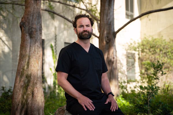
Cold Spring Harbor Laboratory (CSHL) President and CEO Bruce Stillman has been dissecting DNA replication, a critical step in cell division, since the 1980s. His lab studies how Origin Recognition Complexes — ORCs — coordinate DNA duplication. They discovered how our cells assemble and disassemble ORCs during the cell division cycle. One ORC protein is sequestered into small liquid droplets, keeping it apart until the right time to recruit other proteins and initiate DNA replication.
The ORC recognizes where to initiate replication at numerous locations along the long, linear stretches of DNA in our cells’ chromosomes. Fully assembled ORCs recruit other proteins to make precise copies of the chromosomes. This mechanism is necessary to inherit DNA accurately without errors that can lead to disorders such as cancer.
Scientists have studied the structure of ORCs in several species. Stillman explains:
“We’ve previously studied this in baker’s yeast, but it turns out that human cells have a different way of doing things.”
Unlike single-celled yeast, humans have a variety of cells that divide at different times. To choreograph this, the researchers found that one human ORC protein, ORC1, has certain regions that yeast ORC1 lacks. When ORC binds to DNA, ORC1 recruits CDC6, a protein that assembles other DNA replication proteins. Some of the human-specific regions of ORC1 and CDC6 bind other proteins that regulate DNA replication. Manzar Hossain, a research investigator in Stillman’s lab, says:
“We found that ORC1 and CDC6 interact in a very tangential manner. We found a very short time period which allows them to interact.”
DNA-bound ORC1 is sequestered into liquid droplets that briefly change shape, then brings in CDC6. Kuhulika Bhalla, a postdoc in Stillman’s lab, explains:
“So if you can imagine a lava lamp, like you’ve got liquid, but you’ve got other colored liquid within it. And they still managed to stay separated.”
Throughout most of the cell division cycle, ORC1 and CDC6 amounts oscillate in the cell. Stillman explains that “both high and low amounts of ORC1 lead to severe consequences for cell viability. So, you have to have just the right amount” of each protein throughout the cell cycle. Stillman and his colleagues have shown that CDC6 recruits other regulatory proteins that control the activity and levels of ORC1 in both space and time. They published their findings in Molecular Cell.
Story Source:
Materials provided by Cold Spring Harbor Laboratory. Original written by Jasmine Lee. Note: Content may be edited for style and length.

Read more →

Rare genetic variant puts some younger men at risk of severe COVID-19

A study of young men with COVID-19 has revealed a genetic variant linked to disease severity.
The discovery, published recently in eLife, means that men with severe disease could be genetically screened to identify who has the variant and may benefit from interferon treatment.
For most people, COVID-19, the disease caused by the virus SARS-CoV-2, causes only mild or no symptoms. However, severe cases can rapidly progress towards respiratory distress syndrome.
“Although older age and the presence of long-term conditions such as cardiovascular disease or diabetes are known risk factors, they alone do not fully explain differences in severity,” explains first author Chiara Fallerini, Research Fellow in Medical Genetics at the Department of Medical Biotechnologies, University of Siena, Italy. “Some younger men without pre-existing medical conditions are more likely to be hospitalised, admitted to intensive care and to die of COVID-19, which suggests that some factors must cause a deficiency in their immune system.”
Recent research has suggested that genes controlling interferon are important in regulating the immune response to COVID-19. Interferon is produced by immune cells during viral infection. It works alongside molecules on the surface of immune cells called Toll-like receptors (TLR) which detect viruses and kickstart the immune response. “When a recent study identified rare mutations in a TLR gene, TLR7, in young men with severe COVID-19, we wanted to investigate whether this was an ultra-rare situation or just the tip of the iceberg,” says co-senior author Mario Mondelli, Professor of Infectious Diseases at the Division of Clinical Immunology and Infectious Diseases, Fondazione IRCCS Policlinico San Matteo and University of Pavia, Italy.
The team studied a subset of 156 male COVID-19 patients younger than 60 years old, selected from a large multicentre study in Italy, called GEN-COVID, which started its activity on March 16, 2020. GEN-COVID is a network of more than 40 Italian hospitals coordinated by co-senior author Alessandra Renieri, Full Professor of Medical Genetics at the University of Siena, and Director of Medical Genetics at Azienda Ospedaliero-Universitaria Senese, Siena, Italy.
The team first analysed all the genes on the X chromosome of men with both mild and severe cases of COVID-19, and identified the TLR7 gene as one of the most important genes linked to disease severity. They then searched the entire GEN-COVID database, and selected for younger men (less than 60 years old). This identified rare TLR7 missense mutations in five of 79 patients (6.3%) with life-threatening COVID-19 and no similar mutations in the 77 men who had few symptoms. They also found the same mutation in three men aged over 60: two who had severe COVID-19 and one who had few symptoms — although the mutation found in the man with few symptoms had little effect on TLR function.
To link these mutations to the immune cell response, they treated white blood cells from recovered patients with a drug that switches TLR7 genes on. They found that the TLR7 genes were dampened down in immune cells from patients with mutations, compared to the TLR7 activity seen in normal immune cells. They also found lower levels of interferon in the cells containing the mutation compared to normal white blood cells. This confirmed that the mutations identified directly affect the control of interferon as part of the innate immune response.
To confirm the impact of the mutations on COVID-19 response, the team studied two brothers, one with a mutation in an interferon gene and one without. The levels of interferon gene activity were much lower in the man with the missense mutation, compared with his brother. Moreover, the brother with the mutation had severe COVID-19, while his brother with normal interferon genes was asymptomatic.
“Our results show that young men with severe COVID-19 who have lost function in their interferon-regulating genes represent a small but important subset of more vulnerable COVID-19 patients,” says co-senior author Elisa Frullanti, Researcher of Medical Genetics at the University of Siena.
Co-senior author Alessandra Renieri adds: “These mutations could potentially account for disease severity in up to 2% of young men with COVID-19. We believe that screening for these mutations in men who are admitted with severe disease and promptly treating them with interferon could prevent more deaths.”
Story Source:
Materials provided by eLife. Note: Content may be edited for style and length.

Read more →

'Zombie' genes? Research shows some genes come to life in the brain after death

In the hours after we die, certain cells in the human brain are still active. Some cells even increase their activity and grow to gargantuan proportions, according to new research from the University of Illinois Chicago.
In a newly published study in the journal Scientific Reports, the UIC researchers analyzed gene expression in fresh brain tissue — which was collected during routine brain surgery — at multiple times after removal to simulate the post-mortem interval and death. They found that gene expression in some cells actually increased after death.
These ‘zombie genes’ — those that increased expression after the post-mortem interval — were specific to one type of cell: inflammatory cells called glial cells. The researchers observed that glial cells grow and sprout long arm-like appendages for many hours after death.
“That glial cells enlarge after death isn’t too surprising given that they are inflammatory and their job is to clean things up after brain injuries like oxygen deprivation or stroke,” said Dr. Jeffrey Loeb, the John S. Garvin Professor and head of neurology and rehabilitation at the UIC College of Medicine and corresponding author on the paper.
What’s significant, Loeb said, is the implications of this discovery — most research studies that use postmortem human brain tissues to find treatments and potential cures for disorders such as autism, schizophrenia and Alzheimer’s disease, do not account for the post-mortem gene expression or cell activity.
“Most studies assume that everything in the brain stops when the heart stops beating, but this is not so,” Loeb said. “Our findings will be needed to interpret research on human brain tissues. We just haven’t quantified these changes until now.”
Loeb and his team noticed that the global pattern of gene expression in fresh human brain tissue didn’t match any of the published reports of postmortem brain gene expression from people without neurological disorders or from people with a wide variety of neurological disorders, ranging from autism to Alzheimer’s.

Read more →

Containing the coronavirus effects on the nervous system

A number of studies have shown that human coronaviruses, including SARS-CoV-2 which causes COVID-19, appear to attack neurons and the nervous system in vulnerable populations. This neuroinvasion through the nasal cavity leads to the risk of neurological disorders in affected individuals. Research conducted at the Institut national de la recherche scientifique (INRS) has identified ways to prevent the spread of infection within the central nervous system (CNS). The study, led by Professor Pierre Talbot and his research associate Marc Desforges, now at CHU-Sainte-Justine, was published in the Journal of Virology.
Antiviral immunity to human coronaviruses
The research team is the first to make the demonstration of a direct link between neurovirulence, protein S cleavage by cellular proteases and innate immunity. This antiviral immunity arises from the production of interferons, frontline proteins that help to detect early the presence of the virus.
“Using a common cold coronavirus, similar to SARS-CoV-2, we were able to show that cleavage of the S protein and interferon could prevent its spread to the brain and spinal cord in mice,” says Talbot, who has been studying coronaviruses for nearly 40 years.
Two therapeutic approaches
According to Marc Desforges, currently a clinical specialist in medical biology at the CHU-Sainte-Justine virology laboratory, the cleavage of the S protein by various cellular proteases is essential for these viruses to effectively infect cells and spread to various organs and systems in the body, including the central nervous system (CNS).
“Our results demonstrate that interferon produced by different cells, including olfactory receptors and cerebrospinal fluid (CSF) producing cells in the brain, could modulate this cleavage. Thus, it could and does significantly limit the viral spread in the CNS and the severity of the associated disease,” says the specialist who worked for 16 years as a research associate at the Armand-Frappier Health Biotechnology Centre of the IRNS.
Taken together, these results point to two potential antiviral targets: protein S cleavage and effective interferon-related innate immunity. “Understanding the mechanisms of infection and viral propagation in neuronal cells is essential to better design therapeutic approaches,” says Talbot. This is especially important for vulnerable populations such as the elderly and immunocompromised.” This discovery opens the door to new therapeutic strategies.
About the study
The article “Potential differences in cleavage of the S protein and type-1 interferon together control human coronavirus infection, propagation, and neuropathology within the central nervous system,” by Alain Le Coupanec, Marc Desforges, Benedikt Kaufer, Philippe Dubeau, Marceline Côté and Pierre J. Talbot, was published in the Journal of Virology. The study was supported by the Canadian Institutes of Health Research (CIHR).
Story Source:
Materials provided by Institut national de la recherche scientifique – INRS. Original written by Audrey-Maude Vézina. Note: Content may be edited for style and length.

Read more →

Physical activity helps curb low-grade inflammation in children

According to a recent Finnish study, accumulating more brisk and vigorous physical activity can curb adiposity-induced low-grade inflammation. The study also reported that diet quality had no independent association with low-grade inflammation. The findings, based on the ongoing Physical Activity and Nutrition in Children (PANIC) Study conducted at the University of Eastern Finland, were published in the European Journal of Sport Science.
The study was made in collaboration among researchers from the University of Jyväskylä, the University of Eastern Finland, the Norwegian School of Sport Sciences, and the University of Cambridge.
Low-grade inflammation is linked to many chronic diseases, but exercise can curb it
Long-lasting low-grade inflammation increases the risk for type 2 diabetes and cardiovascular diseases. Being overweight and obese contribute to low-grade inflammation, but little is still known about the role of lifestyle in curbing low-grade inflammation since childhood.
“Our study showed that children who were physically more active and less sedentary had a healthier inflammatory profile than children who were physically less active,” explains Dr. Eero Haapala from the Faculty of Sport and Health Sciences at the University of Jyväskylä. “However, our results suggest that the positive effects of high levels of vigorous physical activity and low levels of sedentary time on low-grade inflammation are partly explained by their positive effects on body composition.”
Low physical activity, unhealthy diet quality, and being overweight is the most unfavourable combination
Researchers found unhealthier inflammatory profile particularly in children with the lowest levels of physical activity, poorest diet quality and the highest body fat percentage.
“The key message of our results is that increasing physical activity and reducing sedentary time are key in preventing low-grade inflammation since childhood,” says Haapala. “They would be particularly important for overweight children.”
The study looked at the associations between physical activity, sedentary time, diet quality, body fat content, and low-grade inflammation in 390 children aged 6 to 8 years. Physical activity and sedentary time were measured by a combined heart rate and movement sensor and body composition with a DXA device. Low-grade inflammation was assessed using biomarkers measured from blood samples.
Story Source:
Materials provided by University of Jyväskylä – Jyväskylän yliopisto. Note: Content may be edited for style and length.

Read more →

Exercise boosts blood flow to the brain, study finds

It’s not just your legs and heart that get a workout when you walk briskly; exercise affects your brain as well. A new study by researchers at UT Southwestern shows that when older adults with mild memory loss followed an exercise program for a year, the blood flow to their brains increased. The results were published online today in the Journal of Alzheimer’s Disease.
“This is part of a growing body of evidence linking exercise with brain health,” says study leader Rong Zhang, Ph.D., professor of neurology at UTSW. “We’ve shown for the first time in a randomized trial in these older adults that exercise gets more blood flowing to your brain.”
As many as one-fifth of people age 65 and older have some level of mild cognitive impairment (MCI) — slight changes to the brain that affect memory, decision-making, or reasoning skills. In many cases, MCI progresses to dementia, including Alzheimer’s disease.
Scientists have previously shown that lower-than-usual levels of blood flow to the brain, and stiffer blood vessels leading to the brain, are associated with MCI and dementia. Studies have also suggested that regular aerobic exercise may help improve cognition and memory in healthy older adults. However, scientists have not established whether there is a direct link between exercise, stiffer blood vessels, and brain blood flow.
“There is still a lot we don’t know about the effects of exercise on cognitive decline later in life,” says C. Munro Cullum, Ph.D., professor of psychiatry at UTSW and co-senior author of the study. “MCI and dementia are likely to be influenced by a complex interplay of many factors, and we think that, at least for some people, exercise is one of those factors.”
In the study, Zhang, Cullum, and their colleagues followed 70 men and women aged 55 to 80 who had been diagnosed with MCI. Participants underwent cognitive exams, fitness tests, and brain magnetic resonance imaging (MRI) scans. Then they were randomly assigned to either follow a moderate aerobic exercise program or a stretching program for one year. The exercise program involved three to five exercise sessions a week, each with 30-40 minutes of moderate exercise such as a brisk walk.

Read more →

A divided visual field

Hummingbird hawkmoths are small insects that hover in the air like hummingbirds when drinking nectar from flowers. Dr. Anna Stöckl from the Biocentre of the Julius-Maximilians-Universität (JMU) Würzburg in Bavaria, Germany, is studying the visual performance of these insects. Dr. Stöckl and her doctoral student Ronja Bigge now present their latest findings in the journal Current Biology.
“To control their flight, hummingbird hawkmoths rely on optic flow in the lower half of their visual field,” Ronja Bigge explains. Optic flow is the relative motion that the surrounding image casts on the animals’ retinas when they fly. We experience this phenomenon ourselves when travelling by train — the landscape passing by the train windows allows us to estimate our speed, for example.
For hawkmoths, the optic flow also provides information about their own movement. It helps them to control the straightness and speed of their flight. The JMU researchers have now shown with outdoor measurements that the optic flow components parallel to the direction of flight are always strongest below the hawkmoths’ body. This is where the insects see meadows, gardens and streets that provide a varied texture. For flight control, what happens in the lower visual field is therefore the most reliable parameter.
Previously unknown behaviour discovered
“Surprisingly, we were able to show that the hawkmoths displayed a completely different and novel behaviour when we presented them with visual textures in the upper half of their visual field,” says Anna Stöckl.
The animals then oriented themselves along prominent contours in the patterns. Thus, they did not use the visual information for flight control, but for orientation — although the visual patterns were exactly the same as the ones that were previously presented in the lower half of their visual field.
“Our optical measurements in natural habitats showed a comparable relationship: high-contrast structures that can be used for orientation occur primarily in the upper half of the visual field,” says the JMU researcher. These are, for example, the silhouettes of treetops or bushes that form a strong contrast with the sky.
Visual field is divided in two
The conclusion of the Würzburg biologists: “The flight control system and the orientation system of the hummingbird hawkmoth divide the visual field among themselves and focus on the respective area that provides the most reliable information in their natural habitats.”
In other words, it is not only important what the animals see, but also where they see it.
Story Source:
Materials provided by University of Würzburg. Original written by Robert Emmerich. Note: Content may be edited for style and length.

Read more →

How UK, South Africa coronavirus variants escape immunity

All viruses mutate as they make copies of themselves to spread and thrive. SARS-CoV-2, the virus the causes COVID-19, is proving to be no different. There are currently more than 4,000 variants of COVID-19, which has already killed more than 2.7 million people worldwide during the pandemic.
The UK variant, also known as B.1.1.7, was first detected in September 2020, and is now causing 98 percent of all COVID-19 cases in the United Kingdom. And it appears to be gaining a firm grip in about 100 other countries it has spread to in the past several months, including France, Denmark, and the United States.
The World Health Organization says B.1.1.7 is one of several variants of concern along with others that have emerged in South Africa and Brazil.
“The UK, South Africa, and Brazil variants are more contagious and escape immunity easier than the original virus,” said Victor Padilla-Sanchez, a research scientist at The Catholic University of America. “We need to understand why they are more infectious and, in many cases, more deadly.”
All three variants have undergone changes to their spike protein — the part of the virus which attaches to human cells. As a result, they are better at infecting cells and spreading.
In a research paper published in January 2021 in Research Ideas and Outcomes, Padilla-Sanchez discusses the UK and South African variants in detail. He presents a computational analysis of the structure of the spike glycoprotein bound to the ACE2 receptor where the mutations have been introduced. His paper outlines the reason why these variants bind better to human cells.

Read more →

Telehealth consults increased more than 50-fold among privately insured working-age patients during first phase of the pandemic

A study co-authored by researchers at the Johns Hopkins Bloomberg School of Public Health found that telehealth consults among privately insured working-age patients accounted for almost 24 percent of outpatient consults with health care providers during the early phase of the pandemic, March to June 2020, up from less than 0.3 percent during the same period in 2019.
The dramatic shift occurred as many medical practices halted or curtailed in-person office hours and patients stayed away from doctor’s offices out of fear of transmission during the early months of the pandemic. At the same time, insurance companies and the federal government relaxed policies around telehealth to meet demand for remote medical consults via internet video or telephone.
The study was published online March 23 in JAMA Network Open.
For their study, researchers from Johns Hopkins Bloomberg School of Public Health and Blue Health Intelligence®, an independent licensee of the Blue Cross Blue Shield Association, analyzed anonymized claims data drawn from 36.6 million private insurance plan members who were of working age and continuously enrolled during the study period. The claims data for the study were provided by Blue Health Intelligence.
A total of 15 million telehealth claims were submitted during the March to June 2020 study period, with nearly three-quarters involving video support (74.4 percent) and fewer than one in ten occurring via phone (9.2 percent). Just over 3 percent (3.3 percent) were conducted either by email or chat while 13.1 percent were unspecified.
Mental health consults were far more likely to take place virtually — with 46.1 percent taking place via telehealth. By comparison, 22.1 percent of medical consults were virtual. In COVID-19 “hot spot” states — those with a COVID-19 prevalence at least 1.5 times the national average — 36 percent of all consults were telehealth versus 21.6 percent in areas with lower COVID-19 prevalence. The study also found that the greater the COVID-19 prevalence in a specific ZIP code, the higher the use of telehealth.

Read more →

New strategy for fighting brain cancer

Most people relate cholesterol to heart health, but it is also a critical component in the growth and spread of brain cancer. VCU Massey Cancer Center researcher Suyun Huang, Ph.D., recently discovered how cholesterol becomes dysregulated in brain cancer cells and showed that the gene responsible for it could be a target for future drugs.
The mean survival of patients with the most common and aggressive type of brain cancer, glioblastoma multiforme (GBM), is 14 months. The need to find new, effective treatments is urgent and has driven Huang, a member of the Cancer Biology research program at Massey, to detail the workings of numerous genes, proteins, enzymes and other cellular components that contribute to brain cancer growth. Her studies are revealing a biological “roadmap” showing previously unknown functions of genes.
Huang’s most recent study, published in the journal Nature Communications, pinpoints a gene called YTHDF2 as a crucial link in a chain leading to the development and growth of GBM. It works through a process set in motion by another gene with a well-established reputation for driving cancer progression, EGFR.
“These findings are exciting because we can potentially target YTHDF2 expression by using YTHDF2 small molecule inhibitors to control glioblastoma tumor growth and spread,” says Huang, who is also a professor in the Department of Human and Molecular Genetics at VCU School of Medicine. “Our experiments also showed that we can stop the formation and growth of brain cancer cells by blocking YTHDF2 expression, so it could also be a powerful target for drug development.”
EGFR is frequently overactivated in many aggressive cancers, including GBM. Huang’s team found that EGFR drives the overexpression of TYHDF2, which then sustains increased cholesterol levels for the invasive growth and development of GBM cells through a process that degrades the LXR? and HIVEP2 genes. LXR? is known to regulate cholesterol levels within cells and HIVEP2 is involved in the development of brain tissue.
Huang’s study is the first to describe this cell signaling cascade, and it helps fill in important parts of the “roadmap” leading to GBM. It is also the first study to show that N6-methyladenosine (m6A), a DNA modification found in nearly all cell-based life forms, plays a role in brain tumor growth and cholesterol metabolism. Huang’s team found that the increase in YTHDF2 expression caused m6A modifications in the mRNA of LXR? and HIVEP2, which inhibited their functions.
Next, Huang and her collaborators plan to evaluate different YTHDF2 inhibitors and establish their effects in lab and animal models.
“EGFR inhibition and cholesterol regulation are both promising strategies for GBM treatment,” says Huang. “Our study offers an exciting new approach that could potentially work hand-in-hand with these strategies to regulate and treat GBM.”
Story Source:
Materials provided by Virginia Commonwealth University. Original written by John Wallace. Note: Content may be edited for style and length.

Read more →