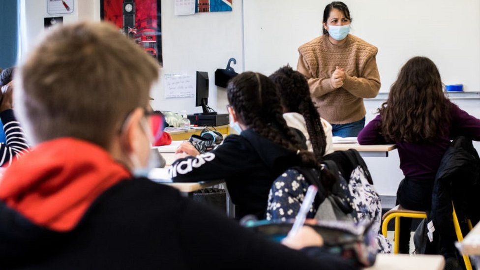
Why It Pays to Think Outside the Box on Coronavirus Tests

Universities and other institutions looking to protect themselves from Covid-19 may benefit from sharing their testing resources with the wider community, a new study suggests.Last year, when the National Football League decided to stage its season in the midst of the coronavirus pandemic, it went all-in on testing. The league tested all players and personnel before they reported for summer training camp, and continued near-daily testing in the months that followed. Between Aug. 1 and the Super Bowl in early February, the N.F.L. administered almost one million tests to players and staff.Many other organizations have sought safety in mass testing. The University of Illinois is testing its students, faculty and staff twice a week and has conducted more than 1.6 million tests since July. Major corporations, from Amazon to Tyson Foods, have rolled out extensive testing programs for their own employees.Now, a new analysis suggests that schools, businesses and other organizations that want to keep themselves safe should think beyond strictly themselves. By dedicating a substantial proportion of their tests to people in the surrounding community, institutions could reduce the number of Covid-19 cases among their members by as much as 25 percent, researchers report in a new paper, which has not yet been published in a scientific journal.“It’s natural in an outbreak for people to become self-serving, self-focused,” said Dr. Pardis Sabeti, a computational biologist at Harvard University and the Broad Institute who lead the analysis. But, she added, “If you’ve been in enough outbreaks you just understand that testing in a box doesn’t makes sense. These things are communicable, and they’re coming in from the community.”The study has “really profound implications, especially if others can replicate it,” said David O’Connor, a virologist at the University of Wisconsin, Madison, who was not involved in the analysis but reviewed a draft of the paper. As the pandemic enters its second year, he said, “We want to start using more sophisticated modeling and probably economic theory to inform what an optimal testing program would look like.”Dr. Sabeti is an epidemic veteran, part of teams that responded to an Ebola outbreak in West Africa in 2014 and a mumps outbreak in the Boston area a few years later. When the coronavirus closed down the country last spring, many colleges and universities sought her advice on how to safely reopen.At a time when testing resources were in short supply, many of these institutions were proposing intensive, expensive testing regimens focused entirely on their own members. Again and again, Dr. Sabeti suggested that universities think more broadly, and allocate some of their tests to people who might be friends, family members or neighbors of their students and employees.“The metaphor I often used on the calls was to say, ‘You’re in a drought in a place with a lot of forest fires, and you have a shortage of fire alarms,’” she recalled. “‘And if you run out and buy every fire alarm and install it in your own house, you’ll be able to pick up a fire the moment it hits your house, but at that point it’s burning to the ground.’”Still, convincing university officials to divert precious testing resources away from their own institutions was a hard sell, Dr. Sabeti said, especially without data on the effectiveness of the approach. So she and her colleagues decided to gather some.“Fundamentally, the paper is about the intersection of kindness and success — how being generous with one’s resources actually is the most effective” strategy, said one of the study’s authors.Pete Kiehart for The New York TimesThey developed an epidemiological model to simulate how a virus might spread through a midsize university, like Colorado Mesa University, one of the schools Dr. Sabeti’s team has been advising. (Several C.M.U. officials and researchers are co-authors of the paper.)Using real-world data from C.M.U., the researchers created a baseline scenario in which 1 percent of people at the school, and 6 percent of those in the surrounding county, were infected by the coronavirus, and the university was testing 12 percent of its members every day. The team assumed that they had a complete list of each university member’s close off-campus contacts, and that if someone tested positive for the virus, they and their contacts would quarantine until they were no longer infectious.Under these conditions, the researchers found, if the university used all of its tests on its own members, it would have roughly 200 Covid-19 cases after 40 days. But if instead it parceled out some of those tests, using them on community members who were close contacts of students and staff, the number of cases dropped by one-quarter.“The optimal proportion of tests to use outside the institution on those targeted, first-degree contacts came out to be about 45 percent,” said Ivan Specht, an undergraduate researcher in Dr. Sabeti’s lab and a co-author of the paper. In short, institutions could reduce their caseloads by one-fourth if they used almost half their tests on people just outside their direct membership. That percentage “is remarkably high considering that most institutions use zero percent of their tests outside of themselves,” Mr. Specht noted.The researchers then tweaked the model’s parameters in various ways: What if the virus were more prevalent? What if students and staff did not report all their contacts? What if they were better about mask-wearing and social distancing? What if the university deployed more tests, or fewer?.css-1xzcza9{list-style-type:disc;padding-inline-start:1em;}.css-rqynmc{font-family:nyt-franklin,helvetica,arial,sans-serif;font-size:0.9375rem;line-height:1.25rem;color:#333;margin-bottom:0.78125rem;}@media (min-width:740px){.css-rqynmc{font-size:1.0625rem;line-height:1.5rem;margin-bottom:0.9375rem;}}.css-rqynmc strong{font-weight:600;}.css-rqynmc em{font-style:italic;}.css-akgeos{margin-bottom:15px;font-family:nyt-franklin,helvetica,arial,sans-serif;font-size:0.75rem;line-height:1rem;color:#787878;}@media (min-width:740px){.css-akgeos{font-size:0.8125rem;line-height:1.125rem;}}.css-yoay6m{margin:0 auto 5px;font-family:nyt-franklin,helvetica,arial,sans-serif;font-weight:700;font-size:1.125rem;line-height:1.3125rem;color:#121212;}@media (min-width:740px){.css-yoay6m{font-size:1.25rem;line-height:1.4375rem;}}.css-1dg6kl4{margin-top:5px;margin-bottom:15px;}#masthead-bar-one{display:none;}#masthead-bar-one{display:none;}.css-1pd7fgo{background-color:white;border:1px solid #e2e2e2;width:calc(100% – 40px);max-width:600px;margin:1.5rem auto 1.9rem;padding:15px;box-sizing:border-box;}@media (min-width:740px){.css-1pd7fgo{padding:20px;width:100%;}}.css-1pd7fgo:focus{outline:1px solid #e2e2e2;}#NYT_BELOW_MAIN_CONTENT_REGION .css-1pd7fgo{border:none;padding:20px 0 0;border-top:1px solid #121212;}.css-1pd7fgo[data-truncated] .css-rdoyk0{-webkit-transform:rotate(0deg);-ms-transform:rotate(0deg);transform:rotate(0deg);}.css-1pd7fgo[data-truncated] .css-eb027h{max-height:300px;overflow:hidden;-webkit-transition:none;transition:none;}.css-1pd7fgo[data-truncated] .css-5gimkt:after{content:’See more’;}.css-1pd7fgo[data-truncated] .css-6mllg9{opacity:1;}.css-coqf44{margin:0 auto;overflow:hidden;}.css-coqf44 strong{font-weight:700;}.css-coqf44 em{font-style:italic;}.css-coqf44 a{color:#326891;-webkit-text-decoration:underline;text-decoration:underline;text-underline-offset:1px;-webkit-text-decoration-thickness:1px;text-decoration-thickness:1px;-webkit-text-decoration-color:#ccd9e3;text-decoration-color:#ccd9e3;}.css-coqf44 a:visited{color:#333;-webkit-text-decoration-color:#333;text-decoration-color:#333;}.css-coqf44 a:hover{-webkit-text-decoration:none;text-decoration:none;}Unsurprisingly, the more testing the university did, and the more information it had about its members’ close contacts, the fewer Covid-19 cases there were. But in virtually every scenario, sharing at least some tests with the broader community led to fewer cases than hoarding them.“The surprising thing is just how robust that finding is in the face of some pretty plausible variations,” said A. David Paltiel, a professor of health policy and management at Yale School of Public Health, who was not involved in the study.Still, he noted, there were plenty of scenarios that the model didn’t test, and the paper still needs to undergo a thorough peer review.Its predictions should also be tested in the real world, Dr. O’Connor said: “It needs to be explored and tested head-to-head with other allocation methods.”But if the findings hold up, it would suggest that schools and other institutions that are trying to reopen safely should think beyond their own walls when they develop testing programs. “Even if your goal is only to protect the students in your care, you will still be doing the maximum to protect those students by taking care of the people in the surrounding community,” Dr. Paltiel said. “That’s a pretty strong argument.”Some universities are beginning to adopt this outlook. C.M.U. now offers free tests to all of its students’ self-reported contacts, whether or not they are affiliated with the university, and runs a testing site that is open to local residents, said Amy Bronson, a co-chair of the university’s Covid-19 task force and an author of the paper.And in November, the University of California, Davis, began offering free coronavirus tests to anyone who lives or works in the city. The Healthy Davis Together program, a partnership with the city, has since administered more than 450,000 tests and identified more than 1,000 people with the virus, said Brad Pollock, an epidemiologist at U.C. Davis who directs the project.“A virus does not respect geographic boundaries,” Dr. Pollock said. “It is ludicrous to think that you can get control of an acute infectious respiratory disease like Covid-19, in a city like Davis that hosts a very large university, without coordinated public health measures that connect both the university and the community.”There are barriers to the more altruistic approach, including internal political pressure to use testing resources in house and concerns about legal liability. But the researchers hope that their model convinces at least some institutions to rethink their strategy, not only during this epidemic but also in future ones.“An outbreak is an opportunity to buy a lot of community good will, or to burn a lot of community good will,” Dr. Sabeti said. “We could have spent an entire year building up that relationship between organizations and institutions and their communities. And we would have done all that hard work together, as opposed to everybody turning inward.”

Read more →

Green leafy vegetables essential for muscle strength

Eating just one cup of leafy green vegetables every day could boost muscle function, according to new Edith Cowan University (ECU) research.
The study, published today in the Journal of Nutrition, found that people who consumed a nitrate-rich diet, predominantly from vegetables, had significantly better muscle function of their lower limbs.
Poor muscle function is linked to greater risk of falls and fractures and is considered a key indicator of general health and wellbeing.
Researchers examined data from 3,759 Australians taking part in Melbourne’s Baker Heart and Diabetes Institute AusDiab study over a 12-year period. They found those with the highest regular nitrate consumption had 11 per cent stronger lower limb strength than those with the lowest nitrate intake. Up to 4 per cent faster walking speeds were also recorded.
Lead researcher Dr Marc Sim from ECU’s Institute for Nutrition Research said the findings reveal important evidence for the role diet plays in overall health.
“Our study has shown that diets high in nitrate-rich vegetables may bolster your muscle strength independently of any physical activity,” he said.

Read more →

As People Reflect on Their Bodies, Museums Turn to Artists for Answers

The pandemic has led to new contemplations of fragility, and sick or disabled artists are using new attention to imagine a more accessible art world.Many artists with chronic illnesses or disabilities feared the worst when the pandemic started. Like those who are immunocompromised or have underlying conditions, accessing care and continuing to work would be tough. And it was. Some artists moved to remote areas to save money and protect themselves; others maintained strict quarantines in their homes.But the creative juices never stopped flowing, at least not for Panteha Abareshi, whose first major solo exhibition opened online, with the Los Angeles Municipal Art Gallery.“It was a massive, frantic crunch,” Abareshi, whose work pulls from a lifetime of experience with chronic pain, said about the three-month planning process.Through videos, performances and sculptures, Abareshi examines the disabled body as a depersonalized object in the medical system. It’s a feeling now understood by more of the general public.“Able-bodied people have never had to think about the politics of their bodies as it pertains to sickness,” said Abareshi, who is 21. “And now they want to experience that subjectivity.”Panteha Abareshi’s art in a first major solo exhibition online, at Los Angeles Municipal Art Gallery. For Abareshi, the nuances of disability and chronic illness are often lost on able-bodied  individuals.Panteha AbareshiAnd, Abareshi said, “There is a real expectation by the public to find some superficial positivity within the disabled experience, a portrayal that follows notions of empowerment or emancipation.” “People want that kind of message because it means they can stop being critical of their own relationships to illness,” Abareshi went on, even when living while sick is more complex.As the public becomes more aware of chronic illness through the coronavirus’s lasting effects on the body, artists who focus on it, like Abareshi, are receiving more inquiries from cultural institutions that are interested in work commenting on the health system. Some of these artists have mixed feelings: happy for the opportunities but painfully aware of how many museums lack accessibility options.Abareshi’s  “Aggregation” (2020), an installation featuring video of the artist connecting to an EKG machine, part of the exhibition at Los Angeles Municipal Art Gallery.Panteha AbareshiIn a normal year, Alex Dolores Salerno might not have had the opportunity to become an artist-in-residence at the Museum of Art and Design, in New York. But virtual programming opened the door, as organizers became more receptive to artists who often have to stay close to home.Salerno has taught audiences about the history of artists who have worked from their beds. Salerno’s own work — sculptures designed from bed frames, linens and mattress toppers — explores interdependency and care. But the artist is still navigating how much to disclose about their disability.“I think about this demand that marginalized groups have to give a diagnosis or explanation to prove their identities,” Salerno said. “Why are marginalized groups always the ones asked to provide the public with an education?”Alex Dolores Salerno’s “At Work (Rest) no. 1” (2019), another example of the artist’s use of bedding materials to examine notions of care and interdependency.Alex Dolores SalernoSalerno’s “Pillow Fight” (2019). Salerno became an artist-in-residence at the Museum of Art and Design in New York in the last year.Object StudiesA similar question had flicked through the mind of Sharona Franklin, who moved to a small border town in Canada to save money after the pandemic shut down businesses associated with her work. Later, several high-profile institutions came calling for her kaleidoscopic jelloid sculptures infused with medicinal herbs and filled with syringes — sculpted shrines based on her experience living with a degenerative disease.“I’m working so much right now and hoping it will pay off,” she said.Since last summer, she has been contacted for various opportunities: a solo exhibition for spring 2022, which would be her first at a major institution, at the List Visual Arts Center at the Massachusetts Institute of Technology; to have her work shown in a gallery in Brussels; and to participate in a group exhibition, which opened March 13, at the Remai Modern, in Saskatchewan, with artists whose work critiques the medical industry.Such artists often find themselves explaining accessibility and how there is no one-size-fits-all situation, as well as navigating a system that wasn’t built for them. Some have created their own advocacy groups in the past year, like the artists behind the Sick in Quarters collective. Many have become impromptu consultants on disability rights, teaching well-intentioned curators how to talk about disease.Franklin, in a self-portrait, “DTES Social Housing,” in her home. The artist is known for creating jelloid sculptures embedded with medicinal herbs and syringes.Sharona FranklinFranklin’s “Mycoplasma Altar,” one of her jelloid sculptures infused with medicinal herbs and filled with syringes.Sharona Franklin and Kings LeapAmanda Cachia, a curator and lecturer at California State University San Marcos, said, “I’m pretty exhausted.” Since the pandemic started, she has received requests to speak with institutions about accessibility, including at the Munch Museum, in Norway, and the USC Pacific Asia Museum, in California.“It’s not just how much labor is demanded of the artists’ bodies,” she tells her audiences, “but how curators communicate their ideas, needs and interests without language that’s offensive.”Bethany Montagano, director of the USC Pacific Asia Museum, said frank conversations about disability have changed her institution’s direction.“Museums need to be far more than A.D.A. compliant,” she said in a statement. “We are working as a staff to lay out strategic priorities, which involve planning programs and planning exhibitions that not only include but buoy the voices of sick and disabled artists.”The museum is also “prioritizing actively acquiring works from sick and disabled artists.”A spokeswoman for the Munch Museum said that Cachia’s talk was inspiring. The museum is planning a variety of new accessibility initiatives, including the creation of a diversity council and plans to translate a contemporary art exhibition into sensory experiences for audiences.Among other institutions that are turning to disabled people for guidance is the Shed, which also created a disability council — on it, a range of people with different disabilities — to advise curators on accessibility for programming. Those types of discussions will help inform curation decisions, said Solana Chehtman, the organization’s director of civic programs. “We wanted to put access and artistry at the center,” Chehtman said, mentioning an ongoing digital commissioning series. “And I think this is a time to recognize what sick and disabled artists have made.”Local governments are backing the efforts. New York City’s Department of Cultural Affairs says that it has invested $400,000 in the current fiscal year to support organizations that help artists, audiences and cultural workers with disabilities. Over the last three years, the agency has devoted $1.68 million for disability access and artistry.“We are committed to fostering a cultural community that is accessible to all,” Gonzalo Casals, the cultural affairs commissioner, said in a statement. He added that the agency was working on being inclusive “by supporting and expanding disability inclusion within the buildings, programming, and hiring practices of our city’s cultural institutions.”Last year, the Ford Foundation and the Andrew W. Mellon Foundation announced the Disability Futures fellowship, a joint initiative to provide 20 artists with $50,000 grants.Emil Kang, the program director of arts and culture at the Mellon Foundation, said, “What we have already done is only a drop in the bucket.”“We wanted to show the world that disabled artists are and have always been making work,” he said. “There just hasn’t been a national program like this before.”Ezra Benus, an artist who also helps administer thefellowship, said, “The world is experiencing illness, so people have turned to us.”“There is also pressure on sick and disabled people to create work only based on our illnesses, which can be difficult to navigate,” he added. As artists are more engaged with cultural institutions, some are now coming prepared with access riders, which outline the terms of their engagement.Christine Sun Kim, an artist who performed the national anthem in American Sign Language at the Super Bowl in 2020, is writing her own document for organizations working with deaf artists like herself, with resources and tip sheets.The pandemic has presented its own challenges for Kim, who said she reduced her workload after attending virtual events on Zoom, where it was difficult to focus on the host and interpreter. “It’s just too much for me,” Kim said. “My deaf friends often FaceTime separately with their own interpreters when on Zoom.”But she also sees an opportunity for institutions to start thinking broadly about accessibility.“There has definitely been a shift in the United States where people are becoming more aware,” she said. Whether or not more accommodating policies survive in the long-term, artists like Franklin feel confident their work will.“Friends think the world is going to forget about us once people aren’t scared for their own lives,” she said. “But the art we make is going to stick around.”

Read more →

A novel marker of adult human neural stem cells discovered

The mammalian center for learning and memory, hippocampus, has a remarkable capacity to generate new neurons throughout life. Newborn neurons are produced by neural stem cells (NSCs) and they are crucial for forming neural circuits required for learning and memory, and mood control. During aging, the number of NSCs declines, leading to decreased neurogenesis and age-associated cognitive decline, anxiety, and depression. Thus, identifying the core molecular machinery responsible for NSC preservation is of fundamental importance if we are to use neurogenesis to halt or reverse hippocampal age-related pathology.
While there are increasing number of tools available to study NSCs and neurogenesis in mouse models, one of the major hurdles in exploring this fundamental biological process in the human brain is the lack of specific NSCs markers amenable for advanced imaging and in vivo analysis. A team of researchers led by Dr. Mirjana Maletic-Savatic, associate professor at Baylor College of Medicine and investigator at the Jan and Dan Duncan Neurological Research Institute at Texas Children’s Hospital, and Dr. Louis Manganas, associate professor at the Stony Brook University, decided to tackle this problem in a rather unusual way. They reasoned that if they could find proteins that are present on the surface of NSCs, then they could eventually make agents to “see” NSCs in the human brain.
“The ultimate goal of our research is to maintain neurogenesis throughout life at the same level as it is in the young brains, to prevent the decline in our cognitive capabilities and reduce the tendency towards mood disorders such as depression, as we age. To do that, however, we first need to better understand this elusive, yet fundamental process in humans. However, we do not have the tools to study this process in live humans and all the knowledge we have gathered so far comes from analyses of the postmortem brains. And we cannot develop tools to detect this process in people because existing NSC markers are present within cells and unreachable for in vivo visualization,” Maleti -Savati said. “So, in collaboration with our colleagues from New York and Spain, we undertook this study to find surface markers and then develop tools such as ligands for positron emission tomography (PET) to visualize them using advanced real-time in vivo brain imaging.”
Typically, antibodies are made against known antigens but the team set out to generate antibodies for unknown target proteins, which made their mission rather challenging. They solved this problem by relying on an age-old method of generating antibodies by injecting mice with whole-cell or membrane preparations. This resulted in 1648 clones out of which 39 reacted with NSCs. Upon closer examination, one potential candidate most strongly labeled NSCs. Mass spectrometric analysis of the human hippocampal tissue identified the target protein as the Brain-Abundant Signal Protein 1 (BASP-1), previously shown to be present in the neurons of the mouse brain but not in NSCs. Interestingly, the specific antibody that recognizes BASP-1 in NSCs did not label neurons or any other cells apart from NSCs, indicating that it could be used to visualize these cells in the live mammalian brain.
“Using our new antibody, we found that BASP-1 is restricted to NSCs in neurogenic niches in the mammalian brains, including humans, during development in utero and after birth. Thus, our work identified membrane-bound BASP-1 protein as a possible biomarker of NSCs that would allow us to examine the mechanisms of adult human neurogenesis as well as to explore its role in the process,” Maleti -Savati concluded.
With this newly discovered biomarker, scientists can better understand the relevance and intricate mechanisms of neurogenesis, which may lead to new future therapeutic approaches to treat and manage neurological and neuropsychiatric disorders associated with diminished neurogenesis. The study was published in the journal, Scientific Reports.
Story Source:
Materials provided by Texas Children’s Hospital. Original written by Rajalaxmi Natarajan, PhD. Note: Content may be edited for style and length.

Read more →

Lung cancer resistance: the key is glucose

Cancers are not only made of tumor cells. In fact, as they grow, they develop an entire cellular ecosystem within and around them. This “tumor microenvironment” is made up of multiple cell types, including cells of the immune system, like T lymphocytes and neutrophils.
The tumor microenvironment has predictably drawn a lot of interest from cancer researchers, who are constantly searching for potential therapeutic targets. When it comes to the immune cells, most research focuses on T lymphocytes, which have become primary targets of cancer immunotherapy — a cancer therapy that turns the patient’s own immune system against the tumor.
But there is another type of immune cell in the tumor microenvironment whose importance in cancer development has been overlooked: neutrophils, which form part of the body’s immediate or “innate” immune response to microbes. The question, currently debated among scientists, is whether neutrophils help or inhibit the tumor’s growth.
Now, a team of researchers led by Etienne Meylan at EPFL’s School of Life Sciences has discovered that the metabolism of neutrophils determines their tumor-supportive behavior in lung cancer development. The study is published in Cancer Research, a journal of the American Association for Cancer Research.
What intrigued the scientists was that cell metabolism in cancer becomes deregulated. Being neutrophil specialists, they considered the possibility that when these cells reside within the tumor microenvironment, their metabolism may also change, and that could affect how they contribute to the cancer’s growth.
Focusing on glucose metabolism in a genetically-engineered mouse model of lung adenocarcinoma, the scientists isolated tumor-associated neutrophils (TANs) and compared them to neutrophils from healthy lungs.
What they found was surprising: the TANs take-up and metabolize glucose much more efficiently than neutrophils from healthy lungs. The researchers also found that TANs express a higher amount of a protein called Glut1, which sits on the cell’s surface and enables increased glucose uptake and use.
To understand the importance of Glut1 in neutrophils during lung tumor development in vivo, we used a sophisticated system to remove Glut1 specifically from neutrophils,” says Pierre-Benoit Ancey, the study’s first author. “Using this approach, we identified that Glut1 is essential to prolong neutrophil lifespan in tumors; in the absence of Glut1, we found younger TANs in the microenvironment.”
Using X-ray microtomography to monitor adenocarcinomas, the researchers found that removing Glut1 from TANs led to lower tumor growth rate but also increased the efficacy of radiotherapy, a common treatment for lung cancer. In other words, the ability of TANs to metabolize glucose efficiently seems to bestow the tumor with the ability to resist treatment — at least in lung cancer.
The scientists think that, because Glut1 loss diminishes the lifespan of TANs, their “age” determines whether they play a pro- or anti-tumor role. “Usually, we don’t know how to target neutrophils, because they are so important in innate immunity,” says Etienne Meylan. “Our study shows that their altered metabolism in cancer could be a new Achilles heel to consider in future treatment strategies. Undoubtedly, we are only beginning to learn about these fascinating cells in cancer.”

Read more →

A better treatment for sickle cell disease

Sickle cell disease is the most prevalent inherited blood disorder in the world, affecting 70,000 to 100,000 Americans. However, it is considered an orphan disease, meaning it impacts less than 200,000 people nationally, and is therefore underrepresented in therapeutic research.
A team led by Abhishek Jain from the Department of Biomedical Engineering at Texas A&M University is working to address this disease.
“I’m trying to create these new types of disease models that can impact health care, with the long-term goal of emphasizing on applying these tools and technologies to lower health care costs,” said Jain, assistant professor in the department. “We strategically wanted to pick up those disease systems which fall under the radar in orphan disease category.”
Jain’s research is in organ-on-a-chip, where cells from humans can be grown on USB-sized devices to mimic the way the organ would work inside the body. This sort of system is ideal for testing new drug treatments, as drugs cannot be tested on humans, and animal models have not shown to be a good representation of how a patient and disease would interact with a treatment. For sickle cell disease patients, the organ-on-a-chip would also be beneficial because patients can present with mild to severe cases.
Jain works with Tanmay Mathur, a fourth-year doctoral student who trained as a chemical engineer in his undergraduate years. His research focused on microfabrication techniques and simulations, skills he said merged well into the organ-on-a-chip research he now performs in Jain’s lab. The team collaborates closely with the Texas Medical Center in Houston.
The work was recently published in the journal Bioengineering & Translational Medicine. Their paper builds off a 2019 publication in the journal Lab on Chip, where the team demonstrated that endothelial cells (cells that line the blood vessels) could be used to model the disease physiology of a patient without having to stimulate the model to perform differently than a healthy vessel.

Read more →

Shame of contracting COVID-19 can prevent individuals declaring infection to authorities

New research from the University of Kent and Leeds Beckett University has found that feelings of shame and stigmatisation at the idea of contracting Covid-19 are linked to lower compliance of social distancing and the likelihood of reporting infection to authorities and potential contacts in Italy, South Korea and the USA.
In contrast, the study found that individuals who trust their Government’s response to the Covid-19 pandemic and feel a mutual solidarity are more likely to report Covid-19 contraction to authorities and acquaintances.
In Italy and South Korea, individuals are also more likely to follow social distancing regulations if they trust their Government’s response to the pandemic, while in the USA, trust does not lead to social distancing compliance. This could be explained by the behaviour of the former administration that emphasised values of deference to authority and an “American First” policy, while signalling contempt for scientific advice and social distancing.
Many governments around the world have responded to the Covid-19 pandemic by implementing lockdown measures of various degrees of intensity. To be effective, these measures must rely on citizens’ cooperation. These findings published by Frontiers in Psychology suggest that values of hierarchy and interdependence from governments shaming people into obedience may backfire and even make authorities less likely to trace and test new cases, while people may be less likely to comply with regulations. In turn this can negatively impact public health.
The research, led by Dr Giovanni Travaglino (Kent) and Dr Chanki Moon (Leeds Beckett), indicates the importance of cooperation and solidarity in explaining people’s compliance with the norms of social distancing.
Dr Travaglino said: “Our research highlights the importance of managing the stigma associated with Covid-19, which may undermine authorities’ efforts to control it. Governments and decision-makers may achieve better transparency and compliance by focusing on the importance of social cohesion and trustworthiness in their attempts to tackle the pandemic and manage public responses.”
Dr Moon said: “In our research, we identified that roles of trust in governments and self-conscious emotions (shame and guilt) were determinant factors for people’s compliance with social distancing and intentions of reporting infections to health authorities or acquaintances. When governments and decision-makers make policies and regulations in relation to Covid-19, they should be aware that stigmatising or blaming people for contracting the infection could potentially backfire. The governments’ efforts to boost trust are probably the key to overcoming the coronavirus crisis.”
Their research paper, ‘Compliance and Self-Reporting During the COVID-19 Pandemic: A Cross-Cultural Study of Trust and Self-Conscious Emotions in the United States, Italy, and South Korea’ is published by Frontiers in Psychology.
Story Source:
Materials provided by University of Kent. Original written by Olivia Miller. Note: Content may be edited for style and length.

Read more →

The world's earliest stone technologies are likely to be older than previously thought

A new study from the University of Kent’s School of Anthropology and Conservation has found that Oldowan and Acheulean stone tool technologies are likely to be tens of thousands of years older than current evidence suggests.
They are currently the two oldest, well-documented stone tool technologies known to archaeologists.
These findings, published by the Journal of Human Evolution, provide a new chronological foundation from which to understand the production of stone tool technologies by our early ancestors. They also widen the time frame within which to discuss the evolution of human technological capabilities and associated dietary and behavioural shifts.
For the study, a team led by Kent’s Dr Alastair Key and Dr David Roberts, alongside Dr Ivan Jaric from the Biology Centre of the Czech Academy of Sciences, used statistical modelling techniques only recently introduced to archaeological science. The models estimated that Oldowan stone tools originated 2.617-2.644 million years ago, 36,000 to 63,000 years earlier than current evidence. The Acheulean’s origin was pushed back further by at least 55,000 years to 1.815-1.823 million years ago.
Early stone tool technologies, such as the Oldowan and Acheulean, allowed early human ancestors to access new food types, and increased the ease of producing wooden tools or processing animal carcasses.
Dr Key, a Palaeolithic Archaeologist and the lead author of the study, said: ‘Our research provides the best possible estimates for understanding when hominins first produced these stone tool types. This is important for multiple reasons, but for me at least, it is most exciting because it highlights that there are likely to be substantial portions of the artifact record waiting to be discovered.’
Dr Roberts, a conservation scientist and co-author of the study, said: ‘The optimal linear estimation (OLE) modelling technique was originally developed by myself and a colleague to date extinctions. It has proved to be a reliable method of inferring the timing of species extinction and is based on the timings of last sightings, and so to apply it to the first sightings of archaeological artifacts was another exciting breakthrough. It is our hope that the technique will be used more widely within archaeology.’
Although it is widely assumed that older stone tool sites do exist and are waiting to be discovered, this study provides the first quantitative data predicting just how old these yet-to-be-discovered sites may be.
Story Source:
Materials provided by University of Kent. Original written by Olivia Miller. Note: Content may be edited for style and length.

Read more →

Study illuminates the molecular details of lung development

Researchers at the Perelman School of Medicine at the University of Pennsylvania have produced a detailed molecular atlas of lung development, which is expected to be a fundamental reference in future studies of mammalian biology and of new treatments for diseases, such as COVID-19, that affect the lungs.
The researchers, who published their study in Science, generated a broad atlas of cell types in the developing and adult mouse lung by measuring the expression of genes in thousands of individual mouse lung cells across the lifespan, covering multiple cell types and stages of maturation, from early development in the womb to adulthood. Analyzing all this data, they predicted thousands of signaling interactions among different cell types in the developing lung, confirmed many of these with functional experiments, and identified several cells and molecular regulators that are critically important for normal lung development.
“This study provides foundational information to guide our understanding of how lung function develops, and how the early postnatal period of life is a time of rapid adjustment in the lungs to optimize gas exchange,” said study senior investigator Edward Morrisey, PhD, the Robinette Foundation Professor of Medicine, a professor of Cell and Developmental Biology, and director of the Penn-CHOP Lung Biology Institute at Penn Medicine.
The trove of new data is likely to be valuable in the development of future treatments for early-life lung problems, including insufficient lung development in premature babies. It may also speed the search for better therapies for pneumonia and chronic obstructive pulmonary disease (COPD), two of the leading causes of death worldwide.
The study focused largely on the developmental steps leading to the maturation of alveoli. These delicate sac-like structures in the lungs contain thin, capillary-rich membranes that orchestrate the exchange of carbon dioxide in the bloodstream for oxygen in inhaled air. There are hundreds of millions of alveoli in an average human lung, and the total surface area of their gas-exchange membranes has been estimated as approximately the same as a tennis court’s.
Many human diseases, from birth to old age, disrupt these vital structures. Yet the details of how cells emerge and signal to each other to bring about the formation of alveoli in early life have remained largely mysterious.
Morrisey’s team used two relatively new techniques called single-cell RNA sequencing and single cell ATAC sequencing to record the expression and accessibility of genes in thousands of individual cells at seven different time-points during lung development in mice. They then analyzed the gene activity in each cell type, at each time point, to predict which cells were making important signaling molecules and which were expressing the receptors that receive those signals. In this way they made a map of predicted interactions among all these cells, from which they could identify key factors in alveolar development. Lastly, they confirmed the activity of two of these pathways, the Wnt and Sonic Hedgehog (Shh) pathway, using genetic mouse models to inactivate their function in specific cell types identified in the single cell experiments.
A novel finding of the study was the identification of a cell type known as the alveolar type 1 epithelial cell (AT1), which was already known to help form alveolar gas exchange interface, as a crucial originator and hub of molecular signals that guide alveolar development. The researchers also determined that another cell type known as the secondary crest myofibroblast (SCMF) plays a key role in guiding the maturation of alveolar structures. Morrisey’s team moreover identified several transcription factor proteins — which regulate gene activity — as crucial for normal alveolar development. Some of these findings were also confirmed to occur in the human pediatric lung. The vast new dataset generated by the researchers should empower many future studies, including deeper studies of human lung development.
The molecular details of how alveoli develop will also inform future research aimed at treating disorders that affect these structures. Babies that are born very prematurely often suffer from respiratory distress because their alveoli are not yet fully developed. Pneumonias, which can be caused by bacteria or viruses — including SARS-CoV-2 — and can affect anyone from childhood to old age, usually feature a storm of alveoli-damaging immune molecules and immune cells, and the destruction of the alveolar gas-exchange interface. Similarly, COPD, which can result from long-term cigarette smoking, involves chronic inflammation and degeneration of alveolar structures.
“We are hopeful that our study will provide a framework for a better understanding of the molecular pathways that could be harnessed to promote lung regeneration after acute or chronic injury,” Morrisey said.

Read more →

DHA supplement may offset impact of maternal stress on unborn males

Neurodevelopmental disorders like autism and schizophrenia disproportionately affect males and are directly linked to early life adversity caused by maternal stress and other factors, which might be impacted by nutrition. But the underlying reasons for these male-specific impacts are not well understood. Researchers from the University of Missouri School of Medicine and the MU Thompson Center for Autism and Neurodevelopmental Disorders have uncovered possible reasons for male vulnerability in the womb, and they’ve learned a specific maternal dietary supplement called docosahexanoic acid (DHA) may guard against the impact of maternal stress on unborn males during early development.
“We believe differences in metabolic requirements for male and female embryos as early as the first trimester, combined with dynamic differences in the way the male and female placenta reacts to environmental factors, contributes to the increased risk for male neurodevelopmental disorders later in life,” said senior author David Beversdorf, MD, a professor of radiology, neurology and psychology at MU.
Beversdorf worked with principal investigator Eldin Jašarevic, PhD, an assistant professor of pharmacology at the University of Maryland School of Medicine and a team of researchers on the study which involved grouping 40 mice into four different cohorts. Group 1 mothers received a standard diet and were not exposed to any early prenatal stress (EPS). Group 2 got the standard diet while being exposed to (EPS), which consisted of restraint, light, noise and predator threat. Group 3 got a diet modified with supplemental DHA but was not exposed to EPS. Group 4 received DHA supplementation and EPS.
The team analyzed the embryos and placentas at 12.5 days of gestation and found exposure to prenatal distress decreased placenta and embryo weight in males but not females. In the DHA groups, they found the supplement reversed the impact of EPS on males.
“This study yielded two results regarding the interaction between maternal stress and dietary DHA enrichment in early stage embryos,” Beversdorf said. “First, stress on the mother during the first week of gestation appeared to influence gene expression pattern in the placenta, and the gender of the offspring determined the magnitude of disruption. Second, a maternal diet enriched with preformed DHA during periods of high stress showed partial rescue of stress-dependent dysregulation of gene expression in the placenta.”
Beversdorf said future studies will be needed to better understand the complex cellular and molecular mechanisms linking maternal diet consumption, chronic stress during pregnancy, placental gene expression and lasting health outcomes in offspring.
In addition to Beversdorf and Jašarevic, the study authors include University of Missouri colleagues Kevin Fritsche, PhD, professor of nutrition and exercise physiology; David Geary, PhD, professor of psychology; and Rocio Rivera, PhD, associate professor of animal science.
Story Source:
Materials provided by University of Missouri-Columbia. Note: Content may be edited for style and length.

Read more →