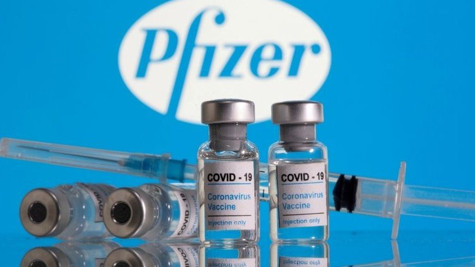
Covid: Canada authorises Pfizer vaccine for children aged 12 to 15

SharecloseShare pageCopy linkAbout sharingimage copyrightReutersCanada has authorised the use of the Pfizer coronavirus vaccine for children between the ages of 12 and 15. It is the first country to do so for that age group. The country’s health ministry made the decision based on data from phase three clinical trials on children that age. “The department determined that this vaccine is safe and effective when used in this younger age group,” an adviser at the ministry said. Pfizer says its jab works well in the age group.Canada has already authorised the use of the Pfizer vaccine in people over 16. The country has recorded more than 1.2 million coronavirus cases and roughly 20% of those have been in people under the age of 19. Children’s risk of becoming very ill or dying with Covid-19 is tiny, and throughout the pandemic they have very rarely needed hospital treatment.’No evidence’ schools spread lots of Covid How will we know Covid vaccines are safe?As part of the vaccine’s approval, Pfizer will have to continue providing information to Canada’s health ministry on the safety, efficacy and quality of the vaccine in those aged 12 to 15. Last March, Pfizer said initial results from trials of its vaccine in this age group showed 100% efficacy and a strong immune response. US President Joe Biden this week laid out plans to roll out vaccines for 12-15 year-olds as soon as possible. US media has reported that the authorisation could come as early as next week. What about other vaccine manufacturers? Pfizer is one of a number of vaccine manufacturers testing jabs on children. The aim of vaccinating them – particularly older children – would be to keep schools open, reduce the spread of coronavirus in the community and protect vulnerable children with conditions which put them at increased risk. Moderna and Johnson & Johnson are currently testing their vaccines on those aged 12-18 with Moderna’s data expected soon. Moderna and Pfizer are also testing their jabs on younger children between six months and 11-years-old. In the UK, AstraZeneca is testing its vaccine on 300 child volunteers. Researchers will assess whether the jab produces a strong immune response in children aged between six and 17.

Read more →

Process for eliminating unneeded cells may also protect against cancer

For all animals, eliminating some cells is a necessary part of embryonic development. Living cells are also naturally sloughed off in mature tissues; for example, the lining of the intestine turns over every few days.
One way that organisms get rid of unneeded cells is through a process called extrusion, which allows cells to be squeezed out of a layer of tissue without disrupting the layer of cells left behind. MIT biologists have now discovered that this process is triggered when cells are unable to replicate their DNA during cell division.
The researchers discovered this mechanism in the worm C. elegans, and they showed that the same process can be driven by mammalian cells; they believe extrusion may serve as a way for the body to eliminate cancerous or precancerous cells.
“Cell extrusion is a mechanism of cell elimination used by organisms as diverse as sponges, insects, and humans,” says H. Robert Horvitz, the David H. Koch Professor of Biology at MIT, a member of the McGovern Institute for Brain Research and the Koch Institute for Integrative Cancer Research, a Howard Hughes Medical Institute investigator, and the senior author of the study. “The discovery that extrusion is driven by a failure in DNA replication was unexpected and offers a new way to think about and possibly intervene in certain diseases, particularly cancer.”
MIT postdoc Vivek Dwivedi is the lead author of the paper, which appears today in Nature. Other authors of the paper are King’s College London research fellow Carlos Pardo-Pastor, MIT research specialist Rita Droste, MIT postdoc Ji Na Kong, MIT graduate student Nolan Tucker, Novartis scientist and former MIT postdoc Daniel Denning, and King’s College London professor of biology Jody Rosenblatt.
Stuck in the cell cycle
In the 1980s, Horvitz was one of the first scientists to analyze a type of programmed cell suicide called apoptosis, which organisms use to eliminate cells that are no longer needed. He made his discoveries using C. elegans, a tiny nematode that contains exactly 959 cells. The developmental lineage of each cell is known, and embryonic development follows the same pattern every time. Throughout this developmental process, 1,090 cells are generated, and 131 cells undergo programmed cell suicide by apoptosis.

Read more →

Stem cells create early human embryo structure in advance for fertility research

Exeter scientists have discovered a simple, efficient way to recreate the early structure of the human embryo from stem cells in the laboratory. The new approach unlocks news ways of studying human fertility and reproduction.
Stem cells have the ability to turn into different types of cell. Now, in research published in Cell Stem Cell and funded by the Medical Research Council, scientists at the University of Exeter’s Living Systems Institute, working with colleagues from the University of Cambridge, have developed a method to organise lab-grown stem cells into an accurate model of the first stage of human embryo development.
The ability to create artificial early human embryos could benefit research into infertility, by furthering understanding of how embryos develop, and the conditions needed to avoid miscarriage and other complications. The embryo models can also be used to test conditions that may improve the development of embryos in assisted conception procedures such as IVF.
The new discovery comes after the team found that a human stem cell was able to generate the founding elements of a blastocyst — the very early formation of an embryo after a fertilised egg divides. Professor Austin Smith, Director of the University of Exeter’s Living Systems Institute, said: “Finding that stem cells can create all the elements of an early embryo is a revelation. The stem cells come from a fully-formed blastocyst, yet they are able to recreate exactly the same whole embryo structure. This is quite remarkable and unlocks exciting possibilities for learning about the human embryo.”
The research has the potential to significantly advance understanding. Few human embryos are available for study, so until now, scientists have largely focussed on animal research, particularly mice, despite the fact that their reproductive systems differ significantly from humans. Around one in seven couples in the UK has difficulty conceiving.
In the research, the team arranged the stem cells into clusters and briefly introduced two molecules known to influence how cells behave in early development. They found that 80 per cent of the clusters organised themselves after 3 days into structures that look remarkably like the blastocyst stage of an embryo — a ball of around 200 cells that forms from the fertilised egg after 6 days. The team went on to show that the artificial embryos have the same active genes as the natural embryo.
The study was directed by Dr Ge Guo, of the University of Exeter’s Living Systems Institute, said: “Our new technique provides for the first time a reliable system to study early development in humans without using embryos. This shouldn’t be seen as a move towards producing babies in a laboratory, but rather as an important research tool that could benefit IVF and infertility studies.”
The next stage for the researchers is to understand how to develop the artificial embryos a few days further to study the critical period when an embryo would implant into the womb, which is when many embryos fail to develop properly.
Story Source:
Materials provided by University of Exeter. Note: Content may be edited for style and length.

Read more →

International study links brain thinning to psychosis

Subtle differences in the shape of the brain that are present in adolescence are associated with the development of psychosis, according to an international team led by neuroscientists at the University of Pittsburgh School of Medicine and Maastricht University in the Netherlands.
In results published today in JAMA Psychiatry, the differences are too subtle to detect in an individual or use for diagnostic purposes. But the findings could contribute to ongoing efforts to develop a cumulative risk score for psychosis that would allow for earlier detection and treatment, as well as targeted therapies. The discovery was made with the largest-ever pooling of brain scans in children and young adults determined by psychiatric assessment to be at high risk of developing psychosis.
“These results were, in a sense, sobering,” said Maria Jalbrzikowski, Ph.D., assistant professor of psychiatry at Pitt. “On the one hand, our data set includes 600% more high-risk youth who developed psychosis than any existing study, allowing us to see statistically significant results in brain structure. But the variance between whether or not a high-risk youth develops psychosis is so small that it would be impossible to see a difference at the individual level. More work is needed for our findings to be translated into clinical care.”
Psychosis is an umbrella term for a constellation of severe mental disorders that cause people to have difficulty determining what is real and what is not. Most often, individuals have hallucinations where they see or hear things that others do not. They also may have strongly held beliefs, or delusions, even when most people do not believe them. Schizophrenia is only one disorder associated with psychosis, and psychotic symptoms can occur in other psychiatric disorders, such as bipolar disorder, depression, body dysmorphic disorder or post-traumatic stress disorder. In people who receive a diagnosis of psychosis, there is a great deal of heterogeneity in outcomes over time.
Diagnosis usually happens in later adolescence and early adulthood, but most often symptoms begin to manifest in the teen years, when clinicians can use psychological assessments to determine a person’s risk of developing full-blown psychosis.
Jalbrzikowsi and Dennis Hernaus, Ph.D., assistant professor in the School of Mental Health and Neuroscience at Maastricht University, are co-chairs of the Enhancing Neuro Imaging Genetics Through Meta-Analysis (ENIGMA) Clinical High Risk for Psychosis Working Group. This group pooled structural magnetic resonance imaging (MRI) scans from 3,169 volunteer participants at an average age of 21 who were recruited at 31 different institutions. About half — 1,792 of the participants — had been determined to be at “clinical high risk for developing psychosis.” Of those high-risk participants, 253 went on to develop psychosis within two years. The co-chairs emphasized that this study would not be possible without the collaborative efforts of the 100-plus researchers involved.
When looking at all the scans together, the team found that those at high risk for psychosis had widespread lower cortical thickness, a measure of the thickness of the brain’s gray matter. In high-risk youth who later developed psychosis, a thinner cortex was most pronounced in several temporal and frontal regions.
Everyone goes through a cortical thinning process as they develop into an adult, but the team found that in younger participants between 12 and 16 years old who developed psychosis the thinning was already present. These high-risk youth who developed psychosis also progressed at a slower rate than in the control group.
“We don’t yet know exactly what this means, but adolescence is a critical time in a child’s life — it’s a time of opportunity to take risks and explore, but also a period of vulnerability,” Jalbrzikowski said. “We could be seeing the result of something that happened even earlier in brain development but only begins to influence behavior during this developmental stage.”
Hernaus stressed that these findings underscore the importance of early detection and intervention in people who show risk factors for developing psychosis, which include hearing whispers from voices that aren’t there and a family history of psychosis.
“Until now, researchers have primarily studied how the brains of people with clinical high risk for psychosis differ at a given point in time,” Hernaus said. “An important next step is to better understand brain changes over time, which could provide new clues on underlying mechanisms relevant to psychosis.”
This research received support from numerous funders listed in the JAMA Psychiatry manuscript. Jalbrzikowski received support from National Institute of Mental Health grant K01 MH112774.

Read more →

Targeted methods to control SARS-CoV-2 spread

At the beginning of the COVID-19 pandemic, intense social distancing and lockdown measures were the primary weapon in the fight against the spread of SARS-CoV-2, but they came with a monumental societal burden. New research from the Center for the Ecology of Infectious Diseases and the College of Public Health at the University of Georgia explores if there could have been a better way.
Published in the journal Proceedings of the Royal Society B, the research analyzes more palatable alternatives to the kind of social distancing mandates that threw a wrench at how businesses, schools and even family gatherings work. The alternatives — widespread testing, contact tracing, quarantines, certification for non-infected people and other public health policy measures — can slow the spread when combined together, but only with significant investments and broad public compliance.
“I understand why government leaders quickly enacted strict social distancing mandates as the COVID-19 pandemic was rapidly spreading in 2020,” said lead author John Drake, director of the Center for the Ecology of Infectious Diseases and Distinguished Research Professor in the Odum School of Ecology. “This was the best that we could do at the time. However, school and workplace closures, gathering limits and shelter-in-place orders have had extreme economic consequences. These are harsh, and we really need to find alternative solutions.”
Drake worked with other researchers to develop two models. One targeted how to find infected people to limit transmission through active case finding (through testing of at-risk individuals), thorough contact tracing when cases arise, and quarantines for people infected and their traced contacts.
The second model focused on a strategy of limiting exposure by certifying healthy individuals.
“Each model was tested independently and in combination with general non-pharmaceutical interventions (NPIs),” said co-author Kyle Dahlin, a postdoctoral associate with the center.

Read more →

Personalized sweat sensor reliably monitors blood glucose without finger pricks

Many people with diabetes endure multiple, painful finger pricks each day to measure their blood glucose. Now, researchers reporting in ACS Sensors have developed a device that can measure glucose in sweat with the touch of a fingertip, and then a personalized algorithm provides an accurate estimate of blood glucose levels.
According to the American Diabetes Association, more than 34 million children and adults in the U.S. have diabetes. Although self-monitoring of blood glucose is a critical part of diabetes management, the pain and inconvenience caused by finger-stick blood sampling can keep people from testing as often as they should. Scientists have developed ways to measure glucose in sweat, but because levels of the sugar are much lower than in blood, they can vary with a person’s sweat rate and skin properties. As a result, the glucose level in sweat usually doesn’t accurately reflect the value in blood. To obtain a more reliable estimate of blood sugar from sweat, Joseph Wang and colleagues wanted to devise a system that could collect sweat from a fingertip, measure glucose and then correct for individual variability.
The researchers made a touch-based sweat glucose sensor with a polyvinyl alcohol hydrogel on top of an electrochemical sensor, which was screen-printed onto a flexible plastic strip. When a volunteer placed their fingertip on the sensor surface for 1 minute, the hydrogel absorbed tiny amounts of sweat. Inside the sensor, glucose in the sweat underwent an enzymatic reaction that resulted in a small electrical current that was detected by a hand-held device. The researchers also measured the volunteers’ blood sugar with a standard finger-prick test, and they developed a personalized algorithm that could translate each person’s sweat glucose to their blood glucose levels. In tests, the algorithm was more than 95% accurate in predicting blood glucose levels before and after meals. To calibrate the device, a person with diabetes would need a finger prick only once or twice per month. But before the sweat diagnostic can be used to manage diabetes, a large-scale study must be conducted, the researchers say.
Story Source:
Materials provided by American Chemical Society. Note: Content may be edited for style and length.

Read more →

Novel CBD analog shows promising pain-halting effects in mice

For patients with chronic pain, ineffective treatments, lowered work productivity, and other factors often coalesce, fueling feelings of hopelessness and anxiety and setting the stage for even bigger problems, including substance use disorders. In 2017 alone, some 18 million Americans misused prescription pain relievers over the course of the previous year. In many of these instances, patients suffering from chronic pain became addicted to prescription opioids.
In addition to being highly addictive, many studies suggest that prescription opioids do not effectively control pain over the long term, and hence researchers have been exploring various alternatives, including cannabidiol (CBD). CBD is a non-psychoactive substance derived from the Cannabis plant.
Studies have shown that while CBD reduces pain sensation in animals, its ability to do so in humans is limited by low bioavailability, the extent to which the drug successfully reaches its site of action. Now, new work by scientists at the Lewis Katz School of Medicine at Temple University suggests this obstacle may be overcome by a novel CBD analog known as KLS-13019.
“In a mouse model of chemotherapy-induced peripheral neuropathy (CIPN), we’ve been able to show for the first time that KLS-13019 works as well as, if not better than, CBD in preventing the development of neuropathy and reversing pain sensitivity after pain has been established,” said Sara Jane Ward, PhD, Assistant Professor of Pharmacology at the Katz School of Medicine and senior investigator on the new study. The findings were published online April 6 in the British Journal of Pharmacology.
KLS-13019, developed by the Pennsylvania-based bio-pharmaceutical and phyto-medical company Neuropathix, Inc., is among the most promising neuroprotective CBD analogs currently under investigation. In previous work in cell models, it was found to be more potent than CBD, and studies in animals suggested it had improved bioavailability.
Encouraged by those initial studies, Dr. Ward and colleagues set out to better understand the pain-relieving capabilities of KLS-13019, relative to CBD, in animals with CIPN. CIPN is a common side effect of certain cancer treatments that damage peripheral nerves, which carry sensory information to the arms, legs, and brain. The severe pain, or peripheral neuropathy, caused by CIPN manifests in different ways in human patients but frequently involves tingling or burning sensations and numbness, weakness, or discomfort in the limbs.

Read more →

Release of drugs from a supramolecular cage

How can a highly effective drug be transported to the precise location in the body where it is needed? In the journal Angewandte Chemie, chemists at Heinrich Heine University Düsseldorf (HHU) together with colleagues in Aachen present a solution using a molecular cage that opens through ultrasonification.
Supramolecular chemistry involves the organization of molecules into larger, higher-order structures. When suitable building blocks are chosen, these systems ‘self-assemble’ from their individual components.
Certain supramolecular compounds are well suited for ‘host-guest chemistry’. In such cases, a host structure encloses a guest molecule and can shield, protect and transport it away from its environment. This is a specialist field of Dr. Bernd M. Schmidt and his research group at the Institute of Organic and Macromolecular Chemistry at HHU.
The chemists in Düsseldorf collaborated with colleagues from the DWI Leibniz Institute for Interactive Materials to find a system that may one day even be able to transport cargo molecules through the human body and release the drug at the desired location.
The solution may be to use discrete ‘Pd6(TPT)4 cages’. These are octahedral cage-like assemblies, bearing polymer chains on each vertex. They are comprised of four triangular panels, palladium atoms and connecting units.
When the individual components are added to an aqueous solution in the correct ratio, the cages self-assemble. If smaller, hydrophobic molecules are added to the cages, they enter the cavities. The researchers demonstrated this effect using pharmaceutically active molecules, like ibuprofen and progesterone.
“The special trick with our system involves the pre-determined rupture points,” explains Dr. Schmidt, last author of the study. “The palladium atoms hold all compounds with a comparatively weak bond. Once you succeed in breaking the atoms out of the compound, the entire octohedral structure breaks apart.”
To break the bonds, the researchers in Aachen use powerful ultrasonification similar to that used medically to break down kidney stones, for example. In water, the ultrasound creates cavitation bubbles that burst and exert huge mechanical shear force on the long polymer chains. The forces are so powerful that the palladium atoms are actually torn from the vertices and thus rupture the octahedral cage. The small drug molecules are agitated in the process but are not damaged.
Dr. Robert Göstl (DWI) says: “Localised ultrasound radiation of the tissue to be treated could mean that the drug transported in the cage is later released at the exact location where the therapy is needed.” The drug molecules used in the study serve merely as examples. In principle, a large number of different hydrophobic molecules can be packed in the cage. Unlike other host-guest systems described, it is not necessary to alter the drug molecules chemically in order to get them in the cage. “To treat tumours, it would be feasible to use cytostatic drugs as the cargo, for example. By releasing them directly at the site of a solid tumour, it may be possible to have chemotherapy that uses much less of the drug and thus has lesser side effects,” explains Schmidt.
This is helped by the fact that the defined cargo volume makes it possible to measure precisely how much of the drug is released at the target site. “The dose administered could even be calculated precisely.”
The study is a Proof of Concept that demonstrated the feasibility of the approach. It also convinced the reviewers and publishers of the journal Angewandte Chemie, who rated the publication as very important.
“The next steps involve determining how real cells respond to our cages. Before any medical use, we need to ensure that they are not toxic.”
Story Source:
Materials provided by Heinrich-Heine University Duesseldorf. Original written by Arne Claussen. Note: Content may be edited for style and length.

Read more →

Microneedles are promising devices for painless drug delivery with minimal side effects

A recent study from the University of Helsinki monitors progress in the development of microneedles for immunotherapy and discusses the challenges regarding their production. Researchers suggest using microneedles for immunotherapy due to the abundance of immune cells under the skin. The aim is to vaccinate or treat different diseases, such as cancer and autoimmune disorders, with minimal invasiveness and side effects.
“Our study addresses the recent achievements in the development of microneedles for immunotherapy of hard-to-treat and chronic diseases to achieve the highest efficiency with minimal side effects,” says Professor Hélder A. Santos, from the University of Helsinki, Faculty of Pharmacy.
As a result of the dynamic nature of the human immune system, the current immunotherapy approaches have mostly been challenged by the lack of adequate long-term responses in patients. On the other hand, the development of microneedles has resulted in the improvement and expansion of immuno-reprogramming strategies since the dermis layer of the skin house a high accumulation of dendritic cells, macrophages, lymphocytes, and mast cells. These cells can transfer generated immunotherapeutic signals to the damaged organs.
According to the paper, microneedles maintain many outstanding properties, such as direct delivery of antibodies, allergens, and therapeutic antigens into the skin, minimal invasiveness, facile fabrication, excellent biocompatibility, convenient administration, and bypassing the first-pass metabolism that allows direct translocation of therapeutics into the systematic circulation. Moreover, transdermal microneedle can improve the biological effect of drugs through adjustable drug release.
Recent advancements of microneedles for immunotherapy of cancer
One of the primary objectives of cancer immunotherapy is the establishment of a broad tumor-targeting T cell repertoire that is able to recognize and destroy heterogeneous tumor cell populations. The paper indicates the suitability of the microneedles for this purpose and their future clinical use, owing to the short treatment time with improvement of patient convenience, and prolonging the overall survival compared to traditional vaccination.

Read more →

3D bioprinting technique controls cell orientation

3D bioprinting can create engineered scaffolds that mimic natural tissue. Controlling the cellular organization within those engineered scaffolds for regenerative applications is a complex and challenging process.
Cell tissues tend to be highly ordered in terms of spatial distribution and alignment, so bioengineered cellular scaffolds for tissue engineering applications must closely resemble this orientation to be able to perform like natural tissue.
In Applied Physics Reviews, from AIP Publishing, an international research team describes its approach for directing cell orientation within deposited hydrogel fibers via a method called multicompartmental bioprinting.
The team uses static mixing to fabricate striated hydrogel fibers formed from packed microfilaments of different hydrogels. In this structure, some compartments provide a favorable environment for cell proliferation, while others act as morphological cues directing cell alignment. The millimeter-scale printed fiber with the microscale topology can rapidly organize the cells toward faster maturation of the engineered tissue.
“This strategy works on two principles,” said Ali Tamayol, coauthor and an associate professor in biological engineering at UConn Health. “The formation of topographies is based on the design of fluid within nozzles and controlled mixing of two separate precursors. After crosslinking, the interfaces of the two materials serve as 3D surfaces to provide topographical cues to cells encapsulated within the cell permissive compartment.”
Extrusion-based bioprinting is the most widely used bioprinting method. In extrusion-based bioprinting, the printed fibers are typically several hundreds of micrometers in size with randomly oriented cells, so a technique providing topographical cues to the cells within these fibers to direct their organization is highly desirable.
Conventional extrusion bioprinting also suffers from high shear stress applied to the cells during the extrusion of fine filaments. But the fine scale features of the proposed technique are passive and do not compromise other parameters of the printing process.
To direct cellular organization, according to the team, extrusion-based 3D-bioprinted scaffolds should be made from very fine filaments.
“It makes the process challenging and limits its biocompatibility and the number of materials that can be used, but with this strategy larger filaments can still direct cellular organization,” said Tamayol.
This bioprinting technique “enables production of tissue structures’ morphological features — with a resolution up to sizes comparable to the cells’ dimension — to control cellular behavior and form biomimetic structures,” Tamayol said. “And it shows great potential for engineering fibrillar tissues such as skeletal muscles, tendons, and ligaments.”
Story Source:
Materials provided by American Institute of Physics. Note: Content may be edited for style and length.

Read more →