Preventing the long-term effects of traumatic brain injury

You’ve been in a car accident and sustained a head injury. You recovered, but years later you begin having difficulty sleeping. You also become very sensitive to noise and bright lights, and find it hard to carry out your daily activities, or perform well at your job.
This is a common situation after a traumatic brain injury — many people experience bad side effects months or years later. These long-term effects can last a few days or the rest of a person’s life.
“No therapies currently exist to prevent the disabilities that can develop after a brain trauma,” says Jeanne Paz, PhD, associate investigator at Gladstone Institutes. “So, understanding how the traumatic brain injury affects the brain, especially in the long term, is a really important gap in research that could help develop new and better treatment options.”
In a new study published in the journal Science, Paz and her team helped close that gap. They identified a specific molecule in a part of the brain called the thalamus that plays a key role in secondary effects of brain injury, such as sleep disruption, epileptic activity, and inflammation. In collaboration with scientists at Annexon Biosciences, a clinical-stage biopharmaceutical company, they also showed that an antibody treatment could prevent the development of these negative outcomes.
A Vulnerable Brain Region
Traumatic brain injuries, which range from a mild concussion to a severe injury, can be the result of a fall, sports injury, gunshot injury, blow to the head, explosion, or domestic violence. Often, soldiers returning from war also suffer head injuries, which commonly lead to the development of epilepsy. Traumatic brain injury affects 69 million people around the world annually, and is the leading cause of death in children and a major source of disability in adults.

Read more →

Discovery about cells' 'batteries' boosts battle against many diseases

A top exercise researcher at the University of Virginia School of Medicine has revealed how our bodies ensure the proper functioning of the powerhouses of our cells. The findings could open the door to better treatments for many common diseases, including Alzheimer’s and diabetes.
The new research from UVA’s Zhen Yan, PhD, and colleagues reveals how our cells sense problems and perform quality control on cellular “batteries” known as mitochondria. Yan has spent many years seeking to better understand the workings of mitochondria, and he calls the new discovery the most exciting of his career.
“Mitochondria are the center of universe to me since literally all cells in our body rely on mitochondria for energy production and must have a bulletproof system to ensure the powerhouses are functioning properly,” said Yan, the director of the Center for Skeletal Muscle Research at UVA’s Robert M. Berne Cardiovascular Research Center. “Chronic diseases, also known as noncommunicable diseases, such as diabetes, heart failure and Alzheimer’s disease that catastrophically impact so many individuals, families and the whole society are caused by problems of the mitochondria in the cells.”
Stress Detectors
Yan and his team discovered special sensors on the outer membrane surrounding the mitochondria in various tissues in both mice and humans. These sensors detect “energetic stress,” such as caused by exercise or fasting, and signal for damaged mitochondria to be degraded and removed. This essential cleanup process is known as “mitophagy,” and its existence was first suggested more than 100 years ago. But how it works has never been fully understood. Yan’s new research offers long-sought answers.
Yan and his colleagues found that the mitochondrial sensors, known as “mitoAMPK,” exist in slightly different forms in different tissues. For example, one type seemed particularly active in skeletal muscle. In a new scientific paper outlining their findings, the researchers describe the variety of sensors as “unexpectedly complex.” They go on to outline how these sensors provide a vital damage-control system that safeguards our cellular energy supply.
One finding of the study that Yan finds extremely exciting: Treating mice with metformin, the most effective, first-line anti-diabetes drug, activates mitoAMPK in skeletal muscles without activating AMPK in the other parts of the cells. The finding is the best illustration of the importance of activating mitoAMPK and mitochondrial quality control in treatment of a common chronic disease that is known to be caused by accumulation of dysfunctional mitochondria in our body. It also explains why regular exercise is so powerful in preventing and treating such diseases.
The new insights gained into mitochondrial quality control will boost efforts to develop new treatments for non-communicable diseases that have reached pandemic proportions and are estimated to cause 71% of all deaths.
Yan, who is part of UVA’s Division of Cardiovascular Medicine, says it will be important for doctors to better understand how specific diseases interfere with mitochondrial function. And his new findings set the stage for that.
“We have developed genetic models for pinpointing the key steps of mitoAMPK activation and are on our way to discover the magic molecules that are controlled by mitoAMPK,” Yan said. “The findings taught us a lot about the beauty of the sensor system in our body. Society should definitely take advantage of these findings to promote regular exercise for health and disease prevention and develop effective exercise-mimetic drugs.”
Story Source:
Materials provided by University of Virginia Health System. Original written by Josh Barney. Note: Content may be edited for style and length.

Read more →

Socializing may improve older adults’ cognitive function in daily life

Socializing with others is important for mental health and wellbeing, and it may help improve cognition, as well — especially for older adults, according to new research.
In a study led by Ruixue Zhaoyang, assistant research professor of the Center for Healthy Aging at Penn State, the researchers found that when adults between the ages of 70 and 90 reported more frequent, pleasant social interactions, they also had better cognitive performance on that day and the following two.
Zhaoyang said the findings — recently published in the journal PLOS ONE — may have special relevance now due to social distancing mitigation measures throughout the COVID-19 pandemic.
“Our study is one of the first to show that whether you have social interactions on one day can immediately affect your cognitive performance that same day and also on the following days,” Zhaoyang said. “The fact that we found that the cognitive benefits of having pleasant social interactions could manifest over such a short time period was a happy surprise and could be a promising area for future intervention studies.”
According to the Alzheimer’s Association, more than six million Americans are currently living with Alzheimer’s disease, and that number is expected to rise to almost 13 million by 2050. Additionally, deaths from Alzheimer’s disease and other dementias have risen by 16 percent during the COVID-19 pandemic.
Zhaoyang said that without reliable drug therapies, it’s critical to find ways to help prevent these conditions before they reach the clinical stage.

Read more →

Enzyme could be major driver of preeclampsia

A new study by UT Southwestern scientists indicates that an enzyme called protein phosphatase 2 (PP2A) appears to be a major driver of preeclampsia, a dangerous pregnancy complication characterized by the development of high blood pressure and excess protein in the urine. The finding, published in Circulation Research, could lead to new treatments for preeclampsia other than premature delivery, which is often the only option.
“Preeclampsia is a regrettably common cause of premature birth, which can be life-threatening for babies and lead to lifelong consequences. Through identifying PP2A’s role in this condition, we may be able to develop treatments for preeclampsia that are far better for both mothers and babies,” said study leader Philip W. Shaul, M.D., Professor and Vice Chair for Research in the Department of Pediatrics at UTSW and Director of the Center for Pulmonary and Vascular Biology. Dr. Shaul co-led this study with Chieko Mineo, Ph.D., Professor of Pediatrics and Cell Biology.
Preeclampsia, which affects 5 to 7% of pregnant women worldwide, can be deadly for gestating mothers and their babies and requires delivery at a premature stage.
Although the causes of preeclampsia aren’t well understood, explained Dr. Shaul, researchers have linked the condition to a variety of risk factors. One is an autoimmune disease known as antiphospholipid syndrome (APS), in which antibodies react to proteins on the surface of some cells. Although APS is relatively rare, affecting only about 5 in every 100,000 people, studies have identified APS antibodies in about 29% of pregnant women with preeclampsia.
To better understand how APS leads to preeclampsia, Dr. Shaul, Dr. Mineo, and their colleagues created an animal model by injecting pregnant mice with APS antibodies. These animals developed high blood pressure and a rise in urine protein, which are characteristics of preeclampsia. In contrast, the APS antibodies didn’t affect blood pressure in nonpregnant mice.
Based on previous work, the researchers knew that a protein called ApoER2 may be related to the harmful actions of APS antibodies on placental cells called trophoblasts. These cells, which normally journey from the fetal side of the placenta to the maternal side to provide the fetus with nutrients, don’t successfully make that connection in preeclampsia. In mice, the APS antibodies prevented trophoblast migration, and growth of the fetus was restricted. When the researchers genetically engineered mice without ApoER2 in trophoblasts, the fetuses developed normally despite APS antibody treatment, and the mothers were protected from developing preeclampsia.
But the scientists knew that ApoER2 didn’t tell the whole story. They found that in the presence of the APS antibodies, ApoER2 triggers the activity of PP2A, an enzyme that regulates protein functions throughout the body. Further experiments showed that in the pregnant mice with APS antibodies, heightened activity in PP2A increased trophoblast production of proteins known to be involved in preeclampsia.
When the researchers gave the pregnant mice a drug that inhibits PP2A, they were protected from preeclampsia, and the treatment had no apparent harmful effects on the mice or their gestating babies.
Hoping to translate these findings to human patients, the scientists examined placentas from women with APS, finding that they too had increased activity of PP2A. However, in a surprising turn, they discovered that compared with placentas from normal pregnancies, those from preeclamptic patients without APS also had increased PP2A activity, suggesting that this mechanism could be operating in a variety of forms of preeclampsia. With further research, Dr. Shaul said, treatments targeting PP2A or its related machinery in the trophoblast may eventually be viable treatments for preeclampsia in pregnant women.
Story Source:
Materials provided by UT Southwestern Medical Center. Note: Content may be edited for style and length.

Read more →

Potential indicator for obesity risk detected during sleep

Throughout the day, we gain energy by breaking down carbohydrates, fats, and proteins in our bodies through the process of metabolism. For example, immediately after eating, most of our energy comes from carbohydrates, while after fasting, most comes from fats. The body’s ability to switch metabolic energy sources in response to changes in nutritional state, such as after meals and during sleep, is called metabolic flexibility. Research has shown that disrupted flexibility is associated with diseases such as obesity and diabetes. Professor Kumpei Tokuyama and his team of researchers at the University of Tsukuba have been studying metabolism during sleep. “We were interested in how metabolism changes during sleep and whether we could detect any metabolic differences in people with inflexible metabolisms,” Professor Tokuyama explains.
The basic method used by the team centers around a measurement called the respiratory quotient, abbreviated as RQ, which measures how much oxygen we use and how much carbon dioxide we breathe out. When the amounts are equal — an RQ equal to 1 — it signals that the energy source is carbohydrates. When the ratio is lower, around 0.8, it indicates that fats or proteins are being used as the energy source. To characterize metabolic changes over time, the researchers measured the carbon dioxide/oxygen ratios from 127 people, every 5 minutes over a 24-hour period.
The first finding was unexpected. Because sleep is like a period of fasting, it could be expected that RQs would decrease all night long, indicating that fat was being burned off more and more as sleep progressed. Instead, they found a different pattern. “We were surprised to find that while RQ values decreased steadily at the beginning of sleep, after reaching a low point, they began to rebound after midnight and continued to increase until people woke up,” says Professor Tokuyama.
Next, the team separated the participants based on how much their RQs varied. High variability means that metabolism is flexible, with RQs values going up and down depending on the body’s need throughout the day. After dividing participants into metabolic flexible and inflexible groups, the team found that even though average RQs over 24 hours were the same between the groups (as were their ages, BMIs, and amounts of body fat), RQs at night were higher for those with less flexible metabolisms, indicating that the participants were burning more carbohydrates than fat.
These findings have the potential for practical use. As Professor Tokuyama explains, “Preventing diseases such as obesity and diabetes is much more preferable to treating them. Yearly checkups that focus on measuring sleeping RQ values could help screen for people at risk for developing metabolic diseases, thus allowing timely interventions.”
Story Source:
Materials provided by University of Tsukuba. Note: Content may be edited for style and length.

Read more →

Newly developed software unveils relationships between RNA modifications and cancers

In a research breakthrough, a team of researchers from the Cancer Science Institute of Singapore (CSI Singapore) at the National University of Singapore has developed a software that can help reveal the relationships between RNA modifications and the development of diseases and disorders.
Led by Professor Daniel Tenen and Dr Henry Yang, the scientists devised ModTect — a new computational software that can identify RNA modifications using pre-existing sequencing data from clinical cohort studies. With ModTect, the team carried out their own novel pan-cancer study covering 33 different cancer types. They found associations between these RNA modifications and the different survival outcomes of cancer patients.
“This work is one of few studies demonstrating the association of mRNA modification with cancer development. We show that the epitranscriptome was dysregulated in patients across multiple cancer types and was additionally associated with cancer progression and survival outcomes,” explained Dr Henry Yang, Research Associate Professor from CSI Singapore.
“In the past decade, the ability to sequence the Human Genome has transformed the study of normal processes and diseases such as cancer. We anticipate that studies like this one, eventually leading to complete sequencing of RNA and detecting modifications directly in RNA, will also have a major impact on the characterisation of disease and lead to novel therapeutic approaches,” commented Prof Tenen, Senior Principal Investigator from CSI Singapore.
The team’s breakthrough was published in the scientific journal Science Advances on 4 August 2021.
What are RNA modifications?
While most people are familiar with DNA, RNA plays just as much of a vital role in the human body’s cellular functions. Unlike DNA, which has the double-helix structure that most people are familiar with, RNA is a family of single-stranded molecules that perform various essential biological roles.

Read more →

Disease genes help developing brains

Holoprosencephaly (HPE) affects around one to four in every 1,000 unborns, and occurs when the cerebral hemispheres of the forebrain fail to divide or only partially divide. It is the most common malformation of the forebrain in humans and is associated with facial disfigurements, such as cleft lip and cleft palate or eyes that are very close together — even to the extent that the two eyeballs merge. The majority of fetuses affected die while still in the womb.
The exact causes of HPE are not yet fully understood. In addition to environmental pollutants and illness of the expectant mother, genetic factors can play a role — including mutations in the genes of the so-called Sonic Hedgehog (SHH) signaling pathway. This pathway controls the embryonic development of organs and the nervous system. Gene defects and a resulting loss of function of LRP2, an SHH co-receptor, result in brain defects that manifest themselves very differently. “We wanted to know why the severity of this disease varies so much,” says Dr. Annette Hammes, head of the Molecular Pathways in Cortical Development Lab at the Max Delbrück Center for Molecular Medicine in the Helmholtz Association (MDC). “While some sufferers have no or only mild symptoms, others have to live with severe deformities — even if the two patients are related to each other and we can therefore assume that the disease is caused by the same gene mutation.”
Disease genes restore the SHH pathway
Researchers have long assumed that there are genes that positively influence this malformation or even prevent it altogether. The Hammes Lab team has now identified two new candidates: “ULK4 and PTTG1, also known as securin,” says co-lead author Dr. Nora Mecklenburg, who was a postdoctoral researcher with Hammes at the time of the study. ULK4 is a gene that has so far been associated with schizophrenia and bipolar disorder, while PTTG1 is mainly researched in connection with cancer. In the journal “Development,” the scientists explain how these proteins can restore an impaired SHH pathway. Nine years of work went into the study, which the journal classifies as a “research highlight.”
But the results were also partly down to chance. Hammes and her team had been studying mice with LRP2 mutations for many years, together with the MDC lab led by Professor Thomas Willnow. “We know that LRP2 influences the formation of the neural tube in early embryogenesis, and it is out of this that the nervous system later develops,” explains the neuroscientist. Without LRP2, the SHH pathway is not sufficiently activated and malformations of the neural tube occur at a very early stage of pregnancy — often resulting in miscarriage. When the scientists crossed the LRP2 mutants of the commonly used black-coated mouse strain (known as “Black 6” for short) with another mouse strain with white coats, the result was very surprising: The white-coated offspring did not show any malformations of the brain or face — despite having a mutation in the SHH co-receptor LRP2. The team concluded that there must be as-yet-unknown factors that influence the SHH pathway, and set out to find them.
RNA analysis with high-throughput sequencing
To do this, Mecklenburg first bred the different mouse strains and examined the animals with regard to their disease characteristics, signaling pathways and genetic makeup. Together with her co-lead authors Franziska Witte, then a doctoral student in Professor Norbert Hübner’s MDC lab, and Izabela Kowalczyk, a doctoral student with Hammes, she sequenced and analyzed the RNA of embryonic cells of the different strains using high-throughput methods. The three scientists discovered that, despite having a similar genome sequence, the cells have completely different transcriptomes — meaning the genes are read very differently. “The extent of the differences even between the wild types of the two mouse strains at this early stage of embryonic development really surprised us,” says Mecklenburg.
The researchers conducted further studies and found that, in the white-coated LRP2 mutants, as well as in the wild types of this strain, certain genes, including ULK4 and PTTG1, are strongly upregulated compared to the Black 6 mice. To see if this affects the SHH pathway, they introduced the genes into cells lacking LRP2 function. “We were able to see that they significantly boost the Sonic Hedgehog signaling pathway,” says Kowalczyk. Their conclusion: “More ULK4 and PTTG1 are produced in the LRP2 mutants with the white mouse ancestors. They compensate for the missing LRP2, restore a sufficiently strong SHH pathway, and prevent the malformations from occurring.” This finding casts the disease genes ULK4 and PTTG1 in a completely new light: While high levels of expression can trigger diseases in the adult organism, they can actually positively influence the development of an embryo. The scientists were also able to pinpoint the location from which these factors amplify the SHH pathway — the antenna-like projections of the neuroepithelial cells, which are the cells that line the inside of the neural tube.
Decoding — and maybe even preventing — genetic diseases
“The fact that we have identified these candidate genes that modulate the SHH pathway in mice takes our knowledge of holoprosencephaly and other genetic diseases one step further,” says Hammes. “With this knowledge, we may even find a way to prevent them.” But there is still a long way to go until then, and the next step for her team is to explore what role the newly discovered SHH pathway modifiers play — not only during embryonic development, but also in the adult brain. The scientists have already located PTTG1 in the cytoskeleton of neurons — a protein network in the cytoplasm that gives the cells stability. The team is currently investigating what role the gene plays in this location.

Read more →

Link between ADHD and dementia across generations

A large study at Karolinska Institutet in Sweden has found a link between ADHD and dementia across generations. The study, published in Alzheimer’s & Dementia: The Journal of the Alzheimer’s Association, shows that parents and grandparents of individuals with ADHD were at higher risk of dementia than those with children and grandchildren without ADHD.
“The findings suggest that there are common genetic and/or environmental contributions to the association between ADHD and dementia. Now we need further studies to understand the underlying mechanisms,” says the study’s first author Le Zhang, PhD student at the Department of Medical Epidemiology and Biostatistics at Karolinska Institutet.
ADHD (attention-deficit/hyperactivity disorder) is a neurodevelopmental disorder characterized by inattention, impulsiveness and hyperactivity. It affects an estimated 3 percent of adults worldwide.
The number of new ADHD diagnoses has increased dramatically in the last decades amid increasing awareness and knowledge about the disorder. However, since the diagnosis is still relatively new, there has only been a limited number of small studies on the development of dementia in people with ADHD, often with conflicting results.
In the current study, the researchers wanted to overcome this by examining to what extent older generations to individuals with ADHD were diagnosed with dementia. The study looked at more than two million people born in Sweden between 1980 and 2001, of whom around 3.2 percent were diagnosed with ADHD. Using national registries, the researchers linked these persons to over five million biological relatives, including parents, grandparents and uncles and aunts, and investigated to what extent these relatives developed dementia.
The researchers found that parents of individuals with ADHD had 34 percent higher risk of dementia than parents of individuals without ADHD. The risk of Alzheimer’s disease, the most common type of dementia, was 55 percent higher in parents of individuals with ADHD. Individuals with ADHD were more likely to have parents with early-onset dementia than late-onset.
The researchers note that the absolute risk of dementia was low for the parent cohort; only 0.17 percent of the parents were diagnosed with dementia during the follow-up period.
The association was lower for second-degree relatives of individuals with ADHD, i.e. grandparents and uncles and aunts. For example, grandparents of individuals with ADHD had 10 percent increased risk of dementia compared to grandparents of individuals without ADHD.
While the study is unable to determine a cause-and-effect relationship, the researchers present several potential explanations that can be explored in future research.
“One could imagine that there are undiscovered genetic variants that contribute to both traits, or family-wide environmental risk factors, such as socioeconomic status, that may have an impact on the association,” says Zheng Chang, researcher at the Department of Medical Epidemiology and Biostatistics at Karolinska Institutet, and the study’s last author. “Another possible explanation is that ADHD increases the risk of physical health conditions, which in turn leads to increased risk of dementia.”
Story Source:
Materials provided by Karolinska Institutet. Note: Content may be edited for style and length.

Read more →

Ann Patchett on Taking a Bad Fall to Find Good Luck

Ann Patchett finds that misfortune in small doses can cast a glittering light on the rest of life.On a Sunday morning in the middle of July, I woke up tired. Who knows why? Maybe, like the dog, I had spent the night chasing rabbits in my sleep. I gave serious consideration to skipping my morning exercises (hadn’t the rabbits been enough?), but then decided to push ahead on the belief that an adherence to routine helps more often than it hurts. Surely gold medalist Sunisa Lee had been tired in Tokyo that morning, but she went flying through the air all the same.When I got to the step-up portion of the 7-Minute Workout, I too was briefly flying. But my liftoff was misaligned, so that coming down I glanced off the edge of my step stool and hit the floor with my full weight on the side of my left foot.Pop!After lying on my back for a few minutes, panting through self-recrimination and the bright crush of pain, I crawled to the phone and called my husband. Karl found me on the floor, foot aloft. He’s a doctor, and he took my tennis shoe off with professional care. “Did you hurt yourself anywhere else?”I said no, thinking my quickly inflating foot was injury enough.“Did you hit your head?” He was gently palpating my foot to see what points made me yelp, while introducing the topic of gratitude into the conversation. I had not hit my head.“That’s how it happens,” he said, helping me to the bed. “You hit your head on the bookcase on the way down. Then it all falls apart.”Karl said we could go to the emergency room right away or wait until tomorrow to see a doctor in the clinic. I opted for the ice pack, the Motrin and the pile of pillows. I opted to wait. Tennessee, the state where we live, is rife with people who decided to pass on the Covid vaccine, which meant that even though we were vaccinated, emergency rooms were no place to sit and wait.The next day the orthopedist showed me the X-rays of my left foot. He told me I had badly sprained it, along with tearing some ligaments. He would get me a walking boot and, in time, all would be well. The doctor was almost to the door when he turned and looked at me again. “Let’s get one more X-ray,” he said.He was smiling when he came back, the bearer of good news. He told me my ankle was fractured. “I’m not going to do surgery,” he said cheerfully. “I could put a screw in there, but I’m not going to do it.” Once immobilized, the bit of bone that had cracked off would mend itself.“Oh,” Karl said, shaking his head after the doctor left us, “are you ever lucky.” He had seen his share of poor outcomes for ankle surgery. In his long career, he had seen pretty much everything.***When I was a child in Catholic school, the nuns never tired of telling us how lucky we were. Of course we were lucky in the obvious ways that should never be taken for granted — lucky for our health, our food, our families, lucky to be able to go to school — but in the face of real disaster, our luck escalated dramatically.At 9, when I came back to school after a car accident, they tallied up my good fortune: a broken nose, a broken wrist, my lip stitched back together, shards of glass still pushing out of my skull — it could have been so much worse! My sister was worse, she was still in the hospital. She would be there for awhile, resting between the white sheets of her astonishing luck. She should have been dead, and she wasn’t.At the time, I thought the nuns were idiots. They simply refused to see how we suffered. But now — 48 years later — I think, man, were we lucky.“If you won’t even complain about being injured and bedridden, I worry that you’re a constitutionally cheerful person who can see the bright side in any situation and this whole thing isn’t going to work out,” a new young friend teased me in an email. I told her not to worry, I am fully capable of misery and complaint, I’m just saving mine.Had I leapt up on a step stool and missed my landing two years ago, I doubt I would have managed the situation with quite so much sagacity. I would have found the boot burdensome (it is). I would have said the timing was impossible (no matter what the timing was). But the pandemic has taught me that my plans are of no importance, that everything can be canceled, that I’m lucky to have a house to live in and a person I love to live with.As is true with most writers, I have a talent for stillness which has only been fortified by the last year and a half. Eight more weeks in the house doesn’t actually constitute a problem. My sprain-ligament-fracture trifecta doesn’t actually constitute a problem. It turns out I know a lot of people who’ve had metal plates screwed into their ankles, and we all know a lot of people who’ve had to deal with things much worse than that.My friend Sister Nena, who taught me to read when I was 6, called to check on me. She’s broken both of her feet before, once the left and once the right. She wanted to know if I had a walking boot. I told her I did. “Oh,” she said, “you’re so lucky.”Bad luck in small doses can cast a glittering light on the rest of life. It shows us just how close we came to smashing our heads on the bookcase, and so makes us look at the bookcase (the room, the house, the street, the town, the life) with a new sense of wonder. Sooner or later, in one form or another, the terrible thing will happen. I didn’t understand that when I was young, no matter how many nuns tried to tell me. Now, I think I do. And I’m grateful that this time I got off easy.Ann Patchett is the co-owner of Parnassus Books in Nashville. Her essay collection “These Precious Days” will be published by HarperCollins in November 2021.

Read more →

Otro riesgo de la covid: problemas persistentes en los riñones

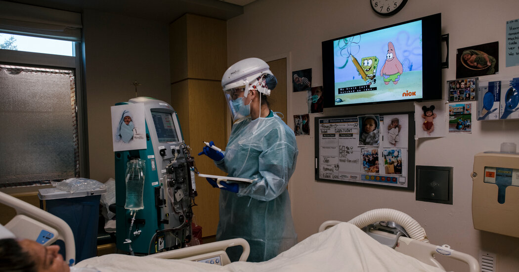
En un gran estudio reciente, los pacientes recuperados de COVID-19 tuvieron 35 por ciento más probabilidades de sufrir daños a largo plazo en los riñones o un deterioro de la función renal.Desde el principio de la pandemia, los médicos han descubierto que la gente que experimenta casos graves de COVID-19 a menudo tiene problemas renales y no solo el daño pulmonar característico de esta enfermedad.Ahora, un estudio muy grande señala que, después de que los pacientes se recuperan de la infección inicial, los problemas renales pueden durar meses y que en algunos pacientes las secuelas pueden originar una reducción considerable de la función renal.En este estudio, publicado el 1 de septiembre en la revista Journal of the American Society of Nephrology, se descubrió que cuanto más enfermos estuvieran los pacientes al principio, más probable era que sufrieran un daño renal prolongado.Pero incluso las personas que contraen el virus y presentan una infección menos grave podrían ser vulnerables.“De manera generalizada, en realidad vemos un mayor riesgo de que se presente una serie de eventos importantes vinculados con el riñón”, señaló F. Perry Wilson, nefrólogo y profesor adjunto de Medicina en la Universidad de Yale, quien no participó en el estudio. “Y lo que más me sorprendió es que estos perduraron”.Los riñones tienen una participación fundamental en el organismo para eliminar las toxinas y el exceso de líquido de la sangre, ayudar a mantener una presión arterial saludable y regular el equilibrio de electrolitos y de otras sustancias importantes. Cuando los riñones no funcionan bien o de manera eficiente, los líquidos se acumulan y provocan inflamación, presión sanguínea alta, debilidad en los huesos y otros problemas.El corazón, los pulmones, el sistema nervioso central y el sistema inmunitario pueden sufrir un deterioro. En la etapa terminal de la insuficiencia renal, quizás se necesite la diálisis o un trasplante de riñón. Esta enfermedad podría provocar la muerte.Este nuevo estudio, el cual se basó en los registros de los pacientes del sistema de salud del Departamento de Asuntos de los Veteranos, analizó la información de 89.216 personas que dieron positivo al coronavirus entre el 1 de marzo de 2020 y el 15 de marzo de 2021, así como la información de 1.637.467 personas que no tuvieron covid.Entre uno y seis meses después de haberse infectado, los sobrevivientes de covid tuvieron aproximadamente un 35 por ciento más de probabilidades de tener un daño renal o un deterioro considerable en la función renal que quienes no tuvieron covid, señaló Ziyad Al-Aly, director del servicio de investigación y desarrollo en el Sistema de Atención a la Salud de San Luis del Departamento de Asuntos de los Veteranos (VA, por su sigla en inglés) y autor sénior del estudio.“Las personas que han sobrevivido a los primeros 30 días de la covid están en riesgo de desarrollar insuficiencia renal”, mencionó Al-Aly, nefrólogo y profesor adjunto de Medicina en la Escuela de Medicina de la Universidad de Washington.Debido a que muchas personas que tienen una función renal disminuida no experimentan dolor ni otros síntomas, “lo que importa en realidad es que la gente sepa que existe un riesgo y que los médicos de los pacientes que tuvieron covid estén muy atentos a la función y a la insuficiencia renal”, explicó.Los dos grupos de pacientes del estudio diferían en que todos los miembros de un grupo se habían infectado con covid y los miembros del otro grupo podían haber tenido una variedad de otros padecimientos. Los especialistas advirtieron que las comparaciones tenían limitaciones.Los investigadores intentaron reducir al mínimo las diferencias a través de análisis detallados que ajustaron a partir de una larga lista de características demográficas, enfermedades preexistentes, uso de medicamentos y si las personas estaban en asilos.Otra limitación es que los pacientes en el estudio del VA eran en su mayoría varones blancos de una edad promedio de 68 años, así que no queda muy claro cuán generalizables son los resultados.Según los expertos, un aspecto sólido de la investigación es que contempla a más de 1,7 millones de pacientes con expedientes médicos electrónicos detallados, lo que la convierte en el estudio más grande hasta ahora sobre problemas renales vinculados a la covid.Aunque lo más probable es que los resultados no sean aplicables para todos los pacientes con covid, muestran que entre los participantes del estudio “existe una afectación muy notable a largo plazo para la salud renal de los sobrevivientes de COVID-19, sobre todo para quienes estuvieron muy enfermos durante la enfermedad aguda”, señaló C. John Sperati, nefrólogo y profesor adjunto de Medicina en la Universidad Johns Hopkins, quien no participó en el estudio.Otros investigadores han descubierto patrones parecidos, “así que este no es el único estudio que indica que estos eventos están ocurriendo después de una infección de COVID-19”, añadió.Sperati y otros especialistas han señalado que incluso si solo un pequeño porcentaje de los millones de sobrevivientes de COVID-19 en Estados Unidos desarrollasen problemas renales prolongados, el impacto para la atención sanitaria sería muy importante.Con el fin de evaluar la función renal, el equipo de investigación evaluó los niveles de creatinina, un producto de desecho que los riñones deben eliminar del cuerpo y también un indicador de la adecuada filtración de la sangre, el cual se denomina índice de filtración glomerular.Wilson explicó que, a lo largo de la vida, los adultos sanos poco a poco pierden la función renal a partir de los treinta y tantos o cuarenta y tantos años a razón de aproximadamente el uno por ciento o menos al año. Las enfermedades y las infecciones importantes pueden provocar una pérdida más significativa o permanente de esta función, la cual puede originar una insuficiencia renal crónica o una insuficiencia renal en etapa terminal.Según Al-Aly, en el nuevo estudio se descubrió que 4757 sobrevivientes de covid habían perdido al menos un 30 por ciento de la función renal en el año posterior a haber tenido la infección.Eso equivale a cerca de “30 años de deterioro en la función renal”, afirmó Wilson.En el estudio se descubrió que existía un 25 por ciento más de probabilidades de que los pacientes que tuvieron covid alcanzaran ese nivel de deterioro que las personas que no contrajeron la enfermedad.Un grupo más pequeño de sobrevivientes de covid presentó deterioro más acentuado. Pero había un 44 por ciento más de probabilidades de que los pacientes con covid perdieran al menos el 40 por ciento de la función renal y era un 62 por ciento más probable que perdieran al menos el 50 por ciento que los pacientes sin covid.Al-Aly informó que en 220 pacientes con covid se detectó insuficiencia renal en etapa terminal, la cual se presenta cuando se pierde al menos el 85 por ciento de la función renal. En el estudio se encontró que los sobrevivientes de covid tenían tres veces más probabilidades de recibir este diagnóstico que los pacientes sin covid.Al-Aly y sus colegas también investigaron un tipo de falla renal repentina llamada insuficiencia renal aguda, la cual otros estudios han encontrado en hasta la mitad de los pacientes hospitalizados con covid. Esta falla puede sanar sin provocar pérdida de la función renal a largo plazo.No obstante, en el estudio del veteranos, se descubrió que meses después de haber tenido la infección, 2812 sobrevivientes de covid sufrieron insuficiencia renal aguda, casi el doble que los pacientes sin covid, aseveró Al-Aly.Wilson señaló que los nuevos datos respaldaban los resultados de un estudio con 1612 pacientes que realizaron él y sus colegas en el cual descubrieron que los pacientes con covid que tenían insuficiencia renal aguda presentaron una función renal significativamente peor en los meses posteriores a su salida del hospital comparados con quienes presentaban daños renales agudos resultantes de otras enfermedades.En el nuevo estudio, los investigadores no compararon de manera directa a los sobrevivientes de covid con las personas infectadas por otros virus, como el de la influenza, lo cual dificultaba saber si “en realidad estás más enfermo que si hubieras tenido otra infección grave”, comentó Sperati.Sin embargo, en un estudio anterior realizado por el equipo de Al-Aly, en el que se analizaron muchos problemas de salud posteriores a la covid, incluidos los problemas renales, las personas hospitalizadas por COVID-19 tenían un riesgo significativamente mayor de desarrollar problemas de salud a largo plazo en prácticamente todas las categorías médicas, incluidas las afecciones cardiovasculares, metabólicas y gastrointestinales, que las personas hospitalizadas por la gripe.Todos los tipos de trastornos renales monitoreados en el nuevo estudio fueron mucho más frecuentes en los pacientes de covid que estuvieron más enfermos al principio, es decir, los que pasaron algún tiempo en cuidados intensivos o los que experimentaron una lesión renal aguda en el hospital.Las personas que estuvieron menos graves durante su hospitalización por covid tenían menos probabilidades de sufrir problemas renales persistentes, pero seguían siendo considerablemente más propensas que los pacientes que no habían tenido covid.“Las personas que corren mayor riesgo son las que realmente lo pasaron mal ya desde el inicio”, dijo Al-Aly. “Pero realmente, nadie se libra del riesgo”.El estudio también descubrió que incluso los pacientes de covid que nunca necesitaron hospitalización tenían un riesgo ligeramente mayor de sufrir problemas renales que la población general de pacientes del VA. Pero el riesgo parecía tan pequeño, dijo Sperati, que “no sé si confiaría” en esos resultados.Wilson observó que algunos pacientes de covid que no necesitaron hospitalización estaban, sin embargo, bastante enfermos y requirieron permanecer en cama durante días. Dijo que es posible que esos fueran los que desarrollaron una disfunción renal a largo plazo, en lugar de las personas en el extremo más leve del espectro de covid.Los médicos no saben por qué la covid puede provocar daño renal. Los expertos explicaron que es posible que los riñones sean especialmente sensibles al aumento de la inflamación o la activación del sistema inmunitario, o que los problemas de formación de coágulos sanguíneos que casi siempre se observan en los pacientes con covid alteren la función renal.Sperati dijo que los pacientes de covid en el hospital parecían tener mayor necesidad de diálisis, y más proteínas y sangre en la orina que los pacientes hospitalizados con otras enfermedades graves.“El coronavirus es probablemente un virus un poco más tóxico para los riñones”, dijo Wilson. “Creo que el síndrome por covid tiene algunos efectos adversos a largo plazo en el riñón”.Pam Belluck es una reportera de ciencia y salud cuyos galardones incluyen un Premio Pulitzer compartido en 2015 y el premio Nellie Bly a la mejor historia de primera plana. Es autora de Island Practice, un libro sobre un doctor peculiar. @PamBelluck

Read more →