Scientists just found 200+ hidden proteins that may drive Alzheimer’s

For decades, the story of Alzheimer’s research has been dominated by a battle between A-beta and tau amyloids, both of which can kill neurons and impact the brain’s ability to function. A new study suggests, however, that these sticky brain plaques may not be operating alone.
Johns Hopkins University researchers have identified more than 200 types of misfolded proteins in rats that could be associated with age-related cognitive decline.
The findings could lead the way to finding new therapeutic targets and treatments in humans that could provide relief for the millions of people over 65 who suffer from Alzheimer’s, dementia, or other diseases that rob them of their memories and independence as they age.
“Amyloids are the buildup of misshapen proteins. They’re big and ugly and easy to see under the microscope, so it makes sense that they catch our attention. But we’re seeing hundreds of proteins misfolding in ways that don’t clump together in an amyloid and yet still seem to impact how the brain functions,” said Stephen Fried, an assistant professor of chemistry and protein scientist who studies how molecules in the brain change during aging. “Our research is showing that amyloids are just the tip of the iceberg.”
The results were published on July 11 in Science Advances.
To understand the molecular differences between older brains that are mentally sharp and those that are experiencing decline, Fried and his team studied 17 2-year-old rats that grew up in the same colony. Seven rats performed poorly on memory and problem-solving tests and were considered cognitively impaired, while 10 performed as well as 6-month-old rats.
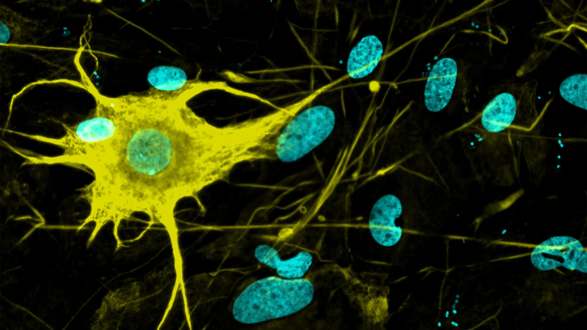
The researchers then measured more than 2,500 types of protein in the hippocampus, the part of the brain associated with spatial learning and memory. For the first time, scientists were able to determine for a large number of proteins whether individual proteins were misshapen or folded incorrectly, allowing the researchers to work out which proteins misfold for all the rats and are associated with aging in general versus which proteins specifically misfold in cognitively impaired rats.

More than 200 proteins were misfolded in the cognitively impaired rats yet maintained their shapes in the cognitively healthy rats. The findings suggest that some of those proteins are contributing to cognitive decline, the researchers said.
Misfolded proteins are unable to carry out tasks necessary for a cell to function properly, so cells have a natural surveillance system that identifies and destroys these misbehaving proteins. Previously, researchers thought misfolded proteins — specifically A-beta and tau proteins — were only disruptive when they clumped into amyloids.
“We think there are a lot of proteins that can be misfolded, not form amyloids, and still be problematic,” Fried said. “And that suggests these misfolded proteins have ways of escaping this surveillance system in the cell.”
But exactly how those misfolded proteins slip past a cell’s security system remains a mystery.
Next, the team plans to look at misfolded proteins under high-resolution microscopes to get a more detailed picture of what their deformities look like at the molecular level.
“A lot of us have experienced a loved one or a relative who has become less capable of doing those everyday tasks that require cognitive abilities,” Fried said. “Understanding what’s physically going on in the brain could lead to better treatments and preventive measures.”

Read more →

It’s never too late: Just moving more could add years to your life

Being consistently physically active in adulthood is linked to a 30–40% lower risk of death from any cause in later life, while upping levels from below those recommended for health is still associated with a 20–25% lower risk, finds a pooled data analysis of the available evidence, published online in the British Journal of Sports Medicine.
The findings prompt the researchers to conclude that switching to a more active lifestyle at any point in adult life may extend the lifespan, and that it’s never too late to start.
Currently, it’s recommended that adults should aim for 150-300 weekly minutes of moderate intensity physical activity, or 75-150 weekly minutes of vigorous intensity physical activity, or a combination of the two, note the researchers.
But while these recommendations were based on the best evidence available, most of it captured measurements of physical activity at only one point in time, which might hide the potential impact of changing patterns during adulthood, they add.
The researchers therefore wanted to find out if differing patterns of physical activity, as well as its cumulative impact during adulthood, might be associated with a lower risk of death from all causes, and specifically from cardiovascular disease and cancer. 
They scoured research databases for relevant studies that assessed physical activity at two or more points in time, and included in their review 85 studies published in English up to April 2024, with sample sizes ranging from 357 to 6,572,984 participants. 
Fifty nine of the studies looked at long term patterns of physical activity across adulthood; 16 looked at the average benefits of different physical activity levels; and 11 explored the potential impact of cumulative physical activity on risk of death.

To overcome the challenges posed by different analytical methods used, the researchers carried out separate analyses for each of them.
Pooled data analysis of the study results showed that, overall, a higher level of physical activity was associated with lower risks of all the included outcomes. 
Consistently active people (32 studies) had around a 30–40% lower risk of dying from any cause, while those who increased their levels of physical activity (21 studies) from below those recommended had a 20-25% lower risk of death from any cause. 
Specifically, participants who switched from being physically inactive to being active were 22% less likely to die from any cause than those who remained inactive, while those who increased their leisure time physical activity levels were 27% less likely to do so.
On the other hand, swapping an active lifestyle for an inactive one wasn’t associated with a lower risk of death from any cause.
Generally, the associations observed between a high level of physical activity and a lower risk of death were more evident for cardiovascular disease than for cancer. 
Compared with participants who were consistently inactive over time, those who were consistently active, overall, or only in their leisure time, were around 40% and 25% less likely to die from cardiovascular disease and cancer, respectively. 

But in general, the evidence for the associations between physical activity patterns and death from a specific cause remained inconclusive, especially for death from cancer.
The pooled data suggested that people who were consistently active or who became active had lower risks of death from any cause, and specifically from cardiovascular disease, when meeting the recommended weekly physical activity levels.
But being consistently physically active and clocking up more than the recommended maximum weekly amount of moderate to vigorous intensity exercise was associated with only a small additional reduction in risk.
Maintaining or increasing physical activity at levels below the recommended weekly amount, however, was associated with appreciable health benefits, indicating that some physical activity is always better than none, say the researchers.  
And an average volume of physical activity that met the recommended weekly amount was also associated with a 30–40% lower risk of death from all causes. But more research is needed to confirm this, they add.
The researchers acknowledge some limitations to their findings, including that most of the studies included in the pooled data analyses relied on subjective assessments of physical activity, which may not always have been accurate. 
And there were only a few studies that looked at cumulative amounts of physical activity, or cancer deaths.
Nevertheless, the findings have important public health implications, insist the researchers.
“First, our results emphasized the importance of [physical activity] across adulthood, indicating that initiating [it] at any point in adulthood may provide survival benefits.” 
They add: “As being consistently active provides greater health benefits than being previously active (ie, no longer maintaining activity), this highlights the importance of sustained [physical activity] over time. 
“Future [physical activity] interventions may not only target inactive people, but also support active people to maintain their activity.”

Read more →

Alzheimer’s has isolated us, says Fiona Phillips’ husband

The husband of TV presenter Fiona Phillips says they have become socially isolated since her Alzheimer’s diagnosis.Phillips, who hosted ITV’s GMTV breakfast programme, announced in 2023 that she had been diagnosed with Alzheimer’s the previous year, at the age of 61.In an interview with the Daily Telegraph, Martin Frizell, a former editor of ITV’s This Morning show, said: “You become almost invisible.”He added: “We still have some close friends. But I think people think, oh gosh, Fiona, maybe she doesn’t look the same, or they don’t know what to say, or it brings into sharp focus their own mortality.”At the time of her diagnosis, Phillips said she had suffered months of brain fog and anxiety – and initially had attributed the symptoms to the menopause.”It’s something I might have thought I’d get at 80,” Phillips explained. “But I was still only 61 years old.”Mr Frizell said he now does not know what to do either with her cookery books or designer clothes – both things she no longer uses.”Fiona hasn’t cooked in two years,” he said.”Part of the heartache now is she’s got this dressing room full of the most amazing clothes but this horrible disease means she’s more than happy just wearing the same T-shirt, the same trousers, the same thing – day in, day out.”Mother-of-two Phillips has written a memoir since her diagnosis, which is due to be released on Thursday.Mr Frizell contributed to the book, saying he had intended to write “a few paragraphs” but ended up writing “24,000 words”.”I started off writing about what a great woman she is and just how horrible it is and dreadfully unlucky that she is the latest in the long line of her family to get it,” he told the Telegraph.”Then I just got very angry as to what little support there is. As a family, we just kind of get through it and at some point we will need more support, but there’s just nothing really.”In 2023, Phillips said the disease had “decimated” her family – with her mother, father and uncle all receiving a diagnosis.She had cared for her parents and made two documentaries about the disease – one in 2009 called Mum, Dad, Alzheimer’s and Me, about her family’s history of dementia, and My Family and Alzheimer’s in 2010.The NHS says the term dementia encompasses “a group of symptoms associated with an ongoing decline of brain functioning”.Alzheimer’s disease is the most common cause of dementia in the UK – but its exact cause is not yet fully understood. There is currently no cure for the disease, though some treatments can temporarily improve symptoms.One in three people born in the UK will be diagnosed with dementia, according to the Alzheimer’s Society charity.Speaking to ITV’s This Morning on Friday, Mr Frizell said: “Society has decided we’re not going to take it as seriously as we should.”The money that’s there for Alzheimer’s research, it’s like buying a Starbucks cup of coffee, basically trying to fight a disease. It’s impossible.”Phillips presented GMTV between 1993 and 2008. She has since led a number of documentaries and episodes of Panorama, and was a columnist for the Mirror.

Read more →

Researchers grow 400+ brain cell types—a leap for Alzheimer’s and Parkinson’s research

Nerve cells are not just nerve cells. Depending on how finely we distinguish, there are several hundred to several thousand different types of nerve cell in the human brain according to the latest calculations. These cell types vary in their function, in the number and length of their cellular appendages, and in their interconnections. They emit different neurotransmitters into our synapses and, depending on the region of the brain – for example, the cerebral cortex or the midbrain – different cell types are active.
When scientists produced nerve cells from stem cells in Petri dishes for their experiments in the past, it was not possible to take their vast diversity into account. Until now, researchers had only developed procedures for growing a few dozen different types of nerve cell in vitro. They achieved this using genetic engineering or by adding signalling molecules to activate particular cellular signalling pathways. However, they never got close to achieving the diversity of hundreds or thousands of different nerve cell types that actually exists.
“Neurons derived from stem cells are frequently used to study diseases. But up to now, researchers have often ignored which precise types of neuron they are working with,” says Barbara Treutlein, Professor at the Department of Biosystems Science and Engineering at ETH Zurich in Basel. However, this is not the best approach to such work. “If we want to develop cell culture models for diseases and disorders such as Alzheimer’s, Parkinson’s and depression, we need to take the specific type of nerve cell involved into consideration.”
Systematic screening was the key to success
Treutlein and her team have now successfully produced over 400 different types of nerve cell. In doing so, the scientists have paved the way for more precise basic neurological research with cell culture experiments.
The ETH researchers achieved this by working with a culture of human induced pluripotent stem cells that had been generated from blood cells. In these cells, they used genetic engineering to activate certain neuronal regulator genes and treated the cells with various morphogens, a special class of signalling molecules. Treutlein and her team took a systematic approach, using seven morphogens in different combinations and concentrations in their screening experiments. This resulted in almost 200 different sets of experimental conditions.
Morphogens
Morphogens are messengers that are known from research into embryonic development. They are not distributed uniformly within an embryo but occur in a variety of concentrations forming spatial patterns. In this way, they define the position of cells within the embryo, for example whether a cell is near the body axis or in the back, abdomen, head or torso. Accordingly, morphogens help to determine what grows where in the embryo.

The researchers used various analyses to prove that they had produced over 400 different types of nerve cell in their experiment. They examined the RNA (and therefore genetic activity) at the level of individual cells, as well as the external appearance of cells and their function: for example, which type of cell appendage they had in which quantities and which electric nerve impulses they emitted.
The researchers then compared their data with information from databases of neurons from the human brain. By doing this, they were able to identify the types of nerve cell that had been created, such as those found in the peripheral nervous system or brain cells and the part of the brain they come from, whether they perceive pain, cold or movement, and so on.
In-vitro neurons for active ingredient research
Treutlein clarifies that they are still a long way off producing all types of nerve cell that exist in vitro. Nonetheless, the researchers now have access to a much larger number of different cell types than they had before.
They would like to use in-vitro nerve cells to develop cell culture models for studying serious neurological conditions, including schizophrenia, Alzheimer’s, Parkinson’s, epilepsy, sleep disorders and multiple sclerosis. Cell culture models of this kind are also of great interest in pharmaceutical research for testing the effects of new active compounds in cell cultures without animal testing, with the ultimate aim of one day being able to cure these conditions.
In the future, the cells could also be used for cell replacement therapy, which involves replacing sick or dead nerve cells in the brain with new human cells.
But there is a challenge to overcome before this can happen: the researchers often produced a mixture of multiple different types of nerve cell in their experiments. They are now working to optimise their method so that each experimental condition only produces one specific cell type. They already have some initial ideas as to how this might be achieved.

Read more →

This tiny brain molecule could hold the key to learning, memory—and Alzheimer’s treatment

Scientists have discovered how a key protein helps maintain strong connections between brain cells that are crucial for learning and memory.
Results of the study, published in the journal Science Advances, could point the way to new treatments for traumatic brain injuries and diseases, such as Parkinson’s and Alzheimer’s, the scientists said.
Their research, led by a Rutgers University-New Brunswick professor, uncovered a previously unknown role for cypin, a brain protein. Members of the research team found that cypin promotes the presence of tags on specific proteins at synapses, namely the tiny gaps where the brain cells, known as neurons, communicate. The marking helps ensure that the right proteins are in the right place, allowing the synapses to work properly.
The researchers said the insight has potentially profound implications for the treatment of brain disorders.
“Our research indicates that developing treatments or therapies that specifically focus on the protein cypin may help improve the connections between brain cells, enhancing memory and thinking abilities,” said Bonnie Firestein, a Distinguished Professor in the Department of Cell Biology and Neuroscience in the School of Arts and Sciences and an author of the study. “These findings suggest that cypin could be used to develop treatments for neurodegenerative and neurocognitive diseases, as well as brain injuries.”
Firestein has been studying cypin for more than two decades. Her latest work uncovered several important aspects of how cypin functions and why it is significant for brain health.
One of the crucial discoveries is that cypin helps add a special tag to proteins in synapses connecting neurons. This tag ensures proteins are correctly positioned and able to send signals effectively. Proper tagging and movement of proteins are essential for the neurons to function correctly.

Another important finding is that cypin interacts with a complex of proteins, known as the proteasome, which is responsible for breaking down proteins. When cypin attaches or binds to the proteasome, it slows down this breakdown process, leading to an accumulation of proteins. This buildup can positively affect various cellular functions, which are important for the communication between neurons.
Firestein’s research also shows that when there is more cypin present, the levels of important proteins in the synapses increase. These proteins are vital for effective communication between neurons, empowering learning and memory.
Additionally, cypin increases the activity of another protein called UBE4A, which also helps with the tagging process. This indicates that cypin’s influence on synaptic proteins is partly because of its effect on UBE4A.
The work highlights the importance of cypin in maintaining healthy brain function and its potential as a target for therapeutic interventions.
“Even though this study is what we call ‘basic research,’ it eventually can be applied in practical, clinical settings,” said Firestein, who already is conducting such “translational” work in parallel. Translational research is a type of research that takes discoveries made in the lab and turns them into practical treatments or solutions to improve human health.
Cypin’s significant role in the workings of the brain’s synapses makes it highly relevant to the potential treatment of neurodegenerative diseases and traumatic brain injury, she said. For example, healthy synaptic function is often disrupted in diseases such as Alzheimer’s and Parkinson’s.

In addition, the protein’s role in promoting synaptic plasticity – the ability of synapses to strengthen or weaken over time – means it may be used to help counteract the synaptic dysfunction seen in neurodegenerative diseases and brain injuries.
The study was supported in part by the National Institutes of Health (NINDS), the Coalition for Brain Injury Research, a charitable foundation dedicated to the memory of Dennis John Benigno, who suffered a traumatic brain injury in junior high school; and private donors Jamuna Rajasingham and Dyan Rajasingham.
Other scientists from Rutgers involved in the study include Kiran Madura, a professor in the Department of Pharmacology at Robert Wood Johnson Medical School; Srinivasa Gandu, Mihir Patel, Ana Rodriguez, former doctoral students in the Department of Cell Biology and Neuroscience.
Jared Lamp and Irving Vega of Michigan State University also contributed to this research.

Read more →

Breakthrough microchip reveals how your body fights viruses—in just 90 minutes

A new microchip invented by Scripps Research scientists can reveal how a person’s antibodies interact with viruses — using just a drop of blood. The technology offers researchers faster, clearer insights that could help accelerate vaccine development and antibody discovery.
“This lets us take a quick snapshot of antibodies as they are evolving after a vaccine or pathogen exposure,” says Andrew Ward, professor in the Department of Integrative Structural and Computational Biology at Scripps Research and senior author of the new paper published in Nature Biomedical Engineeringon June 3, 2025. “We’ve never been able to do that on this timescale or with such tiny amounts of blood before.”
When someone is infected with a virus, or receives a vaccine, their immune system creates new antibodies to recognize the foreign invader. Some antibodies work well against the pathogen, while others attach to it only weakly. Figuring out exactly which parts of the virus the best antibodies stick to is key information for scientists trying to optimize vaccines, since they want to design vaccines that elicit strong, reliable immune responses.
“If we know which particular antibodies are leading to the most protective response against a virus, then we can go and engineer new vaccines that elicit those antibodies,” says Leigh Sewall, a graduate student at Scripps Research and first author of the new paper.
In 2018, Ward’s lab unveiled a technique known as electron microscopy-based polyclonal epitope mapping (EMPEM). This method allowed scientists to visualize how antibodies in blood samples attach to a virus. Although groundbreaking, it had downsides: it took a full week to complete and required relatively large amounts of blood.
“During the COVID-19 pandemic, we began really wanting a way to do this faster,” says Alba Torrents de la Peña, a Scripps Research staff scientist who helped lead the work. “We decided to design something from scratch.”
With the new system, known as microfluidic EM-based polyclonal epitope mapping (mEM), researchers start with four microliters of blood extracted from a human or animal-about one hundred times less than what’s required in original EMPEM. The blood is injected in a tiny, reusable chip where viral proteins are stuck to a special surface. As the blood flow through the chip, antibodies recognize and bind to those. Then, the viral proteins — with any antibodies attached — are gently released from the chip and prepared for imaging using standard electron microscopy. The entire process only takes about 90 minutes.

To test the value and effectiveness of mEM, the research team used the system to map antibodies in humans and mice that had either received a vaccination against or been infected with a virus, including influenza, SARS-CoV-2 and HIV. The new technique was not only fast at mapping out the interactions between antibodies and those viruses, but more sensitive than EMPEM; it revealed new antibody binding sites on both influenza and coronavirus proteins that had not been picked up by EMPEM.
To track how antibodies evolved over time in individual mice after they received a vaccination against one of the pathogens, the team took small blood samples from a mouse at different time points.
“That was something that wouldn’t have been possible in the past, because of the amount of blood needed for EMPEM,” says Sewall. “So to be able to look at an individual over time was really exciting.”
The researchers are now working to automate and multiplex the system, which could eventually allow dozens of samples to be processed in parallel. Ultimately, they envision mEM becoming a widely adopted tool to monitor and guide vaccine development in pathogens ranging from coronaviruses to malaria.
“This technology is useful in any situation where you have really limited sample volume, or need initial results quickly,” says Torrents de la Peña. “We hope this becomes accessible to more researchers as it is simplified and streamlined.”
In addition to Ward, Sewall, and Torrents de la Peña, authors of the study, “Microfluidics Combined with Electron Microscopy for Rapid and High-Throughput Mapping of Antibody-Viral Glycoprotein Complexes,” are Rebeca de Paiva Froes Rocha, Grace Gibson, Michelle Louie, Sandhya Bangaru, Andy S. Tran, Gabriel Ozorowski, Blanca Chocarro Ruiz, Nathan Beutler, Thomas F. Rogers, Dennis R. Burton, and Andrew B. Ward of The Scripps Research Institute; Zhenfei Xie and Facundo D. Batista of the Ragon Institute of MGH, MIT and Harvard; and Subhasis Mohanty and Albert C. Shaw of Yale University School of Medicine.
This work was supported by funding from the National Institutes of Health (AI136621, AI089992, and AI144462), and by the Bill and Melinda Gates Foundation (INV-002916).

Read more →

Some GPs to offer coaching to get sick people back to work

12 minutes agoShareSaveShareSaveGetty ImagesGP surgeries in England can offer advice to patients on getting back to work, including career coaching or exercise classes, as part of a pilot project to reduce the number of people who are signed off work sick. The aim is to help people return to the workplace more quickly to reduce the length of time they need fit notes – better known as sick notes.These are issued by health professionals if a patient is unwell or cannot work for more than seven days.A total of £1.5m is being made available to 15 regions in England, and will be shared between GP practices in these areas to hire coaches or occupational therapists to support patients in their return to work. The Health and Social Care Secretary Wes Streeting said: “It’s about fundamentally changing the conversation from ‘you can’t’ to ‘how can we help you?’ “When someone walks into their doctor’s surgery worried about their job, they should walk out with a plan, not just a piece of paper that closes doors.”Health staff would be offered training to give work and health advice. People could be pointed towards fitness classes or career coaching.In one case quoted by the government, a woman who was off work with a fractured ankle had an assessment with a fitness adviser and was referred for a 12-week exercise programme with the aim of strengthening the ankle.The Department of Health and Social Care says that of 11 million fit notes issued electronically in England last year, 93% simply declared people “not fit for work” with no alternative support offered.This new scheme expands on an initiative launched last October in the same 15 regions called WorkWell, jointly run by the Departments of Work and Pensions and Health and Social Care.It involves NHS staff referring patients to other services. People in work but at risk of quitting have been given advice on mental health in the workplace. In the new scheme, those out of work will be referred by NHS staff to services that offer support for finding a job, such as CV and interview techniques and liaising with employers on appropriate support.Ministers say the policies are part of the move across government to encourage more people back into work with 2.8 million currently out of the workforce due to health conditions. The Royal College of GPs said it recognised the health benefits of being in work and GPs would encourage it where safe to do so, but added that doctors did not issue fit notes without good reason. Professor Kamila Hawthorne, chair of the College, said: “We want to work alongside the Government on this scheme so it’s important that it is not presented as a punitive measure for patients.”Prof Hawthorne also stressed that the new scheme should also not be punitive for “hardworking GPs”, who are “doing their best under enormous pressures, caused by historic underfunding and poor workforce planning”,WorkWell is operating in 15 English regions and the new fit note initiative will provide £1.5m to be shared between them. The regions are Birmingham and Solihull; Black Country; Bristol, North Somerset and South Gloucestershire; Cambridgeshire and Peterborough; Cornwall and the Isles of Scilly; Coventry and Warwickshire; Frimley; Herefordshire and Worcestershire; Greater Manchester; Lancashire and South Cumbria; Leicester, Leicestershire and Rutland; North Central London; North West London; South Yorkshire; Surrey Heartlands.

Read more →

Vaccine roll-outs cut deaths by 60% – study

1 hour agoShareSaveDominic HughesGlobal Health CorrespondentShareSaveGetty ImagesEmergency vaccination programmes – rolled out in response to outbreaks of five major diseases – are believed to have reduced deaths by around 60% over a period of 23 years, according to a new study. A similar number of infections are also thought to have been prevented.Researchers believe that vaccinations stopped much bigger outbreaks of illnesses like Ebola, cholera and yellow fever.The study also points to the significant economic benefits of vaccination programmes, which run into billions of dollars. Researchers say this is the first comprehensive study to assess the impact of emergency vaccination programmes in response to the outbreak of five infectious diseases – Ebola, measles, cholera, yellow fever and meningitis. They studied 210 different incidents from 2000 to 2023, covering 49 different countries. The vaccine roll-outs seem to have had an impressive impact, reducing deaths by nearly 60%.The number of overall cases of theses infections were also reduced by nearly 60%.The swift deployment of vaccines also appears to have halted wider outbreaks.There were economic benefits too – worth an estimated $32 billion. These benefits come mainly from averting deaths and years of life lost to disability.But researchers believe this could be a significant underestimate of overall savings, as it doesn’t take into account the costs of dealing with a wider outbreak, or the economic disruption caused by a more serious health emergency. It’s believed the 2014 Ebola outbreak, which occurred before the existence of an approved vaccine, is estimated to have cost West African countries alone more than $53 billion.The power of vaccinesThe study was backed by Gavi, the vaccine alliance, which was responsible for many of the programmes. Dr Sania Nishtar, the organisation’s CEO, says the study shows just how important quick and effective vaccine roll-outs can be.”For the first time, we are able to comprehensively quantify the benefit, in human and economic terms, of deploying vaccines against outbreaks of some of the deadliest infectious diseases.”This study demonstrates clearly the power of vaccines as a cost-effective counter-measure to the increasing risk the world faces from outbreaks. “It also underscores the importance of fully funding Gavi, so it can continue to protect communities in the next five years to come.”Gavi is currently trying to secure a fresh round of funding in the face of global cuts to foreign aid.

Read more →

The first pandemic? Scientists find 214 ancient pathogens in prehistoric DNA

A research team led by Eske Willerslev, professor at the University of Copenhagen and the University of Cambridge, has recovered ancient DNA from 214 known human pathogens in prehistoric humans from Eurasia.
The study shows, among other things, that the earliest known evidence of zoonotic diseases — illnesses transmitted from animals to humans, like COVID in recent times — dates back to around 6,500 years ago, with such diseases becoming more widespread approximately 5,000 years ago. It is the largest study to date on the history of infectious diseases and has just been published in the scientific journal Nature.
The researchers analyzed DNA from over 1,300 prehistoric individuals, some up to 37,000 years old. The ancient bones and teeth have provided a unique insight into the development of diseases caused by bacteria, viruses, and parasites.
The results suggest that humans’ close cohabitation with domesticated animals — and large-scale migrations of pastoralist from the Pontic Steppe — played a decisive role in the spread of these diseases.
“We’ve long suspected that the transition to farming and animal husbandry opened the door to a new era of disease — now DNA shows us that it happened at least 6,500 years ago,” says Professor Eske Willerslev. “These infections didn’t just cause illness — they may have contributed to population collapse, migration, and genetic adaptation.”World’s oldest trace of the plague
In the study, the researchers found 214 pathogens. A remarkable finding is the world’s oldest genetic trace of the plague bacterium Yersinia pestis, identified in a 5,500-year-old sample. The plague is estimated to have killed between one-quarter and one-half of Europe’s population during the Middle Ages.

Could have implications for future vaccines
The findings could be significant for the development of vaccines and for understanding how diseases arise and mutate over time.
“If we understand what happened in the past, it can help us prepare for the future, where many of the newly emerging infectious diseases are predicted to originate from animals,” says Associate Professor Martin Sikora, the study’s first author.
“Mutations that were successful in the past are likely to reappear. This knowledge is important for future vaccines, as it allows us to test whether current vaccines provide sufficient coverage or whether new ones need to be developed due to mutations,” adds Eske Willerslev.
The study was made possible by funding from the Lundbeck Foundation.

Read more →

TV doctor and IVF pioneer quits BMA over strikes

Lord Robert Winston, a professor and TV doctor who was a pioneer of IVF treatment, has resigned from the British Medical Association (BMA) over planned strikes by resident doctors.Resident doctors, previously known as junior doctors, are planning a walkout for five consecutive days from 25 July until 30 July over a pay dispute with the government. In an interview with The Times, the Labour peer and host of the BBC series Child of Our Time urged against strike action, arguing it could damage people’s trust in the profession.The BMA met with the health secretary earlier this week and said the government had “stated it will not negotiate on pay”.Lord Winston, 84, has been a member of the BMA since 1964.”I’ve paid my membership for a long time. I feel very strongly that this isn’t the time to be striking. I think that the country is really struggling in all sorts of ways, people are struggling in all sorts of ways,” he told The Times.”Strike action completely ignores the vulnerability of people in front of you,” he added.Resident doctors have been awarded a 5.4% pay rise for this financial year – which will go into pay packets from August – following a 22% increase over the previous two years.But the BMA says wages are still around 20% lower in real terms than in 2008.Health Secretary Wes Streeting said the move was “unnecessary and unreasonable”, adding: “The NHS is hanging by a thread – why on earth are they threatening to pull it?”The BMA’s resident doctors committee co-chairs Dr Melissa Ryan and Dr Ross Nieuwoudt said they had “no choice” but to strike without “a credible offer to keep on the path to restore our pay”. Lord Winston’s comments come after doctors and patient groups warned that the NHS in England was struggling to reduce wait times – a top priority for the NHS.”Doctors need to be reminded that every time they have a patient in front of them they have someone who is frightened and in pain. It’s important that doctors consider their own responsibility much more seriously,” he said.

Read more →