A Hairy Truth About Your Sense of Touch

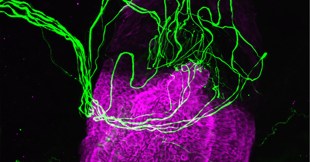
Researchers showed that cells in your hair follicles release important chemical messengers in response to gentle touches to your skin.When someone brushes a hand across your skin, it’s like a breeze blowing through a forest of countless small hairs. Nerves that surround your hair follicles detect that contact, and very far away in your brain, other cells fire. Some of the neurons responding to light contact might make you shiver and give you goose bumps. Some might tell you to move away. Or they might tell you to move closer.Scientists who study the sense of touch have explored which cells bear these messages, and they have made an intriguing discovery: Follicle cells triggered by hair movements release the neurotransmitters histamine and serotonin, chemical messengers linked to biological phenomena as varied as inflammation, muscle contraction and mood changes. The observation, reported in October in the journal Science Advances, lays the groundwork for tracing how gentle touch makes us feel the way it does.Studying hair follicles is challenging, because they begin to decay soon after being removed from the body, said Claire Higgins, a bioengineering professor at Imperial College London and an author of the study. So she and her colleagues went to a hair transplant clinic. There, they were able to look at freshly harvested follicles, which they gently prodded with a very small rod to simulate touch.The scientists knew from work done by other groups that the neurons in the skin surrounding hair follicles are capable of sensing movement.“When you brush your hair, you feel it because the sensory neurons are directly being stimulated,” Dr. Higgins said.But they were curious whether the cells of the follicle itself — the tube from which a hair sprouts — could be contributing to some of the feelings associated with more gentle touch. Not all of the follicle cells had movement sensors, but some did. The researchers identified these and watched them carefully as the rod touched them.“We found that when we stimulated our hair follicle cells, they actually released mood-regulating neurotransmitters serotonin and histamine,” Dr. Higgins said.Blocking the receptors for these neurotransmitters on nearby neurons meant that they no longer fired when the hair was stroked, confirming the link between the follicle cells and the neurons’ response.Just because these neurotransmitters are associated with mood in the brain does not mean that they are linked to emotion elsewhere in the body, Dr. Higgins said. They are messengers, and the nature of the message they carry depends on which cells they are stimulating.But she points to research by Francis McGlone, a neuroscientist at Liverpool John Moores University in England who has studied the rewarding feelings we get from touch. He and his colleagues have identified nerves in the skin that respond to gentle touch, generating that warm glow we get from human contact.Were the neurotransmitters being released by follicle cells in this study stimulating those nerves specifically? No one knows, but Dr. Higgins hopes future work will illuminate the identity of the cells the neurotransmitters target. She is curious how increasing levels of serotonin or histamine in the skin might change what happens in the brain, at the other end of the transmission. In the tiny sheath of cells containing each hair, there may be answers to questions about something as fundamental as human connection.“The follicle never ceases to amaze me,” she said.

Read more →

If You’ve Ever Heard a Voice That Wasn’t There, This Could Be Why

With a ghostly finger in a lab, researchers coaxed people to hear phantom voices.Some years ago, scientists in Switzerland found a way to make people hallucinate. They didn’t use LSD or sensory deprivation chambers. Instead, they sat people in a chair and asked them to push a button that, a fraction of a second later, caused a rod to gently press their back. After a few rounds, the volunteers got the creeping sense of someone behind them. Faced with a disconnect between their actions and their sensations, their minds conjured another explanation: a separate presence in the room.In a new study published in the journal Psychological Medicine, researchers from the same lab used the ghostly finger setup to probe another kind of hallucination: hearing voices. They found that volunteers were more likely to report hearing a voice when there was a lag between the push of the button and the rod’s touch than when there was no delay.The findings suggest that the neurological roots of hallucinations lie in how the brain processes contradictory signals from the environment, the researchers said.Hearing voices is more common than you might think, said Pavo Orepic, a postdoctoral researcher at the University of Geneva and an author of the new paper. In surveys, scientists have discovered that many people without a psychiatric diagnosis — perhaps 5 to 10 percent of the general population — report having heard a disembodied voice at some point in their lives.“There is actually a continuum of these experiences,” Dr. Orepic said. “So all of us hallucinate — at certain times, like if you’re tired, you’ll hallucinate more, for instance — and some people are more prone to do so.”In the new study, as in earlier work, Dr. Orepic and his collaborators had volunteers sit in a chair and push the button that caused the rod to touch their backs. During some sessions, there was no delay between the push and the touch, while others had a half-second delay — enough time to give volunteers that feeling that someone was nearby.During all trials, the volunteers listened to recordings of pink noise, a softer version of white noise. Some recordings contained recorded bits of their own voice, while others had fragments of someone else’s voice or no voice at all. In each trial, the volunteers were asked if they had heard anyone speaking.The study found that when people were already experiencing the peculiar feeling of a ghostly presence, they were more likely to say they had heard a voice when there was none. What’s more, hearing a nonexistent voice was more likely if, earlier in the experiment, they had heard bursts of noise with someone else’s voice in them.That suggests the brain was linking the hallucinated presence and the voice, Dr. Orepic said.Intriguingly, volunteers with no lag between the button-pressing and the rod sometimes reported hearing a nonexistent voice as well, and they were more likely to do so if they had recently been hearing clips of their own voice. If volunteers unconsciously decided they were responsible for the feeling of the finger on their backs, they may have been primed to hear their own voice, the researchers said.Together, the findings support the idea that hallucinations may arise from difficulty in recognizing one’s own actions, as well as being primed to expect a particular outcome, Dr. Orepic said. As time went on, people experiencing a ghostly presence in the trial were increasingly likely to hear voices, implying that the brain was somehow drawing on past experience to build up the impression of someone speaking.Delving more into how the brain builds the impression of a voice when none is there, Dr. Orepic said, may rely on help from healthy people who regularly hear voices — for instance, mediums who feel they can communicate with the dead. He points to ongoing studies at Yale with such people who hear voices as a pathway to understanding how these beliefs arise and how they may be controlled. For mediums, hearing voices is not necessarily unwelcome. But perhaps, with their aid, people whose hallucinations are distressing and disruptive may find some peace.

Read more →

Ultrasound Pulses to Brain Send Mice Into a Hibernation-Like State

Experiments offer an intriguing hint at technology that could induce torpor in humans in the future.For many animals, life is a cycle of scarcity as well as plenty. Hibernating creatures curl up underground in winter, slowing their metabolism so they can make it to spring without food. Even laboratory mice, if deprived of food, can enter a state called torpor, a kind of standby mode that economizes energy.It’s something humans have long fantasized about for ourselves: If we ever leave this planet and travel through space, we will experience our own time of scarcity. Science fiction writers tend to imagine a mysterious technology that keeps humans in stasis, able to survive centuries of silence before emerging into a new life. For now, it’s a technology that’s out of reach.But as scientists work to understand states like torpor and hibernation, tantalizing details about how the brain controls metabolism have emerged. Researchers reported in the journal Nature Metabolism on Thursday that they’ve been able to send mice into a torpor-like state by targeting a specific part of the brain with short bursts of ultrasound. It’s unclear exactly why ultrasound has this effect, but the findings suggest that studying the neural circuits involved in torpor could reveal ways to manipulate metabolism beyond the lab.Ultrasound devices, which generate high-frequency sound waves, are best known for their imaging powers. But they have also been used by neuroscientists to stimulate neurons. Correctly tuned, the soundwaves can travel deep into the brain, said Hong Chen, a professor of biomedical engineering at Washington University in St. Louis and an author of the new paper. In 2014, William Tyler, now at the University of Alabama, Birmingham, and his colleagues applied ultrasound to a sensory region in the brain and found that it enhanced a subject’s sense of touch. A growing body of work is exploring ultrasound as a treatment for disorders like depression and anxiety.Curious about a brain region that regulates body temperature in rodents, Dr. Chen and her colleagues constructed tiny ultrasound mouse caps. The devices trained six bursts, each consisting of 10 seconds of ultrasound, on the selected area of the rodent’s brain (Researchers who study the brain with ultrasound must tune their devices carefully to avoid heat that can damage tissues).The mice, the researchers noticed, stopped moving. Measurements of their body temperature, heart rate and metabolism showed a pronounced dip. The mice stayed in this state for about an hour after the ultrasound bursts, and then returned to normal.Looking closer at neurons involved in this response, the researchers identified a protein in their brain membranes, TRPM2, that appears to be sensitive to ultrasound; when the researchers reduced levels of the protein in mice, the mice became resistant to ultrasound’s effects.That’s an important step toward understanding how ultrasound affects neurons, said Davide Folloni, a researcher at the Icahn School of Medicine at Mount Sinai in New York City, who studies the brain using ultrasound; the details have largely been elusive.But it’s also possible that heat generated by the ultrasound, and not just the ultrasound itself, is affecting TRPM2 in the brains of mice, a point that was raised by Masashi Yanagisawa and Takeshi Sakurai of the University of Tsukuba in Japan, in separate interviews. The two have studied neurons in this brain area, and their connection to states of torpor. Both may be in play, Dr. Chen said.In one of the most tantalizing parts of the study, the researchers checked to see whether animals that don’t typically experience torpor — rats — behaved differently when the brain region was stimulated with ultrasound. Indeed, they seemed to slow down, and their body temperatures dropped.“We have to be careful with the rat data,” Dr. Chen cautions. So far, they only have information about temperature, not metabolic rate and other factors.Could ultrasound be a way to change the metabolism of larger animals with no history of torpor, like humans? It’s an intriguing idea, Dr. Sakurai said.“At this stage,” he said, “it remains an unanswered question.”

Read more →

A Link Between Hearing Voices and Hearing Your Own Voice

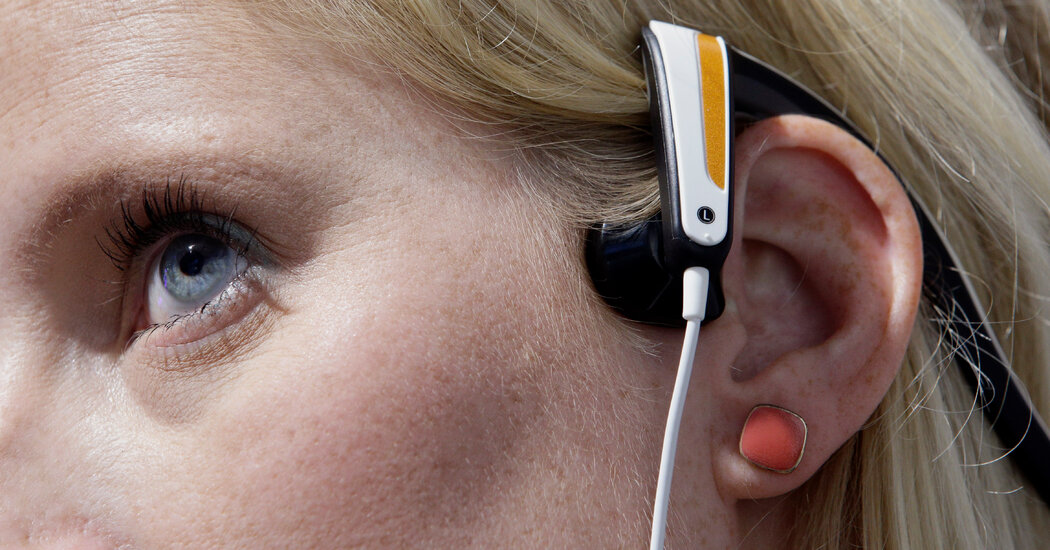
An experiment with bone-conduction headphones suggests a way for neuroscientists to better understand some hallucinations.It is the rare person who likes hearing their own voice on a recording. It sounds fake, somehow — like it belongs to someone else.For neuroscientists, that quality of otherness is more than a curiosity. Many mysteries remain about the origins of hallucinations, but one hypothesis suggests that when people hear voices, they are hearing their own thoughts disguised as another person’s by a quirk of the brain.Scientists would like to understand what parts of the brain allow us to recognize ourselves speaking, but studying this using recordings of people’s own voices has proved tricky. When we talk, we not only hear our voice with our ears, but on some level we feel it as the sound vibrations travel through the bones of the skull.A study published Wednesday in the journal Royal Society Open Science attempted a workaround. A team of researchers investigated whether people could more accurately recognize their voices if they wore bone-conduction headphones, which transmit sound via vibration. They found that sending a recording through the facial bones made it easier for people to tell their voices apart from those of strangers, suggesting that this technology provides a better way to study how we can tell when we are speaking. That is a potentially important step in understanding the origins of hallucinated voices.Recordings of our voices tend to sound higher than we expect, said Pavo Orepic, a postdoctoral researcher at the Swiss Federal Institute of Technology who led the study. The vibration of the skull makes your voice sound deeper to yourself than to a listener. But even adjusting recordings so they sound lower doesn’t recreate the experience of hearing your own voice. As an alternative, the team tried using bone-conduction headphones, which are commercially available and often rest on a listener’s cheekbones just in front of the ear.The team recorded volunteers saying the syllable “ah” and then blended each recording with other voices to produce sounds that were made up of 15 percent of a given person’s voice, then 30 percent, and so on. Then, they had some subjects listen to a series of the sounds with bone-conduction headphones, while others used normal headphones and another group tried laptop computer speakers. The volunteers indicated whether they thought each sound resembled their own voice.People with bone-conduction headphones were more likely to correctly identify their own voices, the team found. When the researchers tried the same experiment using the voices of subjects’ friends — pairs of friends were recruited specifically for the study — they found that the bone-conduction headphones made no difference in helping people identify familiar voices. It was only recognizing their own voices that became easier, suggesting that the devices are recreating some of what we feel and hear as we speak.That opens a door to understanding how one’s brain takes this sensory information and turns it into a recognition of one’s self. In a study published last year, the group recorded the neural activity of people performing these listening tasks and reported the existence of a network of brain regions that are activated as people work to identify themselves.If scientists can understand how the brain builds the concept of self from sound, Dr. Orepic suggests, then perhaps they can unpack what is different in people who hear voices in their heads that are not their own. Perhaps someday listening to recordings of voices, including one’s own, with bone conduction devices could help doctors make diagnoses, if the tool’s performance could be linked to psychiatric disorders.In fact, the team has already begun to study how people who had portions of their brains removed — to treat drug-resistant epilepsy, for instance — perform on the task. The more the brain’s self-recognition network is disturbed by the surgery, the harder the task of self-recognition becomes, Dr. Orepic said, referring to findings in a study that has yet to be peer-reviewed.For one patient, whose personality changed substantially after her surgery and who was eventually diagnosed with borderline personality disorder, the test revealed a surprising pattern.“Every time she heard her voice, she thought it was someone else,” Dr. Orepic said. “And when she hears someone else, she says ‘It’s me.’”

Read more →

How See-Through Glass Frogs Hide Their Red Blood From Predators

The glass frog’s unusual adaptation to bolster its camouflage could offer clues for preventing deadly blood clots in people.Two glass frogs sleeping upside down on a leaf, backlit from the upper side of the leaf.Jesse DeliaAt first glance, you might miss the glass frog of the Costa Rican rainforest. It is, as the name suggests, nearly transparent. Apart from a lime green smear across its back, its skin, muscle and other tissues are see-through. Then there are its tiny organs, which seem to float within this clear flesh, like a pale fruit cocktail in the weirdest Jell-O salad ever to grace a tree branch.As handy as translucence might be for evading predators, it is rare in animals that live on land. Their bodies are full of substances that light can’t penetrate, many of them essential for life. Glass frogs seem to have evolved see-through versions of some of these anatomical features, but they also have some tricks to hide lingering colors when they are at their most vulnerable.In a study published in the journal Science on Thursday, researchers report that when a glass frog falls asleep, almost all of its red blood cells retreat into its liver. They hide in the organ and allow the frog to achieve near invisibility while it rests. In addition to revealing another remarkable adaptation in nature, the discovery could lead to clues for how to prevent deadly blood clots.Like people, glass frogs rely on hemoglobin, a colored protein in red blood cells that delivers oxygen around the body. Jesse Delia and Carlos Taboada, biologists and authors of the new paper, had been spending a lot of time observing the frogs when they realized that sometimes, that red color seemed to disappear.“When they are awake, the circulatory system is red,” Dr. Delia, who works at the American Museum of Natural History in New York, said. “When they are asleep, it’s not.”Where were the red cells going?A glass frog during various levels of activity, showing the change in red blood cell perfusion within its circulatory system. The same animal was filmed backlit across all three conditions.Jesse DeliaTo solve the mystery of the disappearing blood cells, the researchers and their colleagues wanted to take images of the frogs under anesthesia — when the blood cells were clearly visible circulating through their bodies — and asleep, when the cells were nowhere to be seen. To do that, they needed to find a way to peer inside the frog’s organs, which have a mirror-like exterior that helps the frog blend in. Dr. Taboada, a researcher at Duke University, said they suspected the blood would retreat to various organs when not in circulation.The researchers wound up relying not on light but on sound to show them what was inside. They provoked the molecules within the sacs to release ultrasonic waves, which could be used to identify the contents.As soon as they compared the images of sleeping and anesthetized frogs, one big difference jumped out.“All the signal was coming from the liver,” Dr. Taboada said. About 89 percent of the frogs’ red blood cells had packed themselves into that organ.That made sense: The liver, which filters blood, is a logical destination for red blood cells, he said.What was stranger, and what the researchers still don’t understand, was how the frogs could cram all these cells together without dying from blood clots. In most vertebrates, when blood cells bump into each other, it leads to coagulation. The resulting clot can make a scab to seal a wound — or, if the clot is in a blood vessel, it can plug up the circulatory system and kill the creature. In the United States, according to the Centers for Disease Control and Prevention, as many as 100,000 people die from blood clots each year.High-speed photoacoustic imaging of a glass frog recovering from exercise in real time, showing the reduction in circulating red blood cells and the associated increase of red blood cells packing in the liver as the frog falls asleep.. Yao & X. ZhuGlass frogs, the new research suggests, can control when their blood clots. If they are wounded, they will form a scab in the usual way. But when they are asleep, with red blood cells packed like sardines in the liver, no clot forms.The finding implies that glass frogs could have something to teach us about how to prevent clot formation in our own bodies. If future research can illuminate what keeps the frogs safe, it could lead to treatments to reduce deaths from clots in humans.More immediately, the researchers said, the results raised other questions. If 89 percent of the cells that carry oxygen are holed up in the liver while the frog sleeps, how is it breathing? They wonder whether the frogs can shift their metabolisms to a mode that requires barely any oxygen, perhaps akin to what other frogs do when they hibernate for the winter.The new paper is just the beginning of this line of research. The team has already improved their imaging techniques to scan the frogs more quickly and to reveal substances other than blood as they move around in the creatures.“We’re in the lab now,” Dr. Delia said during a phone interview. “There’s literally a frog scanning right now in the system. I have to go check on it in a bit.”

Read more →

How Brains Send a Signal That It’s Time to Vomit

In a study of mice, researchers worked out a neural pathway that could help researchers alleviate nausea symptoms from chemotherapy drugs.Anyone who’s had a shady oyster or a mushroom soup that didn’t sit well remembers the ominous queasiness heralding impending bad times. Bacteria release toxins that start the body’s process of speedily evacuating the contents of the stomach. It’s a protective mechanism of sorts — getting rid of the invaders en masse is probably helpful in the long term, even if it’s unpleasant in the short. But it has remained something of a mystery how the brain gets the alarm signal, then sends another one to tell the stomach to initiate a technicolor yawn.Your next bout of food poisoning isn’t the only reason to understand this particular neural pathway. Figuring out how to counter it could be helpful for people who develop nausea caused by chemotherapy medication and other drugs. As if fighting cancer isn’t painful and scary enough, patients are often so turned off by food that keeping their weight up becomes a major struggle.In a new study, researchers report that both bacteria and chemotherapy drugs appear to trigger the same molecular pathways in the gut. The findings, which were based on experiments with mice and published Tuesday in the journal Cell, showed that a bacterial toxin and a chemo medication both set in motion a cascade of similar neural messages that cause queasiness.Choosing mice for the study was unusual. Mice, it turns out, can’t puke — a little foible that typically makes it difficult to use them to study nausea. Researchers have used cats and dogs in the past, but the biology of mice in general is so much better understood, with much better tools available to scientists to do so.Cao Peng, a professor at Tsinghua University in Beijing, and his colleagues wondered whether mice might still be capable of feeling ill in the way people do after ingesting a chemo drug or a bad salad — or close enough, anyway, that researchers could use the creatures to understand the origins of the sensation.“If we want to get better medications,” Dr. Cao said, “we need to know the detailed mechanism.”The researchers gave the mice a bacterial toxin and watched them closely with high-speed cameras, and they found that the rodents started opening their mouths oddly after the treatment. More tests showed that their abdominal muscles were moving much like the way humans’ stomachs do when they are about to be sick. In effect, the scientists believe the mice were retching, or dry heaving. A chemo drug had the mice behaving similarly, so the scientists delved deeper into which cells were reacting to these triggers and how.They traced the effect to certain neurons in the brain that released neurotransmitters when the drug or the toxin reached the gut. Following those messages back, they discovered cells in the small intestine that reacted to the presence of these noxious substances. A central player in the pathway to nausea and retching was an immune system molecule called interleukin 33, or IL33. Keeping mice from making IL33 significantly reduced their symptoms.It’s possible that drugs that interfere with IL33 or other players in this pathway could help to alleviate the suffering of people having chemotherapy, Dr. Cao said. This paper — identifying behavior in mice that can stand in for vomiting and laying bare the route that signals from the gut take — is a first step in potentially improving the quality of life in chemotherapy patients, if the results hold up in humans.Still, mice given the task of mimicking food poisoning are uncomfortable for about 24 hours after receiving bacterial toxins, Dr. Cao said. After that, they’re back to their active selves. If only we could shake off a turkey sandwich that sat out too long so quickly.

Read more →

Does Your Nose Help Pick Your Friends?

In a small study, researchers in an olfaction lab found that people who had an instant personal connection also had similarities in their body odors.Human beings maintain the polite fiction that we’re not constantly smelling one another. Despite our efforts to the contrary, we all have our own odors, pleasant and less so, and if we are like other land mammals, our particular perfume might mean something to our fellow humans.Some of these, like the reek of someone who hasn’t bathed all month, or the distinctive whiff of a toddler who is pretending they didn’t just fill their diaper, are self-explanatory. But scientists who study human olfaction, or your sense of smell, wonder if the molecules wafting off our skin may be registering at some subconscious level in the noses and brains of people around us. Are they bearing messages that we use in decisions without realizing it? Might they even be shaping whom we do and don’t like to spend time around?Indeed, in a small study published Wednesday in the journal Science Advances, researchers investigating pairs of friends whose friendship “clicked” from the beginning found intriguing evidence that each person’s body odor was closer to their friend’s than expected by chance. And when the researchers got pairs of strangers to play a game together, their body odors predicted whether they felt they had a good connection.There are many factors that shape whom people become friends with, including how, when or where we meet a new person. But perhaps one thing we pick up on, the researchers suggest, is how they smell.Scientists who study friendship have found that friends have more in common than strangers — not just things like age and hobbies, but also genetics, patterns of brain activity and appearance. Inbal Ravreby, a graduate student in the lab of Noam Sobel, an olfaction researcher at the Weizmann Institute of Science in Israel, was curious whether particularly swift friendships, the kind that seem to form in an instant, had an olfactory component — whether people might be picking up on similarities in their smells.The Science of SmellLearn more about our often disregarded, and at times startling, superpower.Perks of Evolution: Genetic changes to our olfactory receptors have altered people’s sensitivities to some odors over time.Lessons From Covid: The loss of smell and parosmia experienced by some have opened new doors to understanding the most neglected sense.The Nasal Ranger: For a half-century, Chuck McGinley has visited society’s stinkiest sites in order to measure, and demystify, smell.The Smell of History: Several scientists, artists and historians are working hard to conserve the smells of our times and revive lost scents.She recruited 20 pairs of so-called click friends, who both characterized their friendship this way. Next she put them through a regimen that’s common in human body odor research: Stop eating foods like onions and garlic, which affect body odor, for a few days. Lay off the after-shave and deodorant. Bathe with an unscented soap provided by the lab. Then put on a fresh, clean, lab-provided T-shirt and sleep in it so it gets good and smelly, before handing it over to the scientists for review.Ms. Ravreby and her colleagues used an electronic nose to assess the volatiles rising from each T-shirt, and they had 25 other volunteers assess the similarity of the smells as well. They were interested to find that, indeed, the friends’ odors were more similar to each other than those of strangers. That could mean that odor was one of the things they picked up on as their relationship began.“It’s very probable that at least some of them were using perfumes when they met,” Ms. Ravreby speculated. “But it did not mask whatever they had in common.”However, there are many reasons friends might smell alike — eating at the same restaurants, having a similar lifestyle and so on — making it difficult to say if the smell or the basis for the relationship came first. To probe this, the researchers had 132 strangers, all of whom stank up a T-shirt first, come into the lab to play a mirroring game. Pairs of subjects stood close to each other and had to mimic the motions of the other as they moved. Afterward, they filled out questionnaires about whether they felt a connection with their partners.The similarities of their odors, strikingly, predicted whether both felt there had been a positive connection 71 percent of the time. That finding implies that sniffing an odor similar to our own generates good feelings. It may be one thing we pick up on when we meet new people, along with things like where they grew up and if they prefer science fiction or sports. But Dr. Sobel cautions that, if this is the case, it is just one factor among many.The Covid pandemic has so far curtailed further research using this design by Ms. Ravreby and colleagues; experiments in which strangers get close enough to smell each other have been difficult to set up.But now, the team is looking into modifying people’s body odor to see whether subjects who’ve been made to smell similarly band together. If scent correlates with their behavior, that’s more evidence that, like other terrestrial mammals, we may be drawing on our sense of smell to help us make decisions.There are many mysteries for them and other researchers to study about how our personal fragrances, in all their complexity, interact with our personal lives. Each puff of air may say more than you know.“If you think of the bouquet that is body odor, it’s 6,000 molecules at least,” Dr. Sobel said. “There are 6,000 that we know of already — it’s probably way more.”

Read more →

Your Body’s Thirst Messenger Is in an Unexpected Place

Scientists traced how a mouse’s brain gets the signal that it had enough to drink. Something similar may happen in humans.Few pleasures compare to a long cool drink on a hot day. As a glass of water or other tasty drink makes its way to your digestive tract, your brain is tracking it — but how? Scientists have known for some time that thirst is controlled by neurons that send an alert to put down the glass when the right amount has been guzzled. What precisely tells them that it is time, though, is still a bit mysterious.In an earlier study, a team of researchers found that the act of gulping a liquid — really anything from water to oil — is enough to trigger a temporary shutdown of thirst. But they knew that gulping was not the only source of satisfaction. There were signals that shut down thirst coming from deeper within the body.In a paper published Wednesday in Nature, scientists from the same lab report that they’ve followed the signals down the neck, through one of the body’s most important nerves, into the gut, and finally to an unexpected place for this trigger: a set of small veins in the liver.The motion of gulping might provide a quick way for the body to monitor fluid intake. But whatever you swallowed will swiftly arrive in the stomach and gut, and then its identity will become clear to your body as something that can fulfill the body’s need for hydration, or not. Water changes the concentration of nutrients in your blood, and researchers believe that this is the trigger for real satiation.“There is a mechanism to ensure that what you’re drinking is water, not anything else,” said Yuki Oka, a professor at Caltech and an author of both studies. To find out where the body senses changes to your blood’s concentration, Dr. Oka and his colleagues first ran water into the intestines of mice and watched the behavior of nerves that connect the brain to the gut area, which are believed to work similarly in humans. One major nerve, the vagus nerve, fired the closest in time with the water’s arrival in the intestines, suggesting that this is the route the information takes on the way to the brain.Then, the researchers went one by one and sliced each of the nerve’s connections to different regions in the gut. To their surprise, nothing changed when they cut off contact to the intestines.Instead, it was the portal veins of the liver — vessels that carry that blood from around the intestine to the filtering organ — whose isolation silenced the messages back to the brain.These veins ferry nutrients and fluid into the liver, so it’s plausible that they could be a monitoring center for thirst, Dr. Oka said. The team found that just running water through the portal veins was not enough to get the nerve to fire, however. Something about the water’s arrival had to trigger another part of the body’s hydration Rube Goldberg machine.The researchers narrowed it down to a hormone called vasoactive intestinal peptide, or VIP. When water reaches the portal veins, VIP levels go up, and it is VIP, rather than the water itself, that causes the vagus to fire, alerting the brain.As intriguing as that is, the scientists don’t know how the water causes this rise. They are hoping to keep following the signals and identify precisely which cells and molecules connect these unassuming veins and the peptide with the grand acronym.“That is the major thing that we are in a good position to tackle next,” Dr. Oka said.And there is probably even more to learn. While VIP causes the vagus nerve to sound off, the signal isn’t as strong as the researchers would expect if it worked alone. Water is so important to the body’s functioning that Dr. Oka and his team think our brains most likely have multiple, redundant ways to monitor it. With every glass of water you drink, you’re putting that system through its paces.

Read more →

Every Pore on Your Face Is a Walled Garden

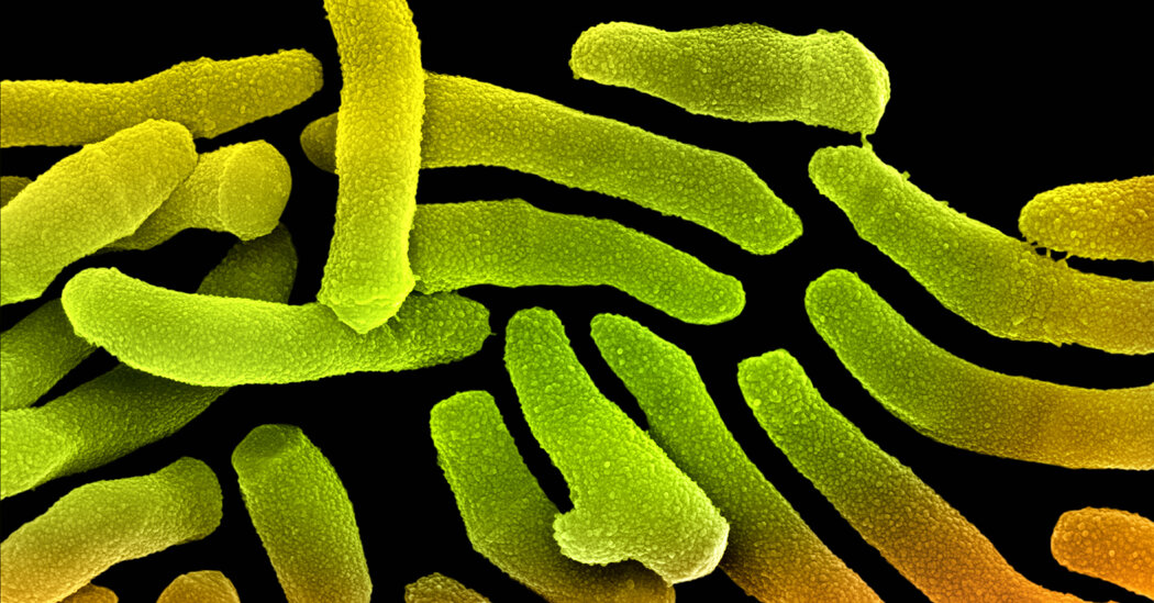
A close examination of human skin found that each pore had a single variety of bacteria living inside.Your skin is home to a thousand kinds of bacteria, and the ways they contribute to healthy skin are still largely mysterious. This mystery may be getting even more complex: In a paper published Thursday in the journal Cell Host & Microbe, researchers studying the many varieties of Cutibacterium acnes bacteria on 16 human volunteers found that each pore was a world unto itself. Every pore contained just a single type of C. acnes.C. acnes is naturally occurring, and the most abundant bacteria on skin. Its link to acne, the skin disease, is not clear, said Tami Lieberman, a professor at M.I.T. and an author of the new paper. If biologists want to unpack the relationship between your face’s inhabitants and its health, it will be an important step to understand whether varying strains of C. acnes have their own talents or niches, and how the strains are distributed across your skin.To collect their samples, Dr. Lieberman and her colleagues used commercially available nose strips and old-fashioned squeezing with a tool called a comedone extractor. They then smeared samples, each a bit like a microscopic glacial core, from within pores on Petri dishes. They did the same with samples from toothpicks rubbed across the surface of participants’ foreheads, cheeks and backs, which picked up bacteria living on the skin’s surface rather than in the pores. They allowed the bacteria to grow, then sequenced their DNA to identify them.Each person’s skin had a unique combination of strains, but what surprised the researchers most was that each pore housed a single variety of C. acnes. The pores were different from their neighbors, too — there was no clear pattern uniting the pores of the left cheek or forehead across the volunteers, for instance.What’s more, judging from the sequencing data, the bacteria within each pore were essentially identical.“There’s a huge amount of diversity over one square centimeter of your face,” said Arolyn Conwill, a postdoctoral researcher who is the study’s lead author. “But within a single one of your pores, there’s a total lack of diversity.”What the scientists think is happening is that each pore contains descendants of a single individual. Pores are deep, narrow crannies with oil-secreting glands at the bottom, Dr. Lieberman said. If a C. acnes cell manages to get down there, it may proliferate until it fills the pore with copies of itself.This would also explain why strains that don’t grow very quickly manage to avoid being outcompeted by speedier strains on the same person. They’re not competing with each other; they’re living side by side in their own walled gardens.Intriguingly, these gardens are not very old, the scientists think. They estimate that the founding cells in the pores they studied took up residence only about one year before.What happened to the bacteria that previously lived there? The researchers don’t know — perhaps they were destroyed by the immune system, fell prey to viruses or were unceremoniously yanked out by a nose strip, clearing the way for new founders.Dr. Lieberman said the finding has implications for microbiome research more broadly. Taking a simple swab of someone’s skin would never hint at the complexity uncovered in this study, for instance. And as scientists consider the possibility of manipulating our microbiomes to help treat disease, the patterns uncovered in this study imply the need for information about the location and arrangement of microbes, not just their identities. In the future, should doctors hope to replace someone’s current skin inhabitants with others, they may need to clean out their pores first.And could it be that another inhabitant on our faces plays a role in how each pore’s bacteria comes and goes?“We have mites on our faces that live in pores and eat bacteria,” Dr. Lieberman said. What role they play in this ecosystem, as far as the maintenance of gardens of C. acnes, has yet to be determined.

Read more →

Cold Tooth Pain's Mysterious Molecular Culprit

Researchers figured out how a jolt of discomfort gets from the damaged outside of your tooth to the nerves inside it.There’s nothing quite like the peculiar, bone-jarring reaction of a damaged tooth exposed to something cold: a bite of ice cream, or a cold drink, and suddenly, that sharp, searing feeling, like a needle piercing a nerve.Researchers have known for years that this phenomenon results from damage to the tooth’s protective outer layer. But just how the message goes from the outside of your tooth to the nerves within it has been difficult to uncover. On Friday, biologists report in the journal Science Advances that they have identified an unexpected player in this painful sensation: a protein embedded in the surface of cells inside the teeth. The discovery provides a glimpse of the connection between the outer world and the interior of a tooth, and could one day help guide the development of treatments for tooth pain.More than a decade ago, Dr. Katharina Zimmerman, now a professor at Friedrich-Alexander University in Germany, discovered that cells producing a protein called TRPC5 were sensitive to cold. When things got chilly, TRPC5 popped open to form a channel, allowing ions to flow across the cell’s membrane.Ion channels like TRPC5 are sprinkled throughout our bodies, Dr. Zimmerman said, and they are behind some surprisingly familiar sensations. For instance, if your eyes start to feel cold and dry in chilly air, it’s a result of an ion channel being activated in the cornea. She wondered which other parts of the body might make use of a cold receptor such as TRPC5. And it occurred to her that “the most sensitive tissue in the human body can be teeth” when it comes to cold sensations.Within the protective shell of their enamel, teeth are made of a hard substance called dentin that’s threaded with tiny tunnels. At the heart of the dentin is the tooth’s soft pulp, where nerve cells and cells called odontoblasts, which manufacture dentin, are intertwined.The prevailing theory for how teeth sense cold had been that temperature changes put pressure on the fluid in dentin’s tunnels, somehow provoking a response in those concealed nerves. But there was little detail about how exactly that could be happening and what could be bridging the gap between them.Dr. Zimmerman and her colleagues looked to see whether mice engineered to lack the TRPC5 channel still felt tooth pain as normal mice did. They were intrigued to find that these mice, when they had damage to their teeth, did not behave as if anything was amiss. They looked, in fact, about the same as if they had been given an anti-inflammatory painkiller, Dr. Zimmerman said.Her co-author Dr. Jochen Lennerz, a pathologist at Massachusetts General Hospital, checked human teeth for signs of the ion channel and found it in their nerves and other cells. That suggested that the channel might have a role in a person’s perception of cold.Over many years, the researchers constructed a way to precisely measure the nerve signals traveling out of a mouse’s damaged molar. They tested their ideas with molecules that could block the activity of various channels, including TRPC5.The picture they slowly assembled is that TRPC5 is active in the odontoblasts. That was a bit of a surprise, as these supporting cells are best known for making and maintaining dentin, not aiding in perception. Within the odontoblasts, Dr. Lennerz said, TRPC5 pops open when the signal for cold comes down the dentin tunnels, and this results in a message being sent to the nerves.As it happens, one substance that keeps TRPC5 from opening is eugenol, the main ingredient in oil of cloves, a traditional treatment for toothache. Though the Food and Drug Administration in the United States is equivocal about eugenol’s effectiveness, if it does lessen the pain for some people, it may be because of its effect on TRPC5.Perhaps the knowledge that this channel is at the heart of cold-induced pain will lead to better treatments for dental pain down the road — better ways to keep that message from becoming overwhelming.

Read more →